


What Is a Bone Density Test and Why It Matters for Your Health
Dr. Juhee Chandra
Strong bones rarely make headlines. Fractures do. A Bone Density Test is one of the few diagnostics that lets you see risk before it turns into a break. You get a precise reading, a clear comparison, and a plan. Not guesswork. In this guide, you will learn which tests exist, whether you should be screened, what your numbers mean, and how to act on them without fuss.
Types of Bone Density Tests Available
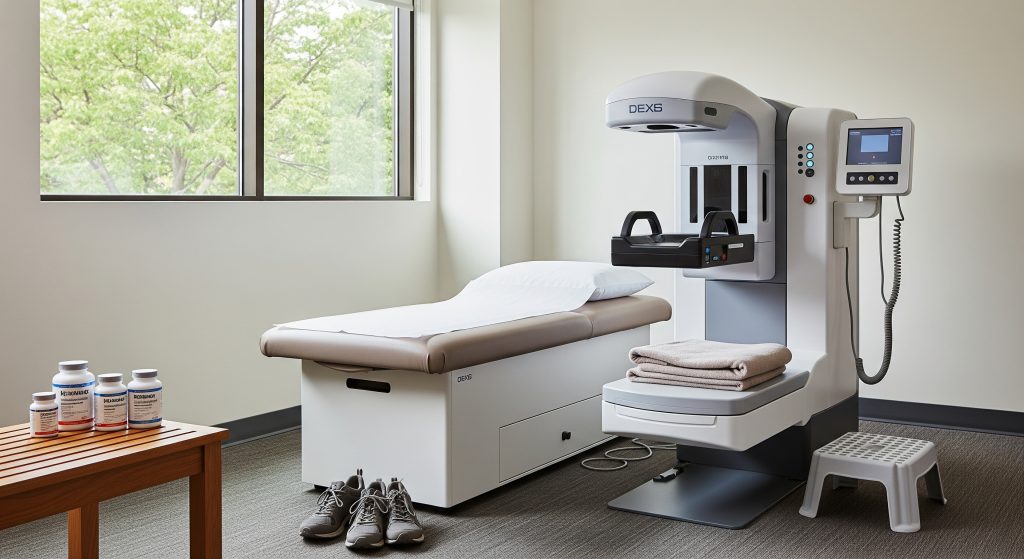
DEXA (DXA) Scan: The Gold Standard
For diagnostic clarity, a DEXA scan is the reference method. As StatPearls explains, DEXA has been FDA approved since 1988, uses two x ray beams to separate bone from soft tissue, and delivers precise bone mineral density values with low radiation. You lie still, the scanner passes over your spine and hips, and you get reliable numbers.
Why does this matter in practice? As Endotext notes, a DEXA scan both classifies osteoporosis status and sets your baseline for monitoring after treatment begins. That baseline is your anchor for future comparisons. It is basically your before photo for bones.
For many, the Bone Density Test means a DEXA of the hip and spine. This is where osteoporotic fractures cluster, and where therapy impact is clearest. As NIH highlights, DEXA helps distinguish osteopenia from osteoporosis so intervention is not delayed.
- Primary outputs: T score, Z score, site specific BMD.
- Typical duration: **10 to 30** minutes.
- Radiation: low dose, far below a CT scan.
If you want one test with minimal ambiguity, choose DEXA. It is the standard others are judged against.
Peripheral Bone Density Testing Options
When central DEXA is unavailable, peripheral devices help you triage risk. As StatPearls observes, heel or wrist measurements can predict fractures and offer access in pharmacies or community clinics. These devices do not replace central DEXA for diagnosis, but they widen the front door for screening.
The method can be practical for outreach or for first pass screening. As Endotext summarises, quantitative ultrasound and other peripheral techniques estimate risk using speed of sound and attenuation. That means you get insight into bone quality as well as density.
Research has also linked these readings with strength related properties. In a technical review, PMC reports that peripheral tools can be cost effective and, in some settings, provide fracture prediction comparable to central methods. Useful, though not definitive. Use them to decide who needs a DEXA.
- Good for triage or when access to hospital imaging is limited.
- Common sites: calcaneus, radius.
- Next step after a high risk result: book a central DEXA.
Quantitative Ultrasound (QUS) Technology
QUS is portable, relatively inexpensive, and radiation free. In a 7 year cohort, OsteoLaus found heel QUS parameters predicted major osteoporotic fractures independently of BMD and clinical risk factors. That independence matters. It shows QUS is capturing aspects of bone quality that density alone misses.
There is nuance. As Endotext cautions, QUS T scores are not interchangeable with DEXA T scores, and QUS should not be used for definitive diagnosis or treatment monitoring. It is a gatekeeper test. If it flags concern, you confirm with DEXA.
QUS continues to improve. One study showed calibration adjustments sharpen accuracy and case finding, which supports wider prescreening use in clinics and community settings, as PubMed outlines. Another analysis in people with diabetes showed strong correlation with DEXA and fracture prediction, as NIH reported.
Bottom line for your Bone Density Test strategy: use QUS where DEXA access is limited, but anchor decisions on DEXA when treatment hinges on the result.
Emerging Technologies: BIA and Optical Densitometry
Bioelectrical impedance analysis is being explored for whole body bone assessment. In a recent evaluation, Scientific Reports described BIA as convenient and fast for broad screening, though reliability in specific groups still needs validation. Consider this an adjunct, not a replacement.
Optical densitometry approaches are also under study. The attraction is speed and safety. The limitation is validation against DEXA across ages and conditions. For now, keep these as research aligned tools that might complement your Bone Density Test workflow in selected settings.
Who Should Get a Bone Density Test
Women 65 and Older: Universal Screening Guidelines
If you are 65 or older, you qualify for screening. As USPSTF states, universal screening in women at this age reduces fracture risk through early identification. The reasoning is straightforward. Bone density declines with age, and prediction improves with a DEXA reading.
This is not niche advice. As NCBI emphasises, older women carry a high baseline risk of fractures, so screening is proportionate to need. If you have not had a Bone Density Test by 65, schedule one.
Postmenopausal Women Under 65 with Risk Factors
Under 65 and postmenopausal, screening depends on risk. As STG Endocrinology advises, consider early testing if you have a previous fracture, a family history, low body weight, or take medicines that accelerate bone loss.
There is no single age cut off that fits everyone. Evidence suggests a tailored approach, as PMC notes. The practical move is to run a clinical risk assessment first, then decide on a Bone Density Test. As USPSTF outlines, risk tools are designed for exactly this task.
Men and Screening Recommendations
Men fracture too, often later, often after silent bone loss. Evidence for routine screening in all men remains mixed, but risk based screening is sensible. As StatPearls indicates, men aged 70 or younger men with risk factors like hypogonadism or long term steroids should be considered for evaluation.
Clinical advice converges around age and risk. As Mayo Clinic notes, a DEXA scan is reasonable for men over 70, or earlier if risk is high. A recent evidence review also underscores targeted screening in men with low body weight or prior fractures, as NCBI summarises.
Lifestyle and history matter. Smoking, family history, and certain medications raise risk, as Mayo Clinic reminds. If that profile matches you, a Bone Density Test is a prudent step.
Medical Conditions and Medications That Require Testing
Some conditions erode bone quietly. Think rheumatoid arthritis, hyperparathyroidism, and chronic inflammatory states. As Endotext notes, these warrant monitoring with periodic DEXA.
Medication exposures also matter. Long term glucocorticoids accelerate bone turnover and loss, increasing fracture risk, as STG Endocrinology explains. Individuals with a history of fragility fractures should be evaluated promptly, as STG Orthopaedics advises. And NCBI reiterates the need for ongoing assessment when corticosteroids are part of long term care.
If you sit in any of these categories, do not delay your Bone Density Test. Earlier detection means earlier risk reduction.
Understanding Your Bone Density Test Results
T-Score Interpretation and Classification
Your T score compares your bone density with a healthy young adult. It guides diagnosis and treatment decisions. Standard cut points are used in clinics worldwide.
|
Classification |
T score |
|
Normal |
-1.0 or higher |
|
Osteopenia |
Between -1.0 and -2.5 |
|
Osteoporosis |
-2.5 or lower |
Two quick points. Site matters, with hip and spine prioritised for clinical decisions. And change over time matters, so keep the same machine where possible for follow up comparisons.
Z-Score Meaning and Age Comparisons
Your Z score compares your bone density with peers of the same age and sex. It is valuable for spotting unusual loss patterns. As PMC explains, Z scores help flag secondary causes in premenopausal women and younger individuals.
Thresholds guide further workup rather than diagnosis. A Z score below **-2.0** suggests bone density lower than expected for your age, as MedicalNewsToday describes. That prompts investigation into endocrine, nutritional, or medication related causes. The concept is straightforward. Atypical loss deserves an explanation.
Think of T for treatment threshold and Z for zebra hunt. As Healthline puts it, Z scores focus on age matched comparisons and signal when to search for underlying conditions.
Osteopenia vs Osteoporosis: Key Differences
Osteopenia and osteoporosis sit on the same spectrum. The difference is degree and fracture risk. As NIH defines, osteopenia is a T score between -1.0 and -2.5, while osteoporosis sits at -2.5 or below. The management aim changes with the category.
The distinction is clinically meaningful. As Mayo Clinic outlines, osteopenia often calls for targeted lifestyle measures, vitamin D and calcium adequacy, and fall prevention. Osteoporosis usually requires pharmacotherapy alongside lifestyle changes.
If you have been searching osteopenia vs osteoporosis and wondering what to do next, begin with a discussion of your fracture risk. Then choose interventions proportionate to that risk.
FRAX Score and Fracture Risk Assessment
FRAX estimates your 10 year risk of a major osteoporotic fracture and of a hip fracture. It integrates clinical risk factors and can be used with or without BMD. As FRAX overviews, the tool has been widely adopted since 2008, with newer versions addressing earlier limitations.
Clinically, FRAX guides when to treat. A common threshold is a T score in the osteopenic range and a major fracture risk at or above **20 percent**, as PMC summarises. It is not perfect. It is directionally useful.
FRAX also aligns with public health priorities. As WHO notes, fragility fractures are a major burden, and risk tools help target prevention. Screening bodies echo this emphasis on assessed risk, as JAMA highlights. Regional validation studies, including Thai data, continue to refine cut offs, as Scientific Reports reports.
FRAX is a decision tool, not a verdict. Use it to calibrate action with risk.
When to Schedule Follow-Up Testing
Follow up timing depends on your baseline result and risk profile. As PubMed states, interval selection should be individualised using the initial T score and clinical risk factors. That is the safest way to track change without overscanning.
Intervals can be long if your bones are robust. As NCBI notes, people with normal baseline results may not need rescreening for up to **15** years. Men with osteoporosis should be monitored similarly to women, with individualised intervals, as IOF advises.
For many, a Bone Density Test every 2 to 3 years after starting therapy is reasonable. Longer if stable. Sooner if risk rises or results change meaningfully.
Preparing for Your Bone Density Test
What to Wear and Avoid Before Testing
Clothing and supplements can interfere with imaging. As Mayo Clinic advises, wear loose clothing without metal zips, belts, or buttons. Metal disrupts X ray paths and can trigger repeat images.
Avoid calcium supplements the day before. As Brown Health notes, hold calcium and multivitamins for 24 hours and leave jewellery at home. Skip underwire bras or any garment with metal.
Flag recent imaging with contrast. As Cleveland Clinic points out, contrast from other tests can temporarily distort readings, so a waiting period may be required.
- Bring prior DEXA reports if you have them.
- List medications and supplements, including steroids and anti epileptics.
- Arrive early to review consent and history.
The Testing Process: What to Expect
Your Bone Density Test is straightforward. As Mayo Clinic explains, X rays measure mineral content at the spine, hip, and sometimes forearm. You will lie on a padded table while the arm of the scanner moves above you.
It is painless and quick. The entire procedure typically takes **10 to 30** minutes. Radiation is low and localised. As CDC notes, DEXA is a low dose modality used specifically for osteoporosis assessment.
Afterwards, results are processed and reported with T and Z scores. Your clinician interprets them with your risk factors and, if helpful, a FRAX estimate.
Safety Considerations and Radiation Exposure
Safety standards in imaging are strict. DEXA uses very low ionising radiation. As PMC details, dose optimisation and clinical justification keep exposure minimal and appropriate, especially important for younger patients.
Facilities follow protection protocols for patients and staff. As WHO guidance outlines, adherence to safety procedures limits unnecessary exposure. Consent processes should align benefits with the small risk, as CDC also notes.
If you are pregnant or might be, tell the team in advance. They will adjust plans accordingly. Prudence first.
Finding a Bone Density Test Near Me
Access is improving across hospitals, imaging centres, and some community clinics. To locate a Bone Density Test near me, use your GP referral network, hospital radiology pages, or national osteoporosis society directories. Many centres allow self referral for screening. Confirm the site offers central DEXA for hip and spine, not only peripheral options.
- Ask about machine model and calibration practices.
- Request same machine for follow ups if possible.
- Check report turnaround time and FRAX integration.
Taking Action After Your Bone Density Test
Numbers should lead to action. Start with a clear view of your baseline, your T score category, and your calculated fracture risk. Then choose interventions sized to that risk. Simple as that, though not always easy.
Consider the practical stack:
- Nutrition and supplementation
- Ensure daily calcium intake through diet first. Supplement if short.
- Maintain adequate vitamin D, especially in winter months.
- Strength, impact, and balance
- Prioritise progressive resistance training 2 to 3 times weekly.
- Add brief impact where safe, and practise balance drills to reduce falls.
- Medication when indicated
- For osteoporosis or high FRAX risk, discuss bisphosphonates, denosumab, or anabolic agents.
- Review adherence, duration, and drug holidays with monitoring.
- Falls prevention at home
- Remove trip hazards, improve lighting, install grab rails in bathrooms.
- Check vision and footwear fit.
- Monitor and adjust
- Repeat your Bone Density Test on an appropriate interval.
- Track meaningful change, not noise. Same machine if possible.
One quick example. A 68 year old with a hip T score of -2.6 and a FRAX major fracture risk of **22 percent** starts a bisphosphonate, adds supervised strength work, and optimises vitamin D. Two years later, DEXA shows stabilisation, and no falls occurred despite a winter slip. That is the aim. Not perfection, but risk brought down to earth.
And yet, there is a caveat. Some individuals will have normal BMD and still fracture due to falls, medications, or secondary disease. Keep a holistic view. The Bone Density Test is a powerful lens, not the entire picture.
How often should I have a bone density test?
Intervals depend on your baseline and risk. With normal BMD, rescreening can stretch to **10 to 15** years. With osteopenia, consider **3 to 5** years. With osteoporosis or on treatment, repeat every **2 to 3** years, or as your clinician advises. Use the same machine when possible to reduce measurement variability.
Can a bone density test detect early osteoporosis symptoms?
Osteoporosis is often asymptomatic until a fracture. A Bone Density Test does not detect osteoporosis symptoms. It quantifies fracture risk before symptoms occur. If you experience height loss, back pain, or a low trauma fracture, seek evaluation promptly.
Is a bone density test covered by insurance?
Coverage varies by insurer and country. Many plans cover DEXA for women aged 65 and older, for men over 70, or for younger individuals with risk factors or qualifying conditions. Confirm with your insurer and your referral pathway before booking.
What’s the difference between central and peripheral bone density tests?
Central tests measure spine and hip with DEXA and underpin diagnosis and treatment decisions. Peripheral tests assess sites like the heel or wrist and help with screening. If a peripheral test is abnormal, you will usually need a central DEXA to confirm.
Can weight loss medications affect my bone density test results?
Some weight loss medicines may influence bone turnover or nutrient absorption to an extent. Rapid weight loss can also reduce mechanical loading on bone. Discuss current medications with your clinician so the Bone Density Test is interpreted in full context.
How accurate are newer bone density testing technologies compared to DEXA?
Newer methods like QUS and BIA show promise for screening and triage. DEXA remains the diagnostic reference for classification and monitoring. Use newer tools to widen access and then confirm crucial decisions with DEXA.