


Duplex Doppler Explained: Everything You Need to Know
Dr. (Prof.) Tarun Kumar
Textbook advice says imaging is either structural or functional. That split no longer holds. Duplex doppler unites crisp anatomy with real-time haemodynamics, so I can see where a vessel is and how it behaves under flow. It is basically ultrasound plus velocity data. Used well, duplex doppler prevents guesswork, speeds decisions, and reduces unnecessary invasive tests. The sections below set out what it is, what it shows, and how I interpret it in practice.
Common Uses and Applications of Duplex Doppler
Vascular Disease Detection and Diagnosis
I use duplex doppler when I need an immediate view of blood flow and vessel structure in one sitting. It shows stenoses, occlusions, and abnormal waveforms without needles or contrast. It also helps me decide urgency. A tight stenosis with a damped waveform and post-stenotic turbulence is handled quickly. Mild plaque with normal velocities is managed conservatively. Duplex doppler gives that nuance in minutes.
- Suspected arterial stenosis or occlusion after reduced pulses or exertional pain.
- Surveillance after bypass grafts or endovascular stents.
- Assessment of pseudoaneurysms and arteriovenous fistulae.
- Pre-operative vascular mapping for grafts, dialysis access, or flap planning.
The practical benefit is simple. Duplex doppler often replaces an immediate CT angiogram when the clinical question is focused and the patient is stable.
Carotid Artery Assessment
Carotid disease is common and uneven. Plaque morphology, lumen narrowing, and velocity patterns jointly matter. Carotid duplex within a comprehensive duplex doppler exam helps me stratify stroke risk and match patients to medical therapy or revascularisation. When grading disease, clinicians often apply percentage bands such as greater than 50 percent and greater than 70 percent stenosis using velocity thresholds and ratios, as NCBI Bookshelf outlines.
- Key measurements: peak systolic velocity, end-diastolic velocity, and internal-to-common carotid ratios.
- Key views: plaque surface, echogenicity, and any ulceration or heterogeneous core.
- Key outcomes: risk stratification and choice of antiplatelet therapy, statins, or referral for endarterectomy or stenting.
One practical point. I re-scan after blood pressure changes or medication adjustments because haemodynamics shift. Small shifts can alter velocity readings and apparent severity.
Peripheral Arterial Disease Evaluation
Claudication is not a diagnosis. It is a symptom with multiple vascular phenotypes. Duplex doppler localises lesions in the aorto-iliac, femoro-popliteal, or tibial segments and shows flow reserve under provocation. I combine rest waveforms with post-exercise velocities when feasible. A triphasic signal that converts to monophasic after walking suggests haemodynamically significant disease.
- Map the lesion length and severity to plan angioplasty or bypass.
- Check inflow and outflow to ensure any intervention will have run-off.
- Surveil grafts at set intervals to catch rising velocities early.
What this means is straightforward. Duplex doppler is both a diagnostic and a planning tool for lower limb disease.
Deep Vein Thrombosis Detection
Venous duplex doppler uses compression, colour, and spectral analysis to detect thrombus and its sequelae. I look for non-compressible segments, echogenic intraluminal material, and absent respiratory phasicity or augmentation. Acute thrombus looks soft and distends the vein. Chronic thrombus appears more echogenic with wall adherence and collateral channels.
- Rule in or out DVT in symptomatic patients with swelling or pain.
- Differentiate acute from chronic findings to guide anticoagulation.
- Assess post-thrombotic changes and valve competence for long-term care.
False positives are rare when technique is sound. But iliac or pelvic thrombus can be challenging, so clinical judgement remains essential.
Renal Artery Doppler Applications
Renal perfusion hinges on inflow and intrarenal resistance. I use duplex doppler to assess suspected renovascular hypertension and to surveil stented renal arteries. The study covers main renal artery velocities and intrarenal indices such as the resistive index. A tardus-parvus waveform distally with delayed systolic upstroke supports a proximal stenosis. For screening refractory hypertension, renal artery doppler is often the safest first test.
- Identify haemodynamically significant main renal artery stenosis.
- Assess intrarenal resistance in medical renal disease.
- Follow grafts after transplantation for stenosis or rejection patterns.
Duplex doppler will not solve every haemodynamic puzzle. But it narrows the differential fast and does so without nephrotoxic contrast.
Pregnancy and Foetal Monitoring
Obstetric duplex doppler focuses on uterine, umbilical, and foetal cerebral vessels. I pay attention to umbilical artery resistance, middle cerebral artery redistribution, and ductus venosus flow. These patterns clarify placental function and foetal adaptation in growth restriction or preeclampsia.
- Track umbilical artery pulsatility and any absent or reversed end-diastolic flow.
- Watch for brain sparing with reduced middle cerebral artery resistance.
- Use serial studies to time delivery when compromise is developing.
The value of duplex doppler here is temporal. Patterns over days matter more than a single reading.
Abdominal Organ Blood Flow Assessment
Duplex doppler extends beyond vessels. I use it to assess hepatic, portal, and mesenteric flow, as well as splenic and pancreatic perfusion in selected cases. Portal vein patency and direction, hepatic artery resistive index, and mesenteric velocities after meals all carry clinical weight.
- Portal hypertension: look for hepatofugal flow or collaterals.
- Mesenteric ischaemia: evaluate fasting and post-prandial velocities and symptom correlation.
- Liver transplant: confirm arterial patency and detect stenosis early.
With experience, duplex doppler becomes a versatile extension of the clinical exam. It answers targeted questions with speed and safety.
Understanding the Technology and Procedure
How Duplex Doppler Combines Two Technologies
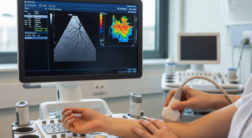
Duplex doppler merges B-mode imaging for anatomy with pulsed-wave Doppler for flow. The first component outlines vessel walls and surrounding tissue. The second measures velocity and direction using the Doppler shift. Add colour coding, and I obtain a fast visual map with quantitative spectra on demand.
- B-mode: defines structure, plaque, lumen, and surrounding landmarks.
- Colour: screens broad regions for flow presence and direction.
- Spectral: samples precise points to quantify velocity and waveform shape.
This triad is the reason duplex doppler answers both the where and the how much in one sitting.
Difference Between Duplex and Standard Doppler Ultrasound
Standard doppler ultrasound often refers to flow assessment without detailed anatomical imaging. Duplex doppler explicitly pairs the flow data with structure, and usually with colour. In practice, duplex is the comprehensive package. It improves localisation, angle correction, and repeatability.
- Standard Doppler: flow only, limited context.
- Duplex: structure plus flow with targeted quantification.
- Colour duplex: rapid screening plus precise sampling.
In clinics with mixed equipment, I request duplex doppler for any decision that depends on both anatomy and haemodynamics.
What to Expect During the Examination
The process is simple. A sonographer or clinician places gel on the skin, then moves a small probe over the target area. The screen shows grey-scale anatomy with colour overlay and audible flow signals. I ask patients to hold their breath or change position for certain samples. The exam is interactive and responsive to what the images reveal.
- Position and exposure of the area under review.
- Grey-scale survey to locate vessels and landmarks.
- Colour sweep to flag disturbed or absent flow.
- Spectral sampling with correct angle and gate size.
- Documentation with images and velocity measurements.
For most indications, duplex doppler is outpatient, quiet, and well tolerated.
Preparation Requirements for Different Scans
Preparation depends on the vascular bed. For carotids and limbs, no special steps are needed. For abdominal work, fasting reduces bowel gas and improves views. Hydration may help for pelvic or renal studies. For dialysis fistula mapping, I review access history and prior interventions before scanning.
|
Scan type |
Preparation |
|
Carotid |
No fasting. Avoid tight collars and jewellery. |
|
Lower limb arterial |
No fasting. Bring comfortable shorts for access. |
|
Abdominal aorta and mesenteric |
Fast 6 to 8 hours if possible. |
|
Renal arteries |
Fast 6 hours. Hydration as advised post-scan. |
|
Venous DVT |
No preparation. Avoid heavy compression garments before the appointment. |
|
Obstetric doppler |
Follow clinic guidance. Usually no fasting. |
Preparation is not ritual. It exists to improve acoustic windows and image quality.
Duration and Comfort Levels
Most duplex doppler studies take between twenty and forty minutes. Complex mapping or post-surgical surveillance can take longer. The probe pressure can feel firm over tender areas or when compressing veins. Pain is uncommon. If a patient is sore or short of breath, I adapt positions and sequence.
- Typical duration: single region, about half an hour.
- Multi-region or graft surveillance: longer by necessity.
- Comfort: gel is cool, pressure is moderate, noise is minimal.
It is a quiet test with real-time feedback. That reduces anxiety for many patients.
Safety Considerations and Contraindications
Ultrasound uses sound waves, not ionising radiation. Duplex doppler is considered safe in routine clinical practice. There is no nephrotoxic contrast. Contraindications are relative and practical. Open wounds, severe pain, or unstable clinical status can limit scanning. For pregnancy, obstetric protocols use conservative output settings.
- No radiation exposure.
- Minimal thermal and mechanical indices under standard settings.
- Care with fresh surgical sites, infection, or severe skin sensitivity.
In practice, the benefits of duplex doppler usually far exceed the very low procedural risk.
Interpreting Results and Clinical Significance
Normal vs Abnormal Blood Flow Patterns
Waveforms are the language of duplex doppler. Normal peripheral arteries show brisk systolic upstroke and early diastolic flow reversal at rest. Central or low-resistance beds such as the carotid or renal arteries maintain forward flow throughout diastole. Abnormal patterns include tardus-parvus waveforms downstream of a tight stenosis, blunted signals with diffuse disease, and absent diastolic flow in high-resistance states.
- Normal peripheral arterial: triphasic at rest, may become monophasic distally with disease.
- Carotid: low resistance forward diastolic flow when normal.
- Renal intraparenchymal: low resistance continuous flow when healthy.
The waveform shape often answers the question before the numbers do.
Understanding Velocity Measurements
Velocities quantify what the waveforms hint at. I pay attention to peak systolic velocity, end-diastolic velocity, and ratios between segments. A focal jump in velocity with post-stenotic turbulence signals a significant narrowing. Ratios reduce the effect of systemic haemodynamics and improve robustness.
|
Term |
Meaning |
|
PSV |
Peak systolic velocity at the sample gate. |
|
EDV |
End-diastolic velocity at the end of the cardiac cycle. |
|
Velocity ratio |
PSV at stenosis divided by PSV proximal to it. |
|
RI |
Resistive index: (PSV – EDV) / PSV for intrarenal assessment. |
Angle correction matters. I keep the Doppler angle near 60 degrees and aligned with flow for accuracy. Small deviations can skew numbers significantly.
Colour Coding and What It Means
Colour is a directional map, not a temperature scale. Red and blue show flow direction relative to the probe, with brightness reflecting velocity magnitude. Turbulence appears as colour mosaic and speckle, warning of disturbed flow. I use colour to find the hotspot, then sample precisely with spectral Doppler.
- Red vs blue indicates direction, not oxygen content.
- Brightness scales with velocity within the chosen range.
- Aliasing signals the need to adjust the velocity scale or angle.
Colour is the scout. Spectral tracing is the proof.
Common Findings and Their Implications
Most duplex doppler reports fall into recognisable patterns. Each pattern suggests a management path, though not without exceptions.
- Carotid plaque with elevated PSV and ulceration: optimise medical therapy and consider referral for revascularisation.
- Short, focal femoral stenosis with good run-off: endovascular treatment is often efficient.
- Diffuse tibial disease with low run-off: prioritise risk factor control and careful wound care.
- Acute femoral DVT: anticoagulation and symptom management with follow-up imaging.
- Renal tardus-parvus waveforms with high main artery velocities: evaluate for renovascular hypertension.
- Portal hepatofugal flow: advanced portal hypertension requiring specialist input.
The contrarian view says imaging overcalls disease. Sometimes it does. But when duplex doppler is performed methodically, it correlates well with clinical outcomes.
When Further Testing Is Needed
Duplex doppler is not always the last step. I escalate when anatomy is complex, when acoustic windows are poor, or when a therapeutic plan demands full mapping.
- CTA or MRA for comprehensive vascular roadmaps before reconstruction.
- Catheter angiography when endovascular treatment is planned.
- Perfusion or functional imaging when flow dynamics remain ambiguous.
Earlier, I noted that duplex doppler can replace CT in focused questions. That still holds. The decision depends on risk, access, and the stakes of being wrong.
Treatment Decisions Based on Results
I channel findings into action. For a carotid lesion with high-grade stenosis and unstable plaque, the referral is urgent. For mild disease, I anchor management in risk factor control and surveillance. For graft surveillance, rising velocities prompt early intervention before failure.
Pros
- Non-invasive, repeatable, and widely available.
- Combines anatomy and haemodynamics for targeted decisions.
- Supports surveillance and early detection of restenosis.
Cons
- Operator dependent and limited by acoustic windows.
- Challenging in obese patients or with heavy calcification.
- May under-visualise deep pelvic or thoracic segments.
Maybe that is the point. Use duplex doppler when the question suits its strengths, and it will serve better than any single-modality test.
Making Sense of Duplex Doppler Technology
Duplex doppler is a practical synthesis. It gives me a real-time conversation with the circulation, not a static picture. The core skill is pattern recognition backed by disciplined measurement. I watch the waveforms, sample attentively, and keep the clinical question front and centre.
Think anatomy, then flow, then context. In that order, every time.
Two brief examples illustrate the workflow. First, a smoker with calf pain and diminished pulses. Duplex doppler shows focal superficial femoral stenosis with a velocity jump and distal turbulence. The plan is targeted angioplasty with surveillance. Second, a patient with resistant hypertension and deteriorating renal function. Duplex doppler shows tardus-parvus intrarenal waveforms and high main renal artery velocities. The plan is confirmatory imaging and, if appropriate, revascularisation.
In clinical operations, I lean on two pieces of insider jargon. First, the PSV ratio or simply the ratio. It stabilises interpretation across blood pressure swings. Second, the RI in intrarenal work. It captures resistance trends in a single number, which is useful for serial assessment. These are not academic ornaments. They shorten time to decision and improve consistency.
There is one more reason duplex doppler endures. It is iterative and safe, and it is fast. I can scan and rescan, and correlate with symptoms, and adjust therapy, and then see the response. That feedback loop is rare in imaging and valuable in everyday care.
Is duplex doppler painful or uncomfortable?
No. Duplex doppler uses a small probe and ultrasound gel. Pressure can feel firm over tender spots, especially during venous compression. The experience is generally comfortable, and any discomfort is brief.
How long does a duplex doppler scan typically take?
Most studies are completed in about thirty minutes. Complex mapping, multilevel arterial disease, or graft surveillance can take longer. The timing depends on the question, the anatomy, and how many segments require detailed sampling.
Can duplex doppler detect all types of vascular problems?
Not all. It excels in accessible arteries and veins and in graft or stent surveillance. Deep thoracic or pelvic vessels can be challenging. When anatomy is complex or the treatment plan is interventional, I add CTA, MRA, or catheter angiography.
What’s the difference between duplex doppler and colour doppler?
Colour is a component, not a separate test. Colour shows flow direction and relative speed across an area. Duplex doppler adds grey-scale anatomy and spectral waveforms for precise velocity measurements. In practice, colour duplex is the full package used in most vascular studies.
How accurate is duplex doppler for diagnosing blood clots?
Venous duplex is highly sensitive for femoral and popliteal DVT when performed with proper technique. Sensitivity is lower in iliac and pelvic segments due to depth and bowel gas. Clinical correlation and, at times, additional imaging are prudent.
Do I need special preparation for a renal artery doppler scan?
Yes. Fasting for about six hours is preferred to reduce bowel gas. This improves the acoustic window and the accuracy of peak velocity measurements. Hydration guidance after the scan varies by clinic, so follow the appointment letter.