


Coronary Angiography Procedure: Preparation, Steps, and Recovery
Dr. (Prof.) Tarun Kumar
Common advice claims that heart tests are either quick or complex. The reality is more nuanced. A coronary angiography procedure is structured, methodical, and safe in experienced hands, yet it deserves precise planning and calm execution. In this guide, I outline what actually happens, how I prepare patients, what to expect in recovery, and where angiography sits next to angioplasty. No drama. Just clarity a smart colleague would want.
What Happens During Coronary Angiography
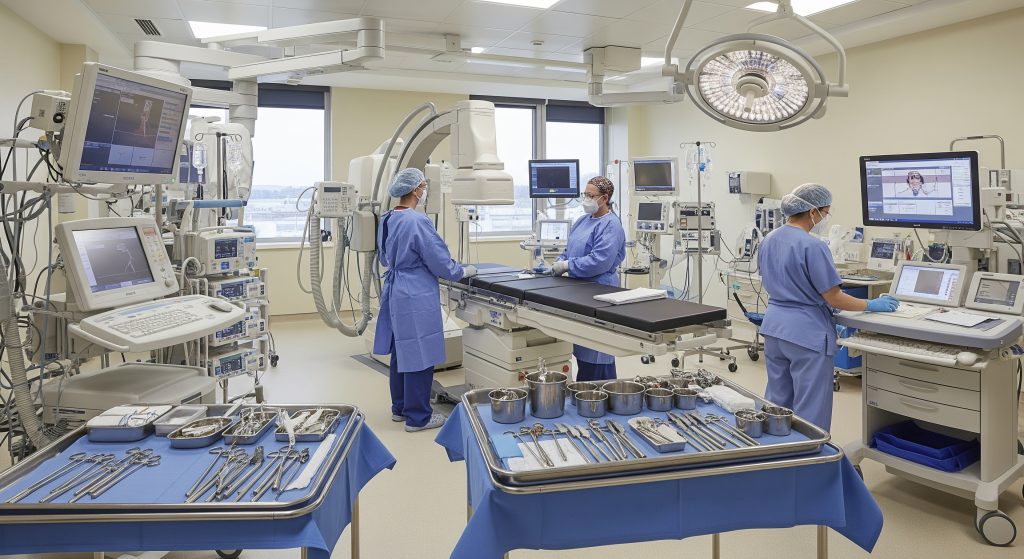
Pre-Procedure Room Setup
I confirm the procedure plan, cross-check allergies, and review renal function and clotting status. The team prepares the sterile field, ultrasound or palpation guides for access, and emergency-readiness checks. Sedation and local anaesthesia are standard. They help patients remain awake yet settled, which preserves communication and safety. I also check catheter sizes, contrast type, and dose planning. Small disciplines, significant outcomes.
- Identity, consent, and site marking verified before entry.
- Monitoring lines placed for heart rhythm, blood pressure, and oxygen saturation.
- Resuscitation equipment and contrast extravasation kits confirmed.
This is not mere ritual. It is how risk stays low and predictability stays high.
1. Local Anaesthesia Administration
I infiltrate local anaesthetic at the access site, usually the wrist or groin. The goal is targeted numbness without motor impairment. I warn about brief stinging, then pressure. Effective analgesia reduces sympathetic responses and improves procedural control. If anxiety runs high, light sedation follows. Nothing excessive. Just enough to maintain comfort and cooperation.
2. Catheter Insertion Through Femoral or Radial Artery
Access is through the radial artery at the wrist or the femoral artery in the groin. I assess pulses, vessel calibre, and prior surgery or grafts. For radial access, I ensure adequate ulnar collateral flow with a quick bedside assessment. The sheath is inserted, flushed, and secured. I advance a guidewire under fluoroscopy, then introduce the diagnostic catheter.
Choice matters. As NCBI reports, transradial access is associated with lower access site complication rates compared with femoral routes. That is why wrist access has become common in routine diagnostic cases. Yet femoral access remains valuable for complex anatomies, larger devices, or when radial pulses are weak.
- Radial access: faster mobilisation and typically less bleeding.
- Femoral access: accommodates larger catheters and complex equipment.
Technique should follow the patient’s anatomy, not a trend.
3. Navigation to Coronary Arteries
I move the catheter to the aortic root and engage the coronary ostia under X-ray guidance. Catheter selection matters. Judkins catheters for most anatomies, Amplatz if the take-off is uncommon. Small torque, small breaths. I watch pressure tracings to avoid damping and prevent ostial trauma. A calm table helps. So does concise communication.
4. Contrast Dye Injection Process
Once seated, I inject iodinated contrast in short bursts while recording. I aim for complete opacification with minimal volume. In patients with renal risk, I track cumulative dose closely and simplify angles. Hydration plans start before the first millilitre. If allergies exist, I use premedication or non-ionic low-osmolality agents. The objective is high diagnostic yield with low physiological burden.
5. X-Ray Imaging and Recording
I record cineangiography in multiple projections to reveal stenoses that may hide in a single view. Cranial and caudal angulations add depth. I minimise radiation time and collimation width. Radiation safety is a discipline, not an afterthought. Every second trimmed matters to staff and patient. I archive images for the multidisciplinary team review and for patients who request copies later.
Duration and Monitoring Throughout
A straightforward coronary angiography procedure typically takes 15 to 45 minutes, depending on access and anatomy. Continuous ECG, oxygen saturation, and non-invasive blood pressure monitoring run throughout. I reassess symptoms in real time. Chest pressure, breathlessness, or sudden hypotension prompt immediate review and manoeuvres. Most cases are wholly uneventful. That is the aim.
Immediate Post-Procedure Assessment
Before removing the catheter, I confirm haemostasis strategy and review the images for completeness. I check the access site, distal pulses, and neurological status in the hand if radial. I calculate contrast volume against renal function and plan hydration or observation accordingly. If results are straightforward, I discuss them briefly in recovery and schedule a detailed consult after full reports are ready. Patients leave the lab informed, not guessing.
Preparation for Cardiac Catheterisation
Medical History Review and Risk Assessment
I review comorbidities, prior strokes or bleeding, kidney disease, and diabetes control. I assess anticoagulants, antiplatelets, and any prior contrast reactions. Symptoms and functional capacity guide pre-test probability and urgency. Risk is never zero. But it can be predicted and reduced with methodical screening.
Required Pre-Procedure Tests
- Blood tests: full blood count, renal profile, coagulation screen.
- ECG and, when indicated, chest X-ray.
- Pregnancy testing where relevant.
If there is heart failure, I review recent echocardiography for left ventricular function. For complex disease or grafts, I study prior reports to anticipate catheter choice. Preparation here saves minutes there.
Medication Adjustments Before Angiography
Antiplatelets usually continue for a diagnostic case. Anticoagulation strategy is individualised. For warfarin, I plan around the INR. For DOACs, a brief pause may be appropriate depending on renal function and bleeding risk. Metformin may be held around the test if renal function is borderline. I confirm allergy premedication protocols where needed. No guesswork, no surprises.
Fasting Requirements and Dietary Restrictions
Clear liquids are usually allowed up to a few hours before, then nothing by mouth for a short window. Policies vary by unit. I prioritise hydration if kidneys are vulnerable. Alcohol is avoided the day before. The fasting plan should balance safety with comfort. Starving patients benefits nobody.
What to Bring on Procedure Day
- Medication list and doses, including over-the-counter products.
- Allergy card and prior stent or surgery information.
- Comfortable clothing and a phone charger.
- Escort information if same-day discharge is planned.
Small practicalities reduce delays and lower stress. It is basically routine risk management.
Consent Process and Questions to Ask
I cover purpose, benefits, alternatives, and reasonably foreseeable risks in plain terms. I invite questions and pause for reflection. Useful prompts include:
- What are the chances this test changes management?
- How will access choice affect recovery today?
- What is the plan if a severe narrowing is found?
Consent is a process, not a signature. It respects autonomy and improves outcomes to an extent.
Immediate Recovery Room Care
On arrival in recovery, I confirm vital signs, access site status, and comfort level. I provide water and light snacks when safe. Written instructions are given before discharge. Clarity matters, especially after sedation. I also ensure a contact pathway for queries at home.
Pressure Application and Bleeding Prevention
Radial access uses a wrist compression device with gradual deflation to maintain patent haemostasis. Femoral access requires longer manual or device-assisted compression and flat bed rest. The aim is haemostasis without occluding the vessel. Good technique reduces bruising and re-bleeding. Precision here avoids an avoidable complication.
Monitoring Vital Signs Post-Procedure
Nursing staff record pulse, blood pressure, and oxygen saturation at regular intervals. I check distal limb perfusion and sensation, plus wrist or groin discomfort. If there is sudden swelling, a firm lump, or pain radiating, I reassess immediately. Early detection prevents escalation. Monitoring is watchful, not intrusive.
When to Resume Normal Activities
|
Activity |
Typical timeframe |
|
Light walking |
Same day with radial access, next day with femoral |
|
Showering |
Usually after 24 hours if dressing stays dry |
|
Driving |
Generally 24 to 48 hours if no intervention was performed |
|
Heavy lifting |
Avoid for 3 to 5 days, especially after femoral access |
These are typical ranges. I tailor advice for work demands, comorbidities, and any access site issues.
Warning Signs to Watch For
- Rapid swelling, persistent bleeding, or a growing bruise at the access site.
- Hand numbness or coldness after radial access.
- Fever, chest pain, or shortness of breath.
- Allergic symptoms such as widespread rash or facial swelling.
If any of these occur, urgent assessment is indicated. Better an early call than a late complication.
Follow-Up Appointments and Results Discussion
I schedule a focused follow-up to review images and the written report. I show the cine loops and explain the functional meaning of each narrowing. If physiology was measured, I explain the values in plain language. Then we discuss treatment choices. Data informs decisions, but values shape priorities.
Long-Term Care Recommendations
Long-term care is twofold. First, secondary prevention: statins, antiplatelets if indicated, blood pressure control, diabetes optimisation, and smoking cessation. Second, lifestyle and rehabilitation: nutrition patterns, supervised exercise, and stress management. I emphasise medication adherence. It is unglamorous and vital. Heart health is a habit, not a headline.
Angiography vs Angioplasty Understanding
Key Differences Between Procedures
The coronary angiography procedure is diagnostic. It maps coronary anatomy and quantifies disease. Angioplasty is therapeutic. It opens a narrowed artery, often with a stent. One shows the problem, the other treats it. The two can occur in the same sitting, but their purposes differ. That distinction focuses decision making and consent.
- Angiography: imaging with contrast and X-ray.
- Angioplasty: balloon dilation with or without stent placement.
When people search for angiography vs angioplasty, they are often weighing timing, risks, and recovery. The right answer depends on the lesion, symptoms, and clinical stability.
When Angiography Becomes Angioplasty
If a severe, culprit lesion is found and consent has been secured, I may proceed directly to angioplasty. This avoids a second vascular puncture and shortens time to relief. In multi-vessel disease or left main disease, a heart team discussion may be preferable. It is not about speed. It is about the right intervention for the right artery.
Treatment Options After Diagnostic Results
- Optimised medical therapy only, if lesions are mild or non-obstructive.
- Angioplasty with stenting for focal, haemodynamically significant disease.
- Coronary artery bypass grafting for complex multi-vessel or left main disease.
Physiology measures, imaging quality, and symptoms guide the choice. And yet, patient preference remains central. Numbers inform. People decide.
Moving Forward After Coronary Angiography
After a straightforward coronary angiography procedure, I focus on practical recovery and long-term prevention. I ensure a written plan covers medicines, activities, and red flags. I recommend a staged return to work with clear limits for the first week, especially after femoral access. If a stent was placed, I emphasise adherence to dual antiplatelet therapy and follow-up timing. A few simple habits underpin success: take medicines, keep moving, eat thoughtfully, and sleep well.
For those wanting visual context, an angiography procedure video can help demystify positioning, contrast injection, and image capture. It will not replace a consultation, but it can reduce uncertainty before the day. If a colleague asks for a succinct takeaway, I offer this: plan carefully, choose access wisely, minimise contrast and radiation, and communicate clearly. Good practice looks simple on the outside. That is the point.
How painful is coronary angiography procedure?
Discomfort is usually limited to the local anaesthetic sting and a feeling of pressure at the access site. Most patients describe the coronary angiography procedure as tolerable rather than painful. Sedation is light and preserves awareness. If pain arises, I address it immediately. Comfort supports safety and image quality.
Can I drive myself home after cardiac catheterisation?
No. Sedation and access site care make solo driving unsafe on the day. Arrange an escort. Driving typically resumes within 24 to 48 hours after a diagnostic coronary angiography procedure, provided there are no complications. Insurance and local guidance may also apply.
What are the risks of coronary angiography?
Risks include bleeding, bruising, vessel spasm, allergic reactions, kidney strain from contrast, and rarely stroke or heart attack. Access-related bleeding is less common with wrist access. As NCBI notes, radial access has lower access site complication rates than femoral in many cohorts. Absolute risk remains low in experienced centres.
How long does complete recovery take?
Many feel normal the next day after radial access. Femoral access may require one to two days of reduced activity. Full recovery from a diagnostic coronary angiography procedure usually occurs within a few days. If stents are placed, activity limits and medicines expand, but most return to routine quickly.
Will I need sedation during the procedure?
Yes, light sedation is typical. I aim for awake cooperation and comfort. Deep sedation is rarely required for a diagnostic coronary angiography procedure. The benefit is fast recovery and a lower risk profile. Clear communication reduces the need for higher doses.
Can angiography and angioplasty be done together?
Yes, if a significant narrowing is identified and consent includes possible treatment, I can proceed in the same sitting. This avoids a second arterial puncture and often shortens symptom duration. Clinical stability and lesion complexity guide the choice.
What happens if blockages are found during angiography?
I discuss the findings and recommend one of three paths: optimised medicines, angioplasty, or bypass surgery. Severity, location, symptoms, and physiology measurements shape the plan. When questions arise, an angiography procedure video can help illustrate the steps involved in the chosen strategy. Decisions are shared, not imposed.
Quick Reference: What to Expect
- Purpose: The coronary angiography procedure maps coronary arteries and grades stenoses.
- Access: Wrist or groin, selected to match anatomy and safety profile.
- Time: Often under an hour for diagnostic studies.
- Recovery: Same day for radial access in many cases.
1 clear plan, 2 safe hands, and the right access choice often decide outcomes. Simple. Effective.