


Knee Cartilage Explained: Causes, Symptoms and Treatments
Dr. Rajeev K Sharma
Stretching and strengthening are often presented as the whole answer to knee problems. That view is incomplete. The fabric that lets the joint glide is knee cartilage, and when it frays, strength alone cannot solve the mechanical and biological issues. I will set out the types of knee cartilage, why it fails, how to recognise damage early, and the options that genuinely help. The aim is straightforward. Clear guidance so decisions are informed, measured, and timely.
Types and Common Causes of Knee Cartilage Damage
Articular Cartilage vs Meniscus
In the knee, two cartilage structures share the workload but serve different roles. Articular cartilage is the smooth lining on the ends of the femur, tibia, and the back of the patella. It minimises friction and distributes load as the joint moves. The meniscus is a C shaped piece of fibrocartilage that acts as a spacer and a shock absorber between femur and tibia. Both matter for stability and force transfer. When either fails, movement becomes painful and less predictable.
Meniscal injury is common with twisting or pivoting movements during sport. A knee cartilage tear of the meniscus often starts with a catch or a pop, then swelling. Articular cartilage damage is quieter at the start. It often presents as stiffness after sitting, or a dull ache with stairs. Over time, roughened surfaces increase friction, and the joint sends pain as a warning. Different tissue, different failure modes, different recovery paths.
-
Articular cartilage: hyaline cartilage, low friction, poor blood supply, limited healing capacity.
-
Meniscus: fibrocartilage, shock absorber, contributes to joint stability and load sharing.
-
Clinical implication: accurate identification guides treatment and prognosis.
1. Trauma and Sports Injuries
Sudden knee valgus, forced rotation, or a direct blow can shear or crush knee cartilage. Football, basketball, and hockey feature frequent pivoting at speed. In those settings, the combination of torque and compression is unforgiving. I see two patterns. Acute focal lesions after a twist or tackle. Or a repeat microtrauma pattern that gradually undermines the surface. The first is dramatic and memorable. The second creeps up and is easy to dismiss as training soreness.
High energy trauma such as road collisions or falls can damage multiple structures at once. Bone bruising, ligament sprain, and knee cartilage damage may overlap. The risk is compounding instability if a meniscal tear is missed. In practice, I prioritise swelling pattern, range loss, and mechanical symptoms to triage imaging. Early load management protects the joint from secondary harm. Speed matters, but so does restraint.
2. Age-Related Wear and Tear
Cartilage thins with age due to cumulative load and a slower repair environment. Chondrocytes reduce matrix production and water content shifts within the tissue. The joint becomes less forgiving under uneven loads. Daily tasks like stairs, squatting, or rising from a chair become testing. Not overnight. Gradually and then more obviously after a flare. Many patients report a rainy day ache. It is a cliché because it is common.
Degenerative change does not mean inevitable decline. Load shaping and muscle conditioning protect the joint. I focus on strength in the quadriceps and gluteal groups, movement quality, and body mass control. The aim is to reduce peak loads and smooth movement. Small changes, accumulated. That is how wear slows.
3. Obesity and Joint Stress
Excess body mass increases compressive forces on the tibiofemoral joint. It also raises inflammatory signalling within the joint environment. Adipose tissue is metabolically active and can amplify cytokines that degrade cartilage. This is a mechanical and a biochemical problem. Both matter. In the clinic, I link weight management with symptom targets, not abstract goals. Patients commit when progress is felt, not only read on scales.
Weight control is a modifiable risk factor for knee osteoarthritis progression. As WHO notes, greater body mass increases joint loading and the likelihood of symptomatic disease. A modest sustained reduction changes forces with every step. It also improves the chance that exercise therapy can be performed without flare ups. The signal is clear enough for me. Weight work sits alongside strengthening from day one.
4. Genetic and Metabolic Factors
Family history can raise susceptibility to cartilage degeneration. So can metabolic issues that affect matrix quality, such as poorly controlled diabetes or lipid disorders. These influences do not determine outcome alone. They shift the baseline, then environment and behaviour do the rest. I ask about family joint history and metabolic health as routine. The history shapes the threshold for imaging and the cadence of follow up.
Risk Factors by Age Group
Risk shifts across a lifetime. In adolescents and young adults, high intensity sport, rapid growth, and alignment issues are common drivers. In midlife, workload and body mass compete with recovery time. After 60, degenerative change and balance deficits elevate risk. As NIH summarises, osteoarthritis incidence rises with age and may affect up to 50% of individuals over 65. That is a large cohort and a clear planning signal.
Regional data vary. In some Asian cohorts, reported prevalence ranges widely between 20.5% and 68.0%, as EMRO reports. Methodology and definitions differ by study, but the direction of travel is consistent. Age and body mass are the dominant levers. That informs prevention and resource allocation. It also sets realistic expectations for recovery timelines in older adults.
Recognising Symptoms and Getting Diagnosed
Early Warning Signs
Early signs are often subtle. A dull ache around the patella after sitting. Puffiness around the joint line in the evening. A sense that the knee lags behind the other when climbing stairs. As SRM Global Hospitals notes in its advisory, persistent pain, swelling, and reduced ease of movement are typical early flags, and ignoring them risks escalation, as SRM Global Hospitals warns. I listen for catching, grinding, or a brief lock. These clues often separate a simple strain from a cartilage issue.
-
Morning stiffness that eases within 30 minutes suggests mechanical irritation rather than severe inflammation.
-
Sharp pain with pivoting suggests a meniscal component.
-
Diffuse ache after load suggests articular surface irritation.
Progressive Symptoms of Knee Cartilage Tear
Untreated lesions tend to escalate. The joint may swell faster after activity. Range reduces, particularly deep flexion. Stairs become a negotiation. A knee cartilage tear in the meniscus may produce episodic locking or giving way. Articular lesions give grating and delayed swelling. There is a cadence to the deterioration. Load, ache, stiffness, then a longer flare. Recognition at this stage can still prevent chronic cycles.
When to See a Doctor
Seek review promptly if the knee locks, gives way, or cannot bear weight after a twist. Sudden swelling within two hours suggests internal bleeding and deserves urgent assessment. Persistent swelling beyond three days also warrants evaluation. If self management fails after two weeks of rest, gentle mobility, and simple analgesia, book an appointment. Early triage prevents secondary joint stress and compensatory gait issues. Better a cautious review than a missed repair window.
Diagnostic Tests and Imaging
Assessment starts with history and physical examination. I test gait, alignment, effusion, joint line tenderness, and mechanical signs such as McMurray and Thessaly. Plain radiographs rule out fracture and reveal joint space narrowing in later disease. MRI visualises articular cartilage and meniscal integrity. Ultrasound helps with effusions and superficial structures. Imaging answers specific questions. It is not a substitute for a careful examination. Choose the scan that will change the plan.
Grading Cartilage Damage Severity
Severity grading helps match treatment to the lesion. Two common systems appear in clinic. The Outerbridge scale and the International Cartilage Repair Society scale. Both categorise surface changes from softening to full thickness loss with exposed bone. Here is a concise reference.
|
Grade |
Description |
|---|---|
|
Grade 1 |
Softening or swelling of cartilage with intact surface. |
|
Grade 2 |
Partial thickness defect. Fissures or fraying less than 50 percent depth. |
|
Grade 3 |
Deep fissures or flap tears greater than 50 percent depth. No exposed bone. |
|
Grade 4 |
Full thickness loss with exposed subchondral bone. |
Meniscal tears are described by pattern and position. Horizontal, vertical, radial, or complex. Anterior horn, body, or posterior horn. Pattern and location predict repairability. Language matters because it shapes the operative plan.
Treatment Options for Knee Cartilage Problems
Conservative Treatment Methods
Most cases start with non operative care. I set a structured plan focused on pain control, swelling reduction, load shaping, and progressive strength. The initial steps are calm and consistent. Rest from aggravating activities, not full rest. Ice or heat based on comfort. Short course analgesia if tolerated. Then movement begins again in a controlled fashion.
-
Activity modification: reduce impact, avoid deep squats and pivots for a defined period.
-
Support: offloading with a cane on the opposite side or a hinged brace as needed.
-
Footwear: stable shoes with cushioning to smooth load through stance.
-
Education: symptom led progression with simple thresholds for flare handling.
Physical Therapy Exercises
A precise programme is the core of conservative care. I emphasise quality of motion over volume. Start with range and control. Then develop strength and endurance. The sequence matters because form protects the joint.
-
Range and mobility
-
Heel slides and stationary cycling with low resistance to nourish knee cartilage through motion.
-
Calf and hamstring stretches to reduce posterior chain tension.
-
-
Neuromuscular control
-
Terminal knee extension with a band to engage quadriceps without joint shear.
-
Step downs from a low box, focusing on alignment over the foot.
-
-
Strength and conditioning
-
Leg press within a safe arc, slow tempo.
-
Hip abductor and extensor work to offload the knee in gait.
-
-
Progressive return to impact
-
Rowing or elliptical, then graduated jogging if symptom stable.
-
I use the term ROM for range of motion and cue it weekly. Gains are small at first. Then the graph bends up. That is the usual pattern with disciplined work.
Medications and Injections
Analgesia should be minimal effective dose. Paracetamol or short course NSAIDs if tolerated. Topical NSAIDs are useful for flares and reduce systemic exposure. For persistent synovitis, corticosteroid injection can reset inflammation and create a window for physiotherapy. Hyaluronic acid injections may improve lubrication for some patients. The evidence is mixed, and selection is key. I reserve them for specific profiles after discussion. Clarity about expected benefit prevents disappointment.
Arthroscopic Procedures
Arthroscopy assists with diagnosis and selected interventions. For meniscal tears, partial meniscectomy removes unstable flaps that catch. Meniscal repair preserves tissue when tear pattern and vascular zone permit. For articular lesions, arthroscopy enables debridement of loose fragments and assessment of defect size. I avoid clean up procedures without a clear mechanical target. Scope for scope’s sake does not serve the joint. Precision first.
Types of Knee Cartilage Surgery
Surgical options depend on defect size, location, depth, and patient factors. The menu is wide, but the logic is simple. Stabilise, replace, or stimulate repair. The choice follows the biology of the tissue and the load the joint must carry. Here is a quick comparison.
|
Procedure |
Best For |
Notes |
|---|---|---|
|
Meniscal repair |
Peripheral tears in vascular zone |
Preserves function. Longer protection phase. |
|
Partial meniscectomy |
Unrepairable flap tears |
Fast recovery. Potential later load increase. |
|
Microfracture |
Small full thickness defects |
Fibrocartilage fill. Strict rehab needed. |
|
Osteochondral autograft transfer |
Small to medium focal defects |
Hyaline like plug from non weight area. |
|
Osteochondral allograft |
Larger defects |
Size matched graft. Availability dependent. |
|
Autologous chondrocyte implantation |
Medium to large defects |
Two stage, cell based repair. |
I discuss knee cartilage surgery when symptoms remain intrusive despite structured conservative therapy. Shared decision making is essential. The goal is durable function, not a quick fix that fades in six months.
Microfracture and Drilling Techniques
Microfracture uses controlled perforations in the subchondral bone to release marrow elements. The clot matures into fibrocartilage, which is less robust than native hyaline cartilage. It can still provide meaningful relief for small defects. Success relies on strict protection and progressive loading during rehab. Drilling is a related technique with similar biology. Size, alignment, and compliance determine outcomes more than surgical theatre alone.
Cartilage Transplant Options
Osteochondral autograft transfer moves small plugs of bone and cartilage from a low load area to the defect. It provides immediate structural fill with hyaline like tissue. The trade off is donor site morbidity and size limits. Allograft transfers tissue from a donor source to reconstruct larger defects. Autologous chondrocyte implantation uses the patient’s cells expanded in a lab and re implanted under a membrane or matrix. This is precise work and best suited to focal defects in otherwise healthy knees. Proper selection is everything.
Post-Surgery Recovery Timeline
Recovery varies by procedure and lesion site. I plan rehab in phases. Protection, reloading, strength, then return to sport or occupational demand. The following timeline is indicative rather than prescriptive.
-
Weeks 0 to 2: protect repair, control pain, reduce swelling, gentle ROM within limits.
-
Weeks 3 to 6: increase ROM, begin closed chain strength, cautious weight bearing progression if allowed.
-
Weeks 7 to 12: build strength and endurance, balance work, introduce low impact cardio.
-
Months 4 to 6: sport specific drills if symptom free and strength symmetric.
-
Beyond 6 months: graded return to full demand once clinical and functional criteria are met.
Milestones trump dates. Swelling, pain scores, strength symmetry, and movement quality guide progression. That approach lowers re injury risk and improves long term confidence.
Knee Cartilage Regeneration and Long-Term Management
Natural Healing Capabilities
Articular cartilage has limited intrinsic healing due to sparse blood supply and low cell turnover. Small superficial lesions may smooth, but they rarely regenerate normal tissue. Meniscus healing depends on tear location. The outer third has better blood supply and a higher repair chance. I set expectations clearly. Improvement is likely. Full biological restoration is less likely except with specific surgical strategies. Hope is essential. Precision is kinder.
Stem Cell Therapy Options
Stem cell based approaches aim to enhance repair by adding progenitor cells to the lesion environment. Sources include bone marrow aspirate concentrate and adipose derived preparations. Evidence continues to evolve. Some patients report improved pain and function, particularly when combined with structured rehab. These therapies are not a universal cure. Indications should be narrow, consent must be thorough, and outcome measures clear. Knee cartilage regeneration is a direction, not a switch.
Platelet-Rich Plasma Treatment
Platelet rich plasma concentrates growth factors from the patient’s blood. The goal is to reduce inflammation and support local repair processes. Protocols vary by preparation and dosing intervals. Results are most promising in earlier degenerative change and as an adjunct to physiotherapy. Selection matters. I do not promise what a modality cannot deliver. I frame PRP as a potential amplifier of the basics, not a replacement.
Dietary Supplements for Cartilage Health
Glucosamine, chondroitin, collagen peptides, and curcumin are commonly used. Responses vary widely. Some patients perceive meaningful benefit. Others do not. I advise a defined trial period with a clear stop date if no change is seen. Omega 3 intake supports general anti inflammatory balance. The fundamentals apply regardless. A balanced diet with adequate protein supports tissue repair and training adaptation.
Lifestyle Modifications
Lifestyle is the quiet force behind joint outcomes. Sleep quality influences pain thresholds and recovery. Smoking impairs microvascular function and healing. Daily movement lubricates the joint and sustains muscle tone. I anchor plans around two elements. Strength sessions twice weekly and consistent low impact cardio, such as cycling or brisk walking. Small, regular inputs beat occasional heroic efforts.
Prevention Strategies
Prevention is specific. Generic advice rarely sticks. I use a practical checklist and revisit it each quarter.
-
Maintain body mass in a healthy range to reduce peak joint loads.
-
Build lower limb strength, especially quadriceps and hip abductors, to stabilise knee tracking.
-
Train movement patterns. Land softly, align knee over second toe, avoid dynamic valgus.
-
Rotate activities. Mix running with cycling or swimming to distribute stress.
-
Update footwear when midsoles compress and lose support.
-
Address workplace ergonomics if standing or kneeling is prolonged.
These actions are simple. They are not easy. Consistency is the lever that moves outcomes.
Managing Chronic Cartilage Issues
Chronic symptoms require a long view and a calm plan. I establish baselines, agree activity ceilings, and define flare rules. Pacing replaces boom and bust patterns. Strength remains central because muscle is protective scaffolding. Analgesia is strategic rather than constant. Bracing or orthoses can assist select cases with alignment issues. Review schedules are proactive. Do not wait for a crisis to recalibrate the plan.
Taking Control of Your Knee Cartilage Health
Effective management starts with clarity. Identify the structure involved, confirm severity, then match treatment to the biology and the demands of daily life. Set measurable goals. Pain at rest, swelling days per month, stairs without hesitation, return to cycling distance. Monitor progress and adapt. If improvement stalls despite disciplined work, escalate evaluation. That may mean imaging or discussing knee cartilage surgery, or it may mean refining physiotherapy. Both paths are valid at the right time.
Control is rarely about a single intervention. It is about the sum of actions across weeks and months. Strength and skill and weight and rest and patience. The knee rewards steady inputs. And yet, setbacks occur. Use them as data, not judgment. The joint is resilient when given a fair environment to heal and adapt.
Frequently Asked Questions
Can knee cartilage grow back naturally?
Articular cartilage has limited natural regenerative capacity. Small superficial defects may smooth over time, but full structural restoration is uncommon. Meniscal tissue can heal if a tear sits in the outer vascular zone. Rehabilitation can improve symptoms and joint function even when tissue does not fully regrow.
How long does knee cartilage surgery recovery take?
Timeframes vary by procedure and lesion size. A partial meniscectomy often allows return to light activity within 2 to 4 weeks. Meniscal repair and cartilage restoration procedures typically require several months before higher impact tasks. I use milestones such as pain control, swelling resolution, and strength symmetry to guide progression safely.
What are the first signs of knee cartilage damage?
Early indicators include a dull ache with stairs or after sitting, mild swelling, and stiffness that fades with gentle movement. Mechanical signs such as catching or brief locking suggest a meniscal tear. If symptoms persist beyond two weeks or escalate after a twist, arrange a clinical review.
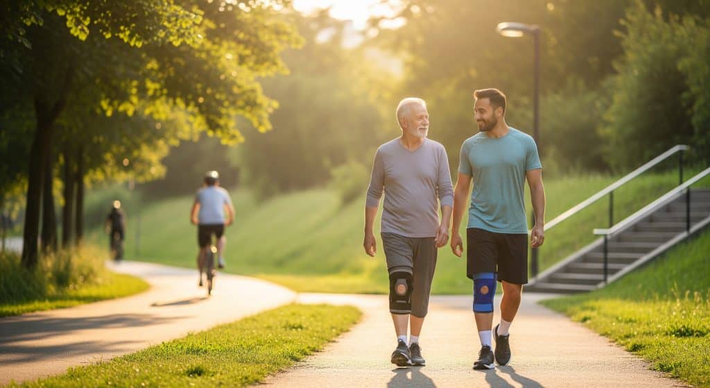
Is walking good for knee cartilage repair?
Yes, controlled walking is beneficial for most patients. It nourishes knee cartilage through cyclical loading and supports conditioning. Start with flat, even surfaces and short durations. Increase time and pace gradually as long as swelling and pain remain stable the next day.
What foods help rebuild knee cartilage?
No food rebuilds cartilage directly. A balanced diet with adequate protein supports tissue repair. Omega 3 rich foods such as oily fish may help with inflammation to some extent. Collagen peptides and vitamin C can support collagen synthesis during training, though responses vary by individual.
Can you prevent knee cartilage damage as you age?
Risk cannot be removed, but it can be reduced. Maintain a healthy body mass, build lower limb strength, and rotate impact with low impact cardio. Train good movement mechanics and replace worn footwear. Regular, moderate activity is protective. It keeps joints mobile and muscles ready to absorb load.