


How Meniscus Tear Is Diagnosed and Treated with Surgery
Dr. Rajeev K Sharma
Conventional wisdom says every knee click needs an MRI and a quick trim in theatre. That blanket approach fails patients. I treat meniscal pathology as a decision tree, not a conveyor belt. Diagnosis flows from the story and the exam. Surgery follows the tear pattern, vascularity, and patient goals. Done well, meniscus tear surgery preserves function today and joint health years from now.
Diagnosing Meniscus Tear: Methods and Key Symptoms
Physical Examination Tests
I start with the basics. Joint line palpation, effusion check, and gait. Then I use targeted manoeuvres such as McMurray and Apley to provoke a reproducible click or pain that matches the history. A positive McMurray with a palpable clunk during tibial rotation often signals a mechanical tear. The Thessaly test on a slightly flexed knee can add confidence, especially when the clinic space allows safe balance. In practice, it is the combination that convinces me, not one dramatic sign. Skilled examination often differentiates a meniscal tear from patellofemoral pain or early osteoarthritis.
-
What I look for: joint line tenderness that localises medially or laterally.
-
Mechanical features: catching, locking, or a painful click on rotation.
-
Confounders: diffuse ache after activity, which points more to degenerative change.
These tests are not simply box ticking. They frame the imaging request and, later, the operative plan.
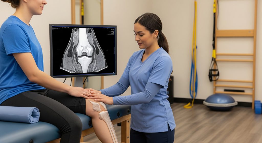
MRI and Imaging Studies
I use MRI to confirm the suspected pattern, define location, and screen for concurrent injuries such as ACL tears. A tear appears as a high signal that reaches an articular surface. Horizontal cleavage, vertical longitudinal, or radial patterns each signal different biomechanics and different options in theatre. Roughly speaking, MRI is reliable but not perfect. As PubMed Central reports, sensitivity for meniscal injury detection ranges from 68% to 87.5%, with advances in modelling improving accuracy.
Plain radiographs still matter when degenerative change is suspected. They help me avoid attributing global knee pain to a small, incidental meniscal signal. Imaging does not replace the history. It refines the working diagnosis.
Arthroscopy for Diagnosis
Arthroscopy is primarily a treatment tool today. I reserve diagnostic arthroscopy for rare, persistently symptomatic cases where examination and MRI conflict and conservative care has failed. When I scope for treatment, I document the tear morphology carefully and probe vascular zones to decide on repair feasibility.
Common Meniscus Tear Symptoms
Typical meniscus tear symptoms include focal joint line pain, swelling after activity, and painful clicking during twisting. Locking suggests a displaced fragment, often a bucket handle. Stiffness after sitting, night pain, or diffuse ache points more toward degenerative tears. Urgent assessment is warranted if the knee cannot fully extend, or if swelling appears rapidly after a twist in sport.
Types of Meniscus Tears
Tear pattern and zone drive treatment. A concise map helps align expectations and surgical choice.
|
Type |
Definition and Implication |
|---|---|
|
Vertical longitudinal |
Runs parallel to the rim. Often repairable, especially in the red zone where blood supply supports healing. |
|
Bucket handle |
Displaced longitudinal tear that flips into the notch and blocks extension. Commonly treated with repair if tissue is viable. |
|
Radial |
Perpendicular to the rim. Disrupts hoop stress. Repair is possible in selected cases, though technically demanding. |
|
Horizontal cleavage |
Splits the meniscus into upper and lower leaves. Frequently degenerative. May respond to targeted trimming if unstable. |
|
Complex |
Mixed patterns across zones. Strategy focuses on preserving any healthy meniscal arc while stabilising symptomatic flaps. |
Degenerative tears develop over time. Traumatic tears follow a twist under load. The distinction matters when discussing recovery and long term risk.
Meniscus Tear Surgery Options and Procedures
Arthroscopic Partial Meniscectomy
When the tear is irreparable yet mechanically unstable, arthroscopic partial meniscectomy removes only the unstable portion and preserves as much rim as possible. I view this as a precision tidy up, not a wholesale removal. Tissue preservation aligns with joint longevity. Evidence has questioned meniscectomy for purely degenerative pain without mechanical symptoms. I discuss the trade off openly, including a possible increased osteoarthritis risk over years.
-
Indications: non repairable flap, complex degenerative segment that repeatedly catches.
-
Goal: stable, smooth meniscal contour with intact peripheral rim.
-
Recovery: walking quickly, structured strengthening within days, sport later by function.
Meniscus tear surgery should aim to preserve shock absorption and load distribution. Removal is a last resort when stability cannot be restored by repair.
Meniscus Repair Surgery
Repair restores continuity and hoop tension. I prefer repair whenever viable tissue and adequate blood supply exist. Modern inside out, all inside, and root repair techniques allow robust fixation while protecting neurovascular structures. The rehabilitation is slower than after trimming, but the joint benefit is meaningful. Weight bearing and flexion are restricted early to protect sutures, then expanded as healing progresses.
Success hinges on the tear pattern, time from injury, and patient factors such as smoking and alignment. Where the posterior root is torn, repairing it reinstates load sharing and may decelerate cartilage wear. That is the rationale. It is basically joint biology meeting biomechanics.
Meniscus Transplant Surgery
In selected younger patients with prior subtotal meniscectomy, persistent compartment pain, and preserved alignment, meniscus allograft transplantation is an option. The aim is symptom relief and load redistribution. I confirm limb alignment, cartilage status, and ligament stability first. Transplant is not a shortcut back to high impact sport. It is a considered salvage for pain and function in the right knee.
Choosing the Right Surgical Option
I match the operation to the patient and the tear, not the other way round. For recent traumatic vertical tears in the vascular zone, repair is usually appropriate. For small, stable degenerative signals with no mechanical features, non operative care takes priority. For large displaced bucket handles in active patients, repair offers the best chance of restoring function.
Where evidence quantifies outcomes, I include it in counselling. In younger patients with reducible traumatic tears, repair success approaches 80% in several series. As PubMed Central summarises, success is higher when surgery occurs soon after injury and criteria for repair are met.
Decision framework I use:
-
Define pattern and zone on exam and MRI.
-
Assess alignment, cartilage, and ligament status.
-
Discuss goals: sport, work demands, and pain tolerance.
-
Choose preservation first. Resort to partial meniscectomy only if repair is not feasible.
Pre-Surgery Preparation Steps
Preparation improves outcomes and reduces risk. I give a clear, itemised plan.
-
Medical optimisation: share a complete medication and supplement list. Temporarily stop anticoagulants and most NSAIDs if advised.
-
Investigations: confirm imaging, arrange any blood tests, and finalise anaesthesia plan.
-
Strength priming: prehabilitation to activate quadriceps and gluteals. Better pre op strength predicts easier early recovery.
-
Logistics: fasting as instructed, arrange transport, prepare a safe home layout with essentials at hip height.
-
Expectation setting: agree weight bearing limits and brace settings for day one.
A well prepared patient spends less energy firefighting small problems and more energy healing.
Post-Surgery Recovery and Rehabilitation
Immediate Post-Operative Care
My priorities are pain control, swelling reduction, and protection of the repair or contour. I use elevation, cryotherapy, and early quadriceps activation. After repair, I often restrict flexion angles in the first weeks and set partial weight bearing with crutches. After partial meniscectomy, I allow weight as tolerated, encouraging symmetrical gait as pain settles. Clear rules prevent over cautious stagnation and reckless overload.
Meniscus Tear Recovery Time Phases
Timelines depend on the procedure and the biology of healing. A repair needs a slower ramp. A trim allows a faster one. Athletes pass through defined checkpoints from pain control to sport specific drills. As New Braunfels Orthopaedic Surgery & Sports Medicine notes, partial meniscectomy patients often return to activity in about 4 to 6 weeks, while repairs may require 3 to 9 months.
-
Phase 1: protect and reduce swelling. Aim for full extension and safe quad firing.
-
Phase 2: restore controlled flexion and begin closed chain strength.
-
Phase 3: build strength, balance, and cardiovascular capacity without irritability.
-
Phase 4: introduce impact and change of direction if the repair is secure and metrics are met.
For non athletes, the structure still applies. It is scaled to daily tasks and work demands.
Essential Meniscus Tear Exercises
I focus on exercises that restore extension, unlock quadriceps, and rebuild hip and calf support. The list below anchors my early and mid stage plan. I progress by criteria, not dates.
-
Early: quad sets, straight leg raises, heel slides within range limits, ankle pumps.
-
Mid: mini squats, step ups, bridges, stationary bike with low resistance.
-
Late: split squats, controlled lunges, single leg balance with reach, resisted hamstring curls if not painful.
Meniscus tear exercises must respect the repair. Slow and steady wins here. I avoid deep loaded flexion early after repair, especially in posterior horn lesions.
Physical Therapy Protocol
A criterion based protocol prevents drift and overreach. We set range targets, swelling thresholds, and strength ratios before advancing. Pain is a guide but not the only guide. I include neuromuscular electrical stimulation when quadriceps lag persists, and I prioritise gait retraining to avoid compensatory patterns that linger.
Progression is earned when swelling remains low, range is symmetrical, and strength reaches safe ratios. Not simply when the calendar flips a week.
Communication between surgeon, therapist, and patient keeps the plan coherent. It also shortens detours.
Return to Activities Timeline
Return is a process, not a date. I use staged exposure and explicit criteria.
-
Daily life: after meniscectomy, most walk unaided within days. After repair, brace and crutches persist for several weeks.
-
Work: desk roles resume early. Manual roles require tested strength and tolerance for uneven ground and kneeling.
-
Sport: linear running arrives before cutting. Cutting arrives before contact. Each step needs pain free control.
For planning, headline numbers can help orient expectations. As New Braunfels Orthopaedic Surgery & Sports Medicine outlines, sedentary work can resume sooner than heavy labour, and adherence to therapy correlates with earlier return.
It sounds strict. It is. The knee rewards method and patience.
Complications and Long-Term Outcomes
Potential Surgical Risks
Every operation carries risk. With arthroscopy, risks include infection, venous thromboembolism, stiffness, and neurovascular injury. Specific to meniscus tear surgery, repair can fail to heal, and trimming too much tissue can accelerate compartment load and cartilage wear. I reduce risk through meticulous technique, haemostasis, and clear rehab boundaries. Patients reduce risk through adherence and early reporting of problems.
-
Short term: bleeding, clots, wound issues, and transient swelling spikes.
-
Medium term: stiffness from under movement or pain avoidance.
-
Long term: re tear, progression of osteoarthritis, or persistent pain from coexistent pathology.
Success Rates by Surgery Type
Success means a stable, pain controlled knee that meets functional goals. Meniscal repair aims for biological healing and protection against later arthritis, though not without exceptions. Partial meniscectomy targets symptom relief with quicker milestones but may trade long term cushioning. Outcomes improve when selection and execution align with the tear pattern. Earlier, I noted repair success approaching 80% in selected younger patients. That context matters when choosing preservation over removal.
When to Seek Medical Attention
Escalate promptly if pain intensifies, swelling persists beyond expected windows, or the knee locks or gives way. A sudden inability to bear weight, fever, or calf pain also warrants contact with the clinical team. Early review prevents small issues from becoming setbacks. As Cleveland Clinic cautions, ignoring persistent symptoms risks further joint damage and prolongs recovery.
Preventing Future Injuries
Prevention is not complicated. It is systematic. Maintain hip and core strength to control knee valgus. Train balance and single leg control. Progress change of direction and landing mechanics, then speed, then contact. Rotate footwear at expected mileage and avoid rapid spikes in weekly training load. After surgery, respect the graduated plan. The best protector of the meniscus is a strong, well coordinated lower limb.
Making Informed Decisions About Meniscus Tear Surgery
Informed consent for meniscus tear surgery goes beyond signing a form. It is a shared, specific plan. I recommend a simple checklist at the decision point.
-
Diagnosis clarity: tear type, location, and associated lesions are understood.
-
Rationale: why repair, trim, or defer. Include alternatives and expected benefits.
-
Risks: immediate and long term, with mitigation steps.
-
Rehabilitation: staged plan, guardrails, and criteria to progress.
-
Return to life: tailored timelines for work, sport, and travel.
One practical example. A 22 year old footballer with an acute bucket handle medial tear and stable ACL. I recommend arthroscopic repair, brace, partial weight bearing, and a measured return over months. Contrast that with a 58 year old walker with diffuse degenerative changes and a horizontal cleavage tear but no locking. I recommend non operative care first with targeted strength, activity modification, and pain control. Two knees. Two paths. Same goal of durable function.
Meniscus tear symptoms guide urgency. Imaging refines choice. Surgery, when indicated, preserves structure and restores mechanics. The rest is disciplined rehabilitation and honest follow up.
Frequently Asked Questions
Can meniscus tears heal without surgery?
Yes, some tears can. Small, stable tears in the outer red zone may settle with structured rehabilitation and load management. Degenerative tears without mechanical catching often improve with strength work, activity modification, and time. I reserve meniscus tear surgery for persistent mechanical symptoms or failure of well executed conservative care.
How long before walking normally after meniscus surgery?
After partial meniscectomy, most patients walk comfortably within a few days as pain and swelling ease. After repair, crutches usually persist for several weeks to protect healing. Normal gait returns once extension is full, swelling is minimal, and quadriceps control is symmetrical.
What happens if meniscus tear surgery is delayed?
For stable, non locking tears, a period of conservative care is appropriate and often beneficial. For displaced bucket handle tears with locking, delay risks cartilage scuffing and stiffness. Timely repair in repairable patterns supports better healing and shorter meniscus tear recovery time overall.
Is meniscus repair better than removal?
When feasible, repair is generally preferable because it preserves meniscal function and may reduce the risk of later osteoarthritis. Removal is suitable when tissue is not repairable or when repeated catching persists. The best procedure is the one that matches the tear and the patient.
Can I drive after meniscus tear surgery?
For right knees, driving is safe once reaction times are normal, pain is controlled without sedating medication, and weight bearing is permitted. After partial meniscectomy, this can be within 1 to 2 weeks. After repair, it often takes longer. Left knees in automatic cars return sooner if safe control is demonstrated.
What activities should be avoided after meniscus surgery?
Early after repair, avoid deep squats, twisting under load, and pivoting. After partial meniscectomy, avoid impact and cutting until swelling is settled and strength is adequate. In both cases, progress is guided by criteria. Meniscus tear exercises prescribed by the therapist are the safest path to activity.