


Iron Overload or Deficiency? How to Read Iron Studies Test Results
Dr. Juhee Chandra
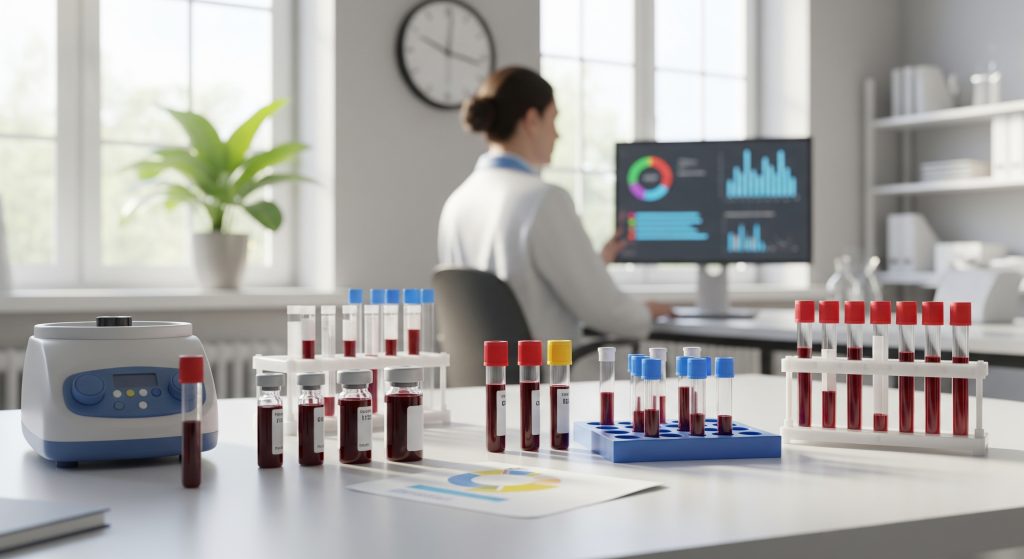
One test result rarely tells the whole story. A single ferritin value can look reassuring or alarming and still mislead if taken in isolation. The smarter approach is to interpret iron studies as a coherent panel, mapped against symptoms and context. I will show how to read each marker with care, then combine them to distinguish deficiency from overload without guesswork.
Essential Components of Iron Studies and Their Normal Ranges
Serum Ferritin Levels and Ferritin Test Normal Range
Ferritin reflects stored iron. It is the closest thing to a fuel gauge for iron reserves. As Mayo Clinic notes, ferritin is also an acute phase protein, so raised values can mirror inflammation, liver disease, or arthritis rather than true iron load; typical adult reference intervals vary by sex, and laboratories often publish ranges such as low tens to a few hundred nanograms per millilitre. Low values suggest depletion that can progress to anaemia if unaddressed.
Here is the practical point. If symptoms fit a deficiency picture and ferritin is below your laboratory’s reference interval, I treat it as iron depletion unless an inflammatory process explains the result. I also cross check the rest of the iron studies to avoid a false conclusion.
- Use the lab’s reference interval rather than a generic threshold.
- Correlate with C-reactive protein or clinical signs of inflammation when ferritin is unexpectedly high.
- Mention the exact term ferritin test normal range when reviewing your report or discussing targets.
Serum Iron and Normal Iron Levels in Blood
Serum iron measures circulating iron bound to transferrin at that moment. It is volatile. Diet, time of day, and recent supplements can swing the result. In deficiency, the value usually sits low. In overload, it can drift higher. Still, serum iron alone is a weak guide to overall status. I pair it with TIBC and transferrin saturation to stabilise the interpretation of normal iron levels in blood.
To reduce noise, I ask patients not to take iron on the morning of the test. Fasting may be requested by your laboratory. The theme remains the same. Treat serum iron as a supporting actor, not the lead.
Total Iron Binding Capacity (TIBC) Values
TIBC estimates how much iron your transferrin could carry if fully loaded. High TIBC often signals that the body is hungry for iron. Low TIBC can reflect chronic inflammation, liver disease, or iron overload. As Cleveland Clinic reports, a commonly cited reference interval is 240 to 450 mcg/dL, with higher values pointing toward deficiency and lower values sometimes seen in overload states.
In practice, I combine TIBC with transferrin saturation. The pairing clarifies whether a high TIBC truly reflects scarcity or whether another process is skewing the result.
Transferrin Saturation Percentage
Transferrin saturation (TSAT) describes the proportion of transferrin that is actually carrying iron. It answers a simple question: is enough iron available to tissues today. As StatPearls summarises, a TSAT below 20% supports iron deficiency, while persistent readings above roughly 50% point toward overload.
Clinically, TSAT is fast to read and useful when ferritin is muddied by inflammation. A very low TSAT with a low ferritin is compelling evidence of iron lack. A high TSAT with a raised ferritin raises concern for iron excess.
Haemoglobin and Haematocrit Levels
Haemoglobin and haematocrit sit outside the classic iron studies, but I always review them. They describe oxygen carrying capacity and overall red cell mass. Low values confirm anaemia. They do not define the cause. Iron deficiency, chronic disease, B12 or folate deficiency, and blood loss can all lower these indices. Hydration status and altitude also influence them. The signal is important, but the differential remains broad.
- Haemoglobin tracks oxygen carrying capacity and clinical impact.
- Haematocrit reflects the proportion of red cells in blood volume.
- Both guide urgency and the need for broader investigation.
Understanding Your Ferritin Levels Chart
Most results portals display a ferritin levels chart with ranges shaded. It looks straightforward. The nuance is that reference intervals differ between laboratories and can be shifted by age, sex, and local population data. I read the chart, then I read the fine print. The correct interpretation sometimes sits one line below the graph.
When plotting serial serum ferritin levels, I look for consistent upward or downward trends rather than single outliers. A gradual rise alongside a high TSAT prompts a review for overload. A steady fall with low TSAT supports depletion. Inflammation can lift the whole series temporarily (for example, after an infection). Context matters.
|
Test |
What it tells you |
|
Serum ferritin |
Iron stores; distorted by inflammation |
|
Serum iron |
Snapshot of circulating iron; variable day to day |
|
TIBC |
Capacity to carry iron; rises in deficiency |
|
Transferrin saturation |
Immediate iron availability; low in deficiency, high in overload |
|
Haemoglobin/Haematocrit |
Anaemia severity; not specific to iron |
Interpreting Iron Studies Results for Deficiency vs Overload
Iron Deficiency Pattern Recognition
The classic deficiency pattern is coherent. Ferritin sits below the reference interval. TIBC is elevated. Transferrin saturation falls below the low threshold described earlier. Serum iron is low or low normal. Haemoglobin may be normal at first and then drift down. I confirm that this is not an inflammatory picture masking true status. In borderline cases, I consider soluble transferrin receptor, which can rise with functional iron lack, though standardisation varies.
- Low ferritin plus low TSAT is compelling for deficiency.
- High TIBC supports the same conclusion.
- Inflammation can blur ferritin; TSAT often cuts through that noise.
Iron Deficiency Anemia Symptoms to Match with Results
Laboratory data must match the clinical story. Iron deficiency anemia symptoms often include fatigue that is disproportionate to effort, exertional breathlessness, and headaches. Some patients report pale skin or cold intolerance. The pattern is usually gradual, not dramatic, and it affects everyday functioning. When the symptom profile and iron studies align, I proceed confidently. When they do not, I widen the search.
A brief example clarifies the point. A distance runner presents with tired legs and low mood. Ferritin is below range and TSAT is low. A realistic plan is iron replenishment and a check for gastrointestinal blood loss. The data and the story agree.
Iron Overload Pattern Identification
Iron overload is a different geometry. Ferritin trends upward and remains elevated on repeat tests. Transferrin saturation often sits in the high range discussed earlier. TIBC may be low or normal. Serum iron can be high. The pattern is most persuasive when sustained over time and not explained by inflammation, alcohol excess, or acute illness.
There is also the question of risk. Persistently high saturation creates a surplus of free iron that can harm tissues. In that setting, I evaluate for haemochromatosis or secondary causes, and I do so before organ damage accumulates. Early clarity is protective.
Haemochromatosis Indicators in Test Results
Hereditary haemochromatosis often features a high ferritin with a raised transferrin saturation and a family history of liver or endocrine disease. I do not rely on one abnormal reading. I repeat the panel, confirm absence of intercurrent illness, and then triage to genetic testing when appropriate. HFE mutations are the usual culprits. Elevated liver enzymes or ultrasound abnormalities nudge me toward imaging for iron in the liver.
Early detection changes outcomes. Phlebotomy can offload iron and prevent damage, but it works best before organs are stressed. The numbers are the start. The follow up is the key.
Anaemia of Chronic Disease vs True Iron Deficiency
Anaemia of chronic disease (also called anaemia of inflammation) can mimic deficiency on symptoms alone. The laboratory pattern diverges. Ferritin is normal or raised because inflammatory signalling increases storage and reduces iron release. Serum iron is low. Transferrin saturation can be low or low normal. TIBC is often low or normal. Compare that with true deficiency, where ferritin falls and TIBC rises. The difference matters for treatment. Iron tablets will not correct anaemia of chronic disease unless deficiency coexists, and the priority is control of the inflammatory condition.
Borderline cases occur. When ferritin sits in the low normal band and symptoms persist, I synthesise all markers and clinical cues rather than chase a single number.
Borderline Results and Grey Areas
Grey areas are common. Ferritin can be mid range in an inflamed patient and still hide a deficit. Haemoglobin can look acceptable while performance and concentration dip. In these situations, I look for internal consistency. TSAT under the low threshold with symptoms and a high TIBC leans toward deficiency despite a ferritin that is not frankly low.
Two practical strategies help:
- Trend the panel. Repeat iron studies after intercurrent illness resolves to remove inflammatory artefact.
- Use a trial of therapy with objective follow up. Rising ferritin and symptom improvement argue for a correct call.
Borderlines are not a failure of testing. They are a reminder to treat the patient and the dataset together.
Clinical Significance and Next Steps Based on Results
When Low Ferritin Requires Treatment
Low ferritin signals exhausted reserves. If symptoms are present, I treat. If a cause is obvious, such as heavy menstrual bleeding or recent blood loss, I address that cause in parallel. Oral iron is the usual first step. I ensure adequate elemental iron intake and advise on timing with food to reduce intolerance. If oral therapy fails or the deficit is severe, intravenous iron becomes appropriate. The goal is not only to raise haemoglobin if low, but to rebuild iron stores and prevent a rapid relapse.
- Treat symptoms and numbers together.
- Replete stores beyond the point where haemoglobin normalises.
- Search for sources of blood loss or malabsorption when the response is slow.
Managing Elevated Serum Ferritin Levels
Raised serum ferritin levels do not always mean iron overload. They frequently reflect inflammation, liver disease, metabolic syndrome, or alcohol intake. I rule out these common drivers first. If transferrin saturation is also high on repeat testing, iron overload moves up the list. In confirmed hereditary haemochromatosis, scheduled phlebotomy is the cornerstone to lower stores and protect organs. Lifestyle adjustments, including moderation of alcohol and attention to liver health, support the medical plan.
In secondary overload, for example after multiple transfusions, chelation may be considered by specialist teams. The unifying principle is to match the intervention to the cause, not merely to the ferritin number.
Additional Tests Your Doctor May Order
Iron studies sit within a wider haematology workup. I usually request a complete blood count with indices, a reticulocyte count, and B12 and folate where relevant. C-reactive protein helps interpret ferritin in inflammatory states. In suspected malabsorption, coeliac screening or gastrointestinal evaluation may be useful. For possible overload, I consider HFE genotyping and liver imaging. These are not reflex tests. They are guided by the pattern you see and the history you take.
- CBC and indices to quantify anaemia and red cell morphology.
- Inflammatory markers to contextualise ferritin.
- Genetics and imaging if overload persists without an obvious secondary cause.
Treatment Options for Abnormal Iron Studies
Options cluster into two groups: replenishment and reduction.
- Replenishment: oral iron salts or newer formulations, dietary optimisation, intravenous iron for intolerance or rapid repletion. I monitor response and adjust dose to balance efficacy and tolerance.
- Reduction: therapeutic phlebotomy for confirmed hereditary haemochromatosis, targeted chelation in transfusional iron overload under specialist care, and lifestyle measures that reduce ongoing hepatic stress.
I add a pragmatic note. Scheduling and adherence often determine success more than the brand of supplement. Clear instructions and realistic follow up improve outcomes.
Monitoring and Follow-up Testing Schedule
Monitoring strategy depends on the starting point and the intervention chosen. After initiating oral iron, I look for a clinical response within weeks and a laboratory response soon after. I continue therapy for a period beyond haemoglobin normalisation to restore stores. In overload, I repeat iron studies at intervals aligned to the phlebotomy schedule, then space out checks once a maintenance target is reached. Roughly speaking, stable patients with normal panels can move to periodic surveillance.
Two rules keep monitoring efficient:
- Do not overtest during acute illness because inflammation distorts ferritin and TSAT.
- Test at consistent times and conditions where possible to reduce variability.
Making Sense of Your Iron Studies Results
Reading iron studies is a pattern exercise. Ferritin shows stores, TSAT shows availability, TIBC shows capacity, and haemoglobin shows impact. Align them with the clinical story and the path becomes clear. Low ferritin with low TSAT and high TIBC is deficiency until proven otherwise. High ferritin with high TSAT is overload until another cause displaces it. Everything else sits in between and asks for context, time, and disciplined follow up.
That is the essence. Use the panel as a whole. Prioritise symptoms and trends. And yet, respect the exceptions. The right diagnosis is often the one that explains both the numbers and the person in front of you.
What ferritin level indicates iron deficiency anaemia?
I diagnose iron deficiency anaemia when ferritin is below the local reference interval and the rest of the iron studies support that conclusion. In inflammatory conditions, a higher ferritin can still hide deficiency, so I lean on transferrin saturation and clinical features. The threshold is therefore contextual rather than a single universal number.
Can you have normal haemoglobin but low ferritin?
Yes. Low ferritin with normal haemoglobin indicates iron depletion without established anaemia. This is common in endurance athletes and in individuals with marginal intake or intermittent losses. Symptoms can still be significant, particularly fatigue and reduced exercise capacity. Early treatment prevents progression to anaemia.
How often should iron studies be repeated after starting treatment?
For oral iron replenishment, I recheck at a sensible early interval to confirm response, then at wider intervals to ensure stores are rebuilt. For maintenance, I reduce frequency to periodic surveillance. In overload managed with phlebotomy, I check more frequently at first, then extend the interval once stable. The schedule is tailored to severity and clinical change.
What causes falsely elevated ferritin levels?
Ferritin rises with inflammation, infection, liver disease, metabolic syndrome, and alcohol use. These states can push ferritin up without reflecting true iron overload. I use transferrin saturation, TIBC, and clinical assessment to separate inflammatory elevation from increased iron stores.
Should I fast before iron studies blood test?
Follow your laboratory’s instructions. Some laboratories request morning sampling and no iron supplements on the day of the test to reduce variability. If fasting is required, it will be specified on the requisition. Consistency between tests is more important than a single rigid rule.
What’s the difference between iron deficiency and iron deficiency anaemia?
Iron deficiency means depleted iron stores, typically shown by a low ferritin and a low transferrin saturation. Iron deficiency anaemia is the later stage when haemoglobin has fallen as well. The first is a warning. The second is a confirmed oxygen carrying problem. Treating the deficiency before anaemia develops prevents the performance and health costs that follow.