


Bone Cancer Causes Explained: Risks, Genetics and More
Dr. Rajeev K Sharma
Advice about bone tumours often starts with a vague catch-all: genetics and chance. That framing is incomplete. I will outline bone cancer causes with precision, show how inherited syndromes alter risk, and clarify patterns that clinicians see every week. The goal is practical clarity. Not speculation.
Primary Causes of Bone Cancer
Genetic Mutations and Inherited Syndromes
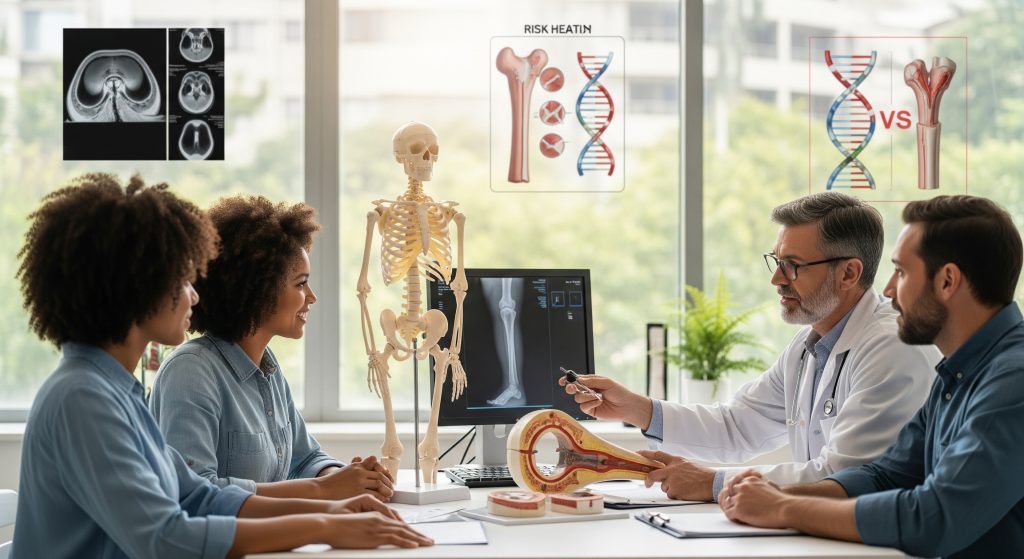
Bone tumours begin with genetic change. Cells accumulate mutations that disrupt growth control, DNA repair, or programmed cell death. Some changes are inherited, others arise in the tumour itself. Both routes matter. In practice, bone cancer causes often converge on a small set of disrupted pathways. That is why similar genes appear across different patients.
Two themes recur. First, germline predisposition in recognised syndromes that raise baseline risk. Second, somatic instability within the tumour, which accelerates additional mutations. I see this pattern in osteosarcoma, chondrosarcoma, and Ewing tumours, though the drivers differ. It is basically genetics plus biology under stress.
-
Disrupted tumour suppressors (for example, TP53 or RB1) remove growth brakes.
-
DNA repair defects increase mutation load and chromosomal breaks.
-
Rapid bone growth during adolescence supplies a fertile microenvironment.
None of this means determinism. It means risk concentrates when several of these forces align.
Li-Fraumeni Syndrome and TP53 Mutations
Li-Fraumeni syndrome sits near the centre of discussion about bone cancer causes. It reflects germline mutations in TP53, the genome’s quality control supervisor. As Inherited TP53 Mutations and the Li-Fraumeni Syndrome reports, such mutations are present in roughly **75%** of clinically diagnosed cases and raise lifetime cancer risk to nearly **100%** for females by age 60 and about **75%** for males. Those figures are stark, though penetrance varies by family and surveillance intensity.
Mechanistically, TP53 loss weakens the cellular response to DNA damage. Cells that should pause or self-destruct continue dividing. The result is more opportunity for malignant transformation in bone and soft tissue. For clinicians, the implication is clear. Families meeting clinical criteria need genetic testing, careful imaging schedules, and prompt assessment of persistent bone pain. Early detection reduces treatment intensity and preserves function.
Retinoblastoma Gene Mutations
RB1 is another tumour suppressor with a defined link to osteosarcoma. Children treated for heritable retinoblastoma carry a germline mutation in RB1. Later-life risk of bone tumours rises because cell cycle control is compromised. In those with a history of heritable retinoblastoma, I maintain a lower threshold for imaging when bone pain persists or local swelling develops. Radiation fields from prior ocular treatment can add risk. The driver here is the same principle. Remove a key brake, and the system over-runs.
Multiple Osteochondromas and Bone Disorders
Hereditary multiple osteochondromas (also called multiple exostoses) create many benign cartilage-capped bony growths. Most remain harmless. A small subset undergoes malignant change into secondary chondrosarcoma. As Multiple osteochondromas notes, the reported risk of malignant transformation ranges from about **0.5%** to **5%**, with risk influenced by lesion number, location, and growth after skeletal maturity.
What does this mean for bone cancer causes? It demonstrates how chronic structural abnormalities can, to a limited extent, seed malignancy when growth signalling drifts. I advise periodic review for new pain, sudden size changes, or neurological symptoms from compressive growth. Targeted MRI is often decisive.
Radiation Exposure and Treatment History
Prior radiotherapy is a recognised contributor in the list of bone cancer causes. Ionising radiation can damage DNA and raise the chance of later sarcomas within or near the radiation field. The effect is stronger at higher doses and with exposure during childhood. This is not a prediction for any individual patient. It is a population risk signal. Key practical points include mapping symptoms against old radiation fields and setting a low threshold for imaging if focal bone pain persists.
Risk Factors and Demographics
Age and Gender Distribution
The incidence of primary bone cancer is bimodal. It rises in adolescence, then again in later adulthood. As Bone Cancer Research Trust outlines, risk clusters in ages **10 to 25** and again over **50**, with males showing a modestly higher incidence than females. This pattern aligns with biology. Pubertal growth spurts and later-life bone remodelling create windows of vulnerability.
Type also varies by age. Osteosarcoma often arises in the long bones of adolescents. Chondrosarcoma features more in older adults. Ewing tumours sit predominantly in teens and young adults. These are general patterns, not hard rules. But they shape suspicion levels in clinic and inform imaging priorities.
Environmental and Chemical Exposures
Environmental contributions to bone cancer causes remain under active study. Heavy metals, certain industrial chemicals, and ionising radiation have been discussed in research settings. The signal is mixed and, in parts, method-limited. Roughly speaking, the clearest environmental association is radiation. Chemical links are suggestive in some cohorts and weak in others. I advise evidence-based caution: reduce avoidable exposures in high-risk workplaces and follow safety standards rigorously.
Previous Cancer Treatment
Therapies that saved a child’s life can raise later sarcoma risk. Alkylating agents and topoisomerase inhibitors have long been examined for their secondary malignancy profiles. Radiotherapy adds a location-specific effect. The absolute risk for any one person is still small. But when a survivor presents with persistent, focal bone pain, I accelerate workup. Prior treatment history is not a footnote. It is a decision variable.
Paget’s Disease and Pre-existing Conditions
Paget’s disease of bone increases focal turnover and architectural distortion. In a small minority of patients, that environment supports sarcomatous change. Clinically, new pain, swelling, or warmth over a known Paget’s site deserves attention. Other pre-existing conditions, including chronic bone infection and certain inherited dysplasias, can also raise risk to some extent. The common thread is persistent dysregulated remodelling.
Growth Patterns and Height Factors
A long-observed feature of adolescent osteosarcoma is its preference for the metaphyseal regions of rapidly growing long bones. Taller stature and rapid growth velocity have been described in some patient groups. The causal chain is not settled. Growth plates present a busy cellular environment, and proliferating osteoblasts are, arguably, more vulnerable to accumulating mutations. It is a plausible link, though not a diagnostic criterion.
Recognising Bone Cancer Symptoms
Early Warning Signs
Early clues are frequently subtle. Pain is the commonest first signal. It often starts intermittent and activity related. Over weeks, it becomes more constant and disturbs sleep. Local swelling may follow. Reduced function creeps in next. In clinic, I listen for the story arc: a nagging ache that stops responding to rest or simple analgesics. That narrative matters more than any one descriptor.
-
Persistent, focal bone pain, often worse at night.
-
Swelling or a palpable lump over bone.
-
Unexplained limitation of motion near a joint.
-
Occasional fever, fatigue, or weight loss in advanced cases.
None of these confirms a tumour. Together, they justify imaging.
Ewing Sarcoma Specific Symptoms
Ewing tumours can present with bone pain, soft tissue swelling, and tenderness. Systemic features are more common here than in other primary bone cancers. Low-grade fever, malaise, and anaemia sometimes appear. I flag night pain that wakes a young person, particularly with a tender mass over a long bone or pelvis. Add unexplained fractures and raised inflammatory markers, and suspicion rises. These patterns align with typical ewing sarcoma symptoms seen in practice.
Pain Patterns and Characteristics
Pain deserves careful parsing. Tumour pain tends to be localised and progressive. It interrupts sleep. It persists despite rest. Sports injuries usually improve over two to four weeks. Tumour pain generally does not. I ask three questions. Does it wake you at night. Is it localised to one spot you can point to. Has it escalated over a month. Three yes answers are a prompt to image.
In metastatic bone pain, patients often describe deep, aching discomfort that worsens with weight bearing. Analgesic response varies. Quality of life suffers. That experience calls for urgent, structured pain control and diagnostic clarity. Quickly.
Swelling and Physical Changes
Swelling reflects the mass effect of the tumour and local inflammation. In long bones, it may be firm and tender with stretched skin and increased warmth. Neurological symptoms emerge when lesion expansion compresses nerves. Think tingling, numbness, or weakness distal to the site. Any new deformity or limb alignment change is a red flag. So is a pathologic fracture after minimal trauma.
When to Seek Medical Attention
Do not wait on persistent bone pain that is getting worse, especially at night. Prolonged swelling, a steadily enlarging lump, or an unexplained fracture should be assessed promptly. Imaging and, when appropriate, biopsy establish the diagnosis. Early referral improves options and function. Delays often occur when symptoms are dismissed as growing pains or overuse injuries. I would rather investigate and reassure than miss a tumour that could have been treated earlier.
Survival Rates and Prognosis
Overall 5-Year Survival Statistics
Interpreting bone cancer survival rates requires nuance. Figures vary by tumour type, stage at diagnosis, primary site, and response to therapy. Broad numbers found online often hide those differences. In specialist centres, outcomes improve with multidisciplinary care, optimised chemotherapy protocols, limb-sparing surgery, and advanced imaging. The direction of travel is positive. The spread of results is wide.
Survival by Cancer Type
Osteosarcoma, Ewing sarcoma, and chondrosarcoma do not share a single outlook. Low-grade chondrosarcoma resected completely can have favourable long-term control. Ewing sarcoma with good chemotherapy response and local control does well in localised cases. High-grade osteosarcoma outcomes depend on necrosis rates after neoadjuvant chemotherapy and the completeness of resection. I stress individual prognostic factors over headline numbers. They are more truthful for the person in front of me.
Localised vs Metastatic Disease
Stage at diagnosis is decisive. Localised disease with clear margins delivers the best outcomes. Metastatic disease at presentation lowers long-term control. That is not a reason for pessimism. It is a reason for precise staging, swift systemic therapy, and surgical planning where feasible. Pulmonary metastasectomy can be considered in selected cases. Biology steers decisions as much as imaging.
Factors Affecting Prognosis
Prognosis reflects several interacting elements. I look at histologic grade, tumour size and site, response to chemotherapy, surgical margins, and whether metastases are present. Genetic features may also matter, including specific rearrangements in Ewing tumours or complex karyotypes in osteosarcoma. Treatment adherence and timely management of complications influence outcomes more than many realise. Supportive care is not an accessory. It is core therapy.
Recent Treatment Advances
Progress is steady rather than spectacular, but it is real. Surgical technique and planning have improved limb function while maintaining margins. Chemotherapy remains foundational in osteosarcoma and Ewing tumours, with ongoing trials testing risk-adapted intensification or de-escalation. Proton therapy offers precise radiation dosing to protect growth plates and joints in selected cases. On the biologics front, targeted agents and immunomodulators are being studied for molecular subsets. Small gains add up.
Understanding Your Bone Cancer Risk
Risk is not destiny. It is information to calibrate attention and action. Here is how I summarise bone cancer causes and personal risk into something useful.
|
Factor |
What it means in practice |
|---|---|
|
Inherited syndromes |
Higher baseline risk. Genetic counselling, structured screening, and a low threshold for imaging. |
|
Previous radiotherapy |
Location-specific risk. Monitor symptoms within prior radiation fields. |
|
Multiple osteochondromas |
Small malignancy risk. Watch for growth after maturity, new pain, or neurological signs. |
|
Adolescent growth |
Higher vigilance for persistent, focal pain near long bones. |
|
Occupational exposures |
Follow safety controls and reduce avoidable ionising radiation or toxin exposure. |
One final perspective. The most actionable step is still symptom-aware behaviour. A persistent, focal pain that escalates over weeks deserves evaluation. If I had to prioritise, I would choose timely imaging over speculative screening for most people without a known syndrome.
Frequently Asked Questions
Can bone cancer be prevented if you have genetic risk factors?
Prevention in a strict sense is not guaranteed. Risk reduction is possible. For those with strong hereditary risk, I recommend genetic counselling, structured surveillance, and prompt evaluation of new symptoms. Lifestyle refinements help general health but do not remove syndromic risk. Early detection shifts outcomes by enabling less extensive surgery and more conservative therapy. That is a tangible benefit.
What are the first symptoms of bone cancer that shouldn’t be ignored?
Persistent, focal bone pain that worsens at night should not be ignored. A swelling or lump over bone that is growing, unexplained reduced function near a joint, or a fracture after minimal trauma also warrants assessment. Taken together, these are classic symptoms of bone cancer in its early phases. An X-ray is usually the first step. MRI and definitive biopsy come next if suspicion remains.
How do bone cancer survival rates compare between children and adults?
Comparisons depend on tumour type, stage, and response to therapy. Children with localised Ewing sarcoma or osteosarcoma treated in specialist centres can achieve strong outcomes, particularly with good chemotherapy response and clear margins. Adults face a different mix of tumours, including more chondrosarcoma, with prognosis tied closely to grade and resectability. Age alone is less important than biology, stage, and treatment quality.
Is bone cancer hereditary in all cases?
No. Only a minority of cases arise from identifiable hereditary syndromes such as Li-Fraumeni or heritable retinoblastoma. The majority appear sporadic, driven by somatic mutations without a family pattern. Family history, early onset, multiple tumours, or syndromic features should still prompt genetic assessment. The absence of a known syndrome never rules out cancer. It narrows the search for cause.
What lifestyle factors might increase bone cancer risk?
There is no single lifestyle factor with a proven, large effect. Avoidable ionising radiation and certain industrial toxins are reasonable targets for reduction. Maintain bone health through balanced nutrition, vitamin D sufficiency, and strength training. These choices do not eliminate risk. They support overall resilience and recovery if treatment is needed.
How quickly does bone cancer typically progress without treatment?
Growth rates vary widely. Ewing tumours can advance over weeks to months. Some chondrosarcomas evolve more slowly. Osteosarcoma often sits between those extremes. As far as current data suggests, progression without treatment risks structural compromise and metastasis. The safer assumption is timely action once a lesion is suspected.
bone cancer causes, bone cancer causes, bone cancer causes, symptoms of bone cancer, ewing sarcoma symptoms, bone cancer survival rates