


What Is an MRI Brain Procedure and Why You Might Need One
Dr. Arunav Sharma
Common advice says brain imaging should be a last resort. That approach risks delay when speed and clarity matter. An MRI Brain Procedure offers detailed, radiation-free imaging that can rapidly direct care when symptoms are uncertain or worsening. You get high-resolution pictures, functional insights, and safe repeat testing. In practice, it is the standard tool for many neurological questions. Here is why.
Common Reasons for Needing an MRI Brain Procedure
1. Persistent Headaches and Migraines
Most headaches do not require imaging. Yet patterns can change. If your headaches escalate in severity, frequency, or character, an MRI Brain Procedure helps exclude structural causes. Red flags include sudden onset, neurological deficits, and headaches that wake you from sleep. The scan can assess the brain tissue, blood vessels, and sinuses in one sitting.
-
Rule out secondary causes such as masses or inflammation.
-
Map anatomy for targeted treatment planning.
-
Provide baseline images if symptoms persist.
2. Unexplained Seizures or Epilepsy
Following a first seizure, the priority is identifying a cause. An MRI Brain Procedure can detect scarring, developmental anomalies, prior bleeds, or small tumours. For established epilepsy, imaging refines the focus for therapy. It guides medication choices and, in selected cases, supports surgical planning.
Where appropriate, a functional mri brain study may complement standard images to localise language or motor regions. Precision reduces risk in any intervention.
3. Suspected Brain Tumours or Masses
When a tumour is suspected, defining location and boundaries is vital. An MRI Brain Procedure delineates soft tissues and surrounding oedema with clarity. It also assists in grading suspicion, although biopsy confirms the diagnosis. Serial scans allow careful monitoring after treatment.
-
Characterise lesion size and effects on nearby structures.
-
Assess mass effect, shift, or compression of ventricles.
-
Support radiotherapy planning with accurate targeting.
4. Signs of Stroke or Brain Bleeding
Time matters in stroke care. An MRI Brain Procedure can identify acute ischaemia and subtle haemorrhage. Diffusion sequences highlight areas at risk. This helps differentiate reversible tissue from established injury. It guides reperfusion decisions and secondary prevention.
If symptoms are transient, imaging can still show small infarcts. That information changes management. For the better.
5. Memory Loss and Cognitive Changes
When cognition declines, the differential is broad. An MRI Brain Procedure evaluates for atrophy patterns, vascular injury, normal pressure hydrocephalus, and treatable causes. The aim is clarity. Imaging supports earlier diagnosis and safeguards against missing reversible conditions.
-
Helpful in distinguishing types of dementia to an extent.
-
Offers structural context for neuropsychological testing.
6. Multiple Sclerosis Diagnosis
Multiple sclerosis often requires evidence of lesions separated in time and space. An MRI Brain Procedure provides that evidence. It shows plaques typical of demyelination and tracks new activity on follow up. Contrast may highlight active inflammation.
This imaging anchors the diagnosis and helps gauge disease activity. It also informs therapy escalation decisions.
7. Head Injury or Trauma Assessment
Computed tomography is common in acute trauma. Yet an MRI Brain Procedure detects diffuse axonal injury, small contusions, and microhaemorrhages with higher sensitivity. It is valuable in persistent post-concussion symptoms. Results can explain ongoing headaches, dizziness, or concentration problems.
Care decisions improve when the injury pattern is visible, not inferred.
8. Dizziness and Balance Problems
Dizziness is usually benign. Still, posterior circulation strokes and cerebellar issues can present subtly. An MRI Brain Procedure evaluates the posterior fossa, brainstem, and auditory pathways. It excludes structural causes when bedside manoeuvres are inconclusive.
-
Useful when symptoms are persistent or progressive.
-
Clarifies central versus peripheral patterns.
9. Vision or Hearing Changes
Visual loss, double vision, or hearing shifts deserve careful assessment. An MRI Brain Procedure reviews the optic pathways and cranial nerves. It examines the pituitary region and internal auditory canals. For pulsatile tinnitus or progressive hearing loss, imaging is a prudent step.
The goal is to rule out compressive or inflammatory causes with confidence.
10. Pre-surgical Brain Mapping
When surgery is planned near eloquent cortex, risk must be quantified. An MRI Brain Procedure defines anatomy and adjacent white matter tracts. Functional mapping can mark speech and movement areas. This allows safer resections and more precise consent discussions.
Better maps. Safer margins.
How the MRI Brain Procedure Works
Understanding Magnetic Resonance Technology
Magnetic resonance uses a strong field and radio waves to align and perturb hydrogen nuclei. During an MRI Brain Procedure, sensors capture signals as the nuclei relax. These signals are reconstructed into detailed images. Tissue contrast arises from differences in relaxation times.
Anatomy is displayed in multiple planes without ionising radiation. That is the fundamental advantage.
Sequence choice tailors what you see. T1, T2, FLAIR, diffusion, and susceptibility provide complementary information.
Contrast vs Non-Contrast Brain Scans
Some studies require contrast enhancement. In an MRI Brain Procedure, a gadolinium agent may be injected to highlight inflammation, breakdown of the blood brain barrier, or tumour vascularity. Many questions do not need contrast. Your clinician orders the appropriate protocol.
-
Contrast helps define active lesions and postoperative changes.
-
Non-contrast studies answer common queries safely and quickly.
If renal function is reduced, alternatives are considered. Risk is assessed, not assumed.
Functional MRI Brain Capabilities
Standard imaging shows structure. A functional mri brain assesses activity by tracking blood oxygen level dependent signals during tasks. It maps regions used for language, motor function, and memory. In context, this complements a standard MRI Brain Procedure before complex surgery.
It is not a mind reader. It is a risk reducer.
Duration and Scanning Process
Expect the visit to last about one hour. The time varies by sequences and whether contrast is used. The MRI Brain Procedure itself often takes between 20 and 45 minutes. Children and complex protocols can take longer.
-
Screening and safety checks.
-
Positioning and coil placement.
-
Motion minimisation and scan acquisition.
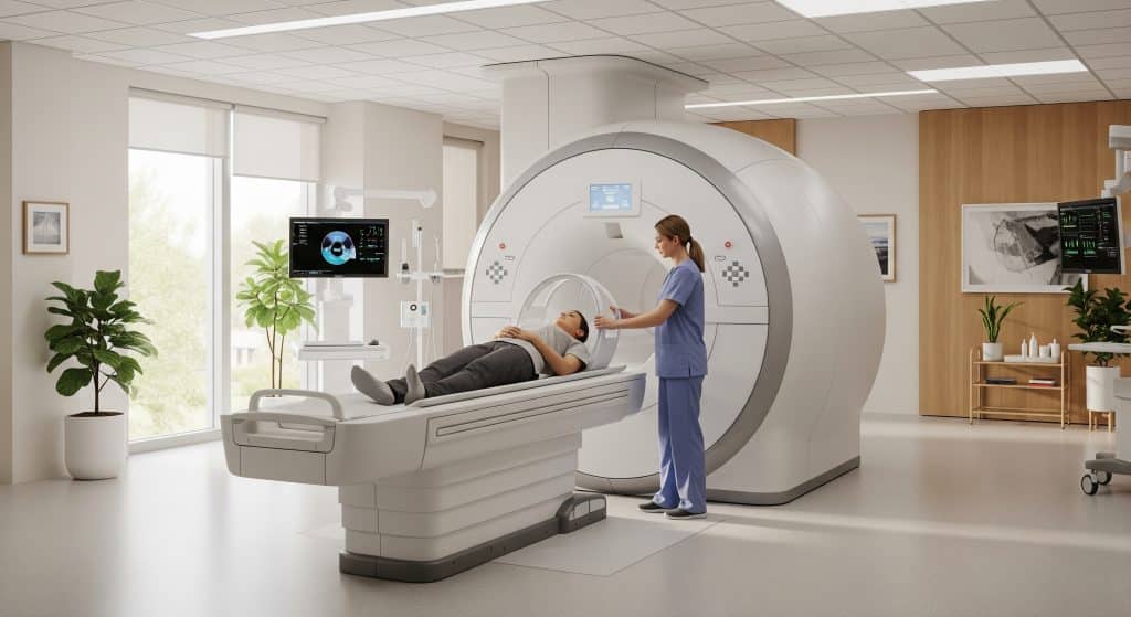
What Happens During the Scan
After checks, you lie on a padded table. A head coil sits around your skull like a helmet. During the MRI Brain Procedure, the machine makes rhythmic tapping noises. Ear protection reduces sound exposure. A panic button is available if you need immediate communication.
Staying still preserves image quality. Gentle coaching helps reduce motion. Breathing remains normal throughout.
Image Processing and Results
Raw data are reconstructed into images and reviewed by a radiologist. The report describes findings, limitations, and recommendations. An MRI Brain Procedure typically produces axial, coronal, and sagittal views for comparison. Clinicians correlate these with your history and examination.
If urgent concerns are seen, escalation is prompt. That is the safety net you want.
MRI Brain Scan Cost and Insurance Coverage
Average Costs Without Insurance
Prices vary widely by region and provider. As current data suggests, a brain mri scan without cover may cost a few hundred to several thousand pounds. The MRI Brain Procedure price reflects scanner quality, staffing, and protocol complexity.
|
Item |
Typical range |
|---|---|
|
Standard non-contrast scan |
£300 to £900 |
|
Scan with contrast |
£450 to £1,400 |
|
Functional mapping add-on |
£350 to £800 |
Insurance Coverage Requirements
Insurers usually require clinical justification and approved referral. Pre-authorisation is often necessary. The MRI Brain Procedure is generally covered when symptoms or guidelines support imaging. Documentation should state the indication clearly.
-
Referrals from an appropriate specialist help approval.
-
Contrast use may need separate authorisation.
Factors Affecting Price Variations
Several elements influence the mri brain scan cost. Location, scanner strength, subspecialty reporting, and urgency all matter. Additional sequences and sedation increase cost. Private hospital settings may charge higher facility fees.
-
Peak time bookings can be priced differently.
-
Follow up scans are sometimes discounted.
Hospital vs Imaging Centre Costs
Hospitals provide comprehensive support and on-site escalation. This can raise fees. Independent imaging centres often offer competitive rates and shorter waits. The MRI Brain Procedure quality depends on protocol, staff expertise, and how results are communicated.
Balance cost, speed, and clinical complexity when choosing a site.
Ways to Reduce Your Expenses
-
Seek a written estimate before scheduling.
-
Ask about self-pay bundles and off-peak pricing.
-
Confirm whether contrast and reporting are included.
-
Verify network status with your insurer in advance.
A clear plan prevents surprise bills after an MRI Brain Procedure.
Preparing for Your Brain MRI Scan
Pre-Scan Instructions and Guidelines
Arrive early for screening and consent. Bring prior imaging and your medication list. The MRI Brain Procedure requires stillness, so comfortable clothing helps. Remove cosmetics with metallic particles if possible.
-
Follow fasting advice if contrast is planned.
-
Share any history of allergies or renal issues.
Metal Objects and Safety Screening
The magnet is always on. Declare implants, pacemakers, aneurysm clips, pumps, or shrapnel. Most dental work is safe. The team will check device models carefully. Safety is non negotiable during an MRI Brain Procedure.
-
Lockers are provided for jewellery and watches.
-
Credit cards and phones must stay outside.
Managing Claustrophobia Concerns
Claustrophobia is common. Open or wider bore scanners can ease anxiety. The MRI Brain Procedure can include short breaks between sequences. Practice calm breathing and keep eyes softly closed.
Some sites offer visualisation aids or music. Discussion ahead of time helps.
Sedation Options Available
When anxiety persists, mild sedation is considered. This requires an escort and short recovery. The MRI Brain Procedure may also use paediatric sedation when children cannot keep still. Risks and benefits are reviewed beforehand.
Plan transport and fasting if sedation is chosen.
What to Expect on Scan Day
Check in, complete forms, and change if needed. Staff position you and provide ear protection. During the MRI Brain Procedure, you will hear knocks and hums. Communication remains open via intercom.
Most people complete the scan without difficulty. Preparation makes the difference.
Post-Scan Care and Follow-up
You may leave immediately after a standard scan. If contrast was given, drink water as advised. The MRI Brain Procedure report is sent to your clinician. You should discuss results and next steps at a scheduled visit.
Keep copies of your images for future comparisons.
Making Informed Decisions About Your Brain MRI
Good decisions balance urgency, clarity, and safety. An MRI Brain Procedure offers detailed structural and, when planned, functional information. It helps confirm diagnoses and narrow differentials. It also helps avoid unnecessary treatments.
-
Ask what specific question the scan must answer.
-
Confirm whether contrast or task based mapping is required.
-
Clarify how results will change your management plan.
One practical approach is simple. State your symptom, your goal, and your concern in one sentence. Then verify how the MRI Brain Procedure will address each point. This aligns expectations and avoids repeat imaging.
A final word on jargon. Clinicians often mention DWI, SWI, and FLAIR. These are sequences that emphasise restricted diffusion, susceptibility effects, and fluid attenuation. They are tools, and the protocol is chosen for your question.
Frequently Asked Questions
Is an MRI brain scan painful or dangerous?
No. The scan is non invasive and painless. An MRI Brain Procedure uses magnetic fields and radio waves, not ionising radiation. Ear protection reduces loud sounds. Some people feel warm due to energy deposition. This is expected and monitored.
Complications are rare. Contrast reactions are uncommon and usually mild. Staff are trained to manage them promptly.
How long does it take to get MRI brain results?
Turnaround varies by site and urgency. Routine reports are often ready within one to two working days. An MRI Brain Procedure performed for acute symptoms may be prioritised. Reports can be delivered sooner when clinical teams request escalation.
Ask the imaging centre for expected timelines at booking.
Can I have an MRI if I have dental implants or fillings?
Most dental fillings and implants are safe. Movement is unlikely. You may feel minor tugging if a piece is ferromagnetic, which is rare. The MRI Brain Procedure team reviews device materials in advance.
Provide implant cards or manufacturer details when available.
What’s the difference between CT and MRI brain scans?
CT uses X rays and is fast, which is ideal for many emergencies. MRI offers superior soft tissue contrast without radiation. An MRI Brain Procedure is particularly strong for detecting demyelination, small tumours, and posterior fossa disease.
CT is often first in trauma. MRI refines the answer. Both have roles.
Will I need contrast dye for my brain MRI?
Not always. Many indications do not require enhancement. Your clinician will match protocol to the question. In an MRI Brain Procedure, contrast is useful for tumours, inflammation, and postoperative assessment.
Renal function and prior reactions inform the final decision.
Can children have MRI brain procedures?
Yes. The technique is safe for children because it avoids radiation. The challenge is motion. Paediatric teams use distraction, short sequences, or light sedation when necessary. An MRI Brain Procedure in children follows the same safety rules with extra support.
Parents can usually stay nearby during preparation and recovery.