


What Is a Berry Aneurysm? Understanding Symptoms and Risks
Dr. Arunav Sharma
Conventional advice says every cerebral aneurysm demands urgent surgery. That instinct is understandable. Yet with a Berry Aneurysm, a measured, evidence-led approach often achieves the safest outcome for you. This guide explains what a Berry Aneurysm is, how to recognise brain aneurysm symptoms, and which choices reduce risk without adding unnecessary procedures.
Types and Characteristics of Berry Aneurysms
Berry Aneurysm vs Other Cerebral Aneurysm Types
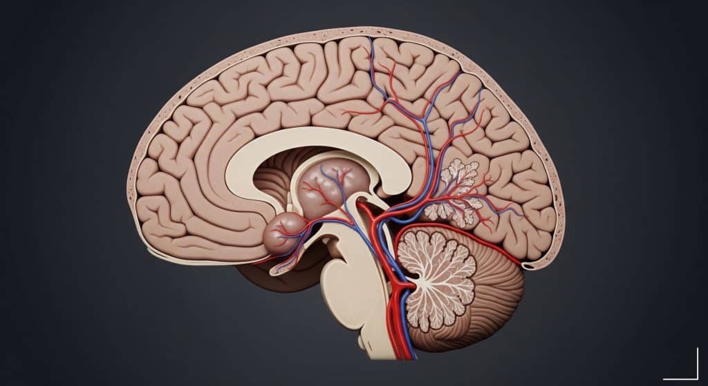
A Berry Aneurysm is a saccular outpouching on a brain artery, usually arising where vessels branch. It differs from fusiform aneurysms, which widen an artery along its length, and from dissecting aneurysms, which result from a tear in the vessel wall. In practice, the saccular shape is the defining feature. As NINDS explains, cerebral aneurysms are grouped by type and rupture status, with saccular aneurysms comprising most cases.
Why this distinction matters. A saccular Berry Aneurysm has a neck that can be clipped or filled with coils. That geometry often influences both risk and treatment selection. It also aligns with where turbulent flow hits arterial bifurcations. That is not a small detail. It is the pattern you and your team plan around.
Common Locations in Brain Arteries
The Berry Aneurysm most often forms on arteries in the circle of Willis. As StatPearls notes, these aneurysms account for about 90% of intracranial aneurysms and cluster at junctions, especially the anterior communicating artery, posterior communicating artery, and middle cerebral artery bifurcation. Roughly speaking, about 85% of cerebral aneurysms sit in the anterior circulation, particularly at branch points, as Cerebral Aneurysm reports.
-
High frequency sites: AComA, PComA, and MCA bifurcation.
-
Less frequent but significant: basilar bifurcation and posterior circulation junctions.
-
Pattern: saccular lesions at bifurcations where flow dynamics are complex.
This clustering is not random. As Radiopaedia describes, saccular lesions at bifurcation points contribute substantially to subarachnoid hemorrhage cases.
Size Classifications and Growth Patterns
Size closely tracks management strategy. As NINDS outlines, common categories are small (less than 11 mm), large (11 to 25 mm), and giant (more than 25 mm). Most small lesions remain asymptomatic. Larger or growing aneurysms can press on nearby structures and trigger neurological symptoms.
|
Size Class |
Typical Considerations |
|---|---|
|
Small (< 11 mm) |
Often monitored; low annual rupture risk if stable. |
|
Large (11-25 mm) |
Higher rupture risk; intervention more likely. |
|
Giant (> 25 mm) |
High mass effect and rupture risk; complex treatment. |
Risk is not size alone. Location matters. As research shows, posterior circulation aneurysms carry higher rupture risk, and even small ruptured aneurysms appear in anterior communicating artery locations. Growth itself is a key warning. As data suggest, growth correlates with age above 50, female sex, smoking, and baseline size, with an approximate growth rate of 2% per year.
Connection to Subarachnoid Hemorrhage Risk
The single most feared complication of a Berry Aneurysm is subarachnoid hemorrhage. As StatPearls notes, ruptured cerebral aneurysms account for about 85% of nontraumatic subarachnoid hemorrhage. The probability rises with aneurysm size and certain locations, particularly in the posterior circulation, as StatPearls also highlights. Precipitating factors can be transient surges in blood pressure. In a focused study, PubMed reports that 3.8% to 14.5% of patients with aneurysmal subarachnoid hemorrhage had sexual activity shortly before rupture.
Prevalence sets context. As AHA journals reports, intracranial aneurysms affect roughly 3% to 6% of the general population, with rupture risk increasing as size exceeds 10 mm and across higher risk groups. The implication is precise. Risk is cumulative and contextual, not binary.
Recognising Brain Aneurysm Symptoms
Warning Signs Before Rupture
You will not always feel a Berry Aneurysm. Some remain silent. Yet a subset produce a warning leak. As PubMed notes, about 15.4% of aneurysmal subarachnoid hemorrhage patients reported sudden headache, vomiting, neck pain, dizziness, or drowsiness before rupture. These events are sometimes misdiagnosed. Timely recognition can change outcomes.
Unruptured lesions may also press on structures, producing pain behind or above an eye, double vision, or facial numbness, as Brain Aneurysm Foundation explains. Treat such changes as a signal. Not a certainty, but a signal.
Emergency Symptoms During Rupture
Rupture is abrupt and dangerous. As StatPearls outlines, classic subarachnoid hemorrhage symptoms include a thunderclap headache, nausea, diplopia, and signs of meningeal irritation. The clinical picture can include vomiting, neck stiffness, and altered consciousness. As PubMed describes, the presentation is a neurological emergency requiring immediate intervention.
-
Sudden severe headache, often the worst ever experienced.
-
Nausea or projectile vomiting.
-
Neck stiffness and light sensitivity.
-
Confusion, seizures, or loss of consciousness.
-
Weakness or acute neurological changes.
As Mayo Clinic notes, many patients describe the pain as the worst headache of their life, sometimes with seizures or collapse. That is not hyperbole. It is a red flag you must act on immediately.
Thunderclap Headache Characteristics
A thunderclap headache peaks within one minute. As StatPearls defines, it is a severe, abrupt headache that often points to a serious cause. Subarachnoid hemorrhage and reversible cerebral vasoconstriction syndrome are among the most common aetiologies. Patients often describe the pain as overwhelming and explosive.
Rapid imaging is essential. As Mayo Clinic outlines, clinical history and urgent neuroimaging help separate benign from dangerous causes. You should treat any first thunderclap headache as an emergency until proven otherwise.
Neurological Changes to Monitor
After a suspected leak or a confirmed rupture, you should monitor cognition, speech, and focal deficits. As StatPearls notes, cognitive impairment can follow a Berry Aneurysm rupture and requires structured assessment. Watch for visual symptoms, sensory changes, or weakness.
Raised intracranial pressure and vasospasm are critical early risks after subarachnoid hemorrhage. As India Clinical Guidelines advise, clinical trajectories and tools such as Transcranial Doppler support early detection of deterioration. Communication changes matter too. As NICE Evidence highlights, post-bleed behavioural and communication shifts can reveal complications that deserve prompt review.
When to Seek Immediate Medical Care
Call emergency services if you experience a thunderclap headache, especially with nausea, vomiting, or neurological deficits. As StatPearls emphasises, severe new headaches with fever, confusion, slurred speech, or focal signs mandate urgent evaluation. This is not a watch-and-wait scenario.
As research shows, warning headaches and projectile vomiting correlate with mortality in aneurysmal subarachnoid hemorrhage, which reinforces the need for immediate care. Recognising combined features like a severe headache with a stiff neck or visual disturbance should prompt an emergency visit, as Mayo Clinic notes. Put simply. Do not delay.
Risk Factors and Causes
Genetic and Family History Connections
Family history changes your baseline risk for a Berry Aneurysm. Clustering in families suggests a genetic component that raises subarachnoid hemorrhage risk, as PubMed indicates. Heritable disorders like Ehlers-Danlos and autosomal dominant polycystic kidney disease also increase risk, as StatPearls notes.
The strongest predictor is a first degree relative with an intracranial aneurysm, as review data explain. Screening discussions are reasonable if multiple relatives are affected, or if associated syndromes are present.
Lifestyle Factors That Increase Risk
Several modifiable behaviours raise the likelihood of a Berry Aneurysm forming or rupturing. Smoking and uncontrolled blood pressure lead the list. As Brain Aneurysm Foundation notes, quitting smoking and controlling hypertension are foundational preventive steps.
-
Stop smoking. The single most effective change you can make.
-
Control blood pressure with medication and lifestyle.
-
Maintain a balanced diet and regular exercise plan.
-
Avoid illicit drugs and heavy daily alcohol use.
As far as current data suggests, daily alcohol intake increases risk, though nuance remains. A Mendelian randomisation study found daily alcohol and smoking increase aneurysm risk, while moderate red wine intake showed a possible protective signal, as ScienceDirect reports. Moderation and blood pressure control are the practical priorities.
Medical Conditions Linked to Berry Aneurysms
Systemic hypertension, connective tissue disease, and polycystic kidney disease are associated with higher risk of a Berry Aneurysm. As AHA journals shows, hypertension increases rupture rates in a dose dependent fashion, and treatment reduces events. Familial patterns also link aortic and cerebral aneurysms, suggesting shared biology, as PubMed indicates.
There is nuance. Diabetes may correlate with aneurysm stabilisation in older patients, as evidence suggests. The mechanism is not fully settled. The actionable point remains steady blood pressure management and tailored surveillance.
Age and Gender Considerations
Risk rises after age 40 and increases again post menopause. Women appear more likely than men to develop a Berry Aneurysm, with risk after menopause rising further, as MedicalNewsToday summarises. Hormonal changes may reduce vascular elasticity, as HBA Support notes. Biology and blood pressure combine here. Screening decisions should reflect both.
Treatment Options for Berry Aneurysms
Monitoring Small Unruptured Aneurysms
Small, stable Berry Aneurysms are often monitored. As review data show, unruptured intracranial aneurysms under 7 mm carry a low annual rupture risk, around 0.7% in some cohorts, and observation is common. Imaging intervals are individualised, typically yearly at first, then spaced if stable.
-
Regular MRI or CTA to track size and morphology.
-
Strict blood pressure control and smoking cessation.
-
Review of new symptoms between scans.
-
Re evaluate if growth exceeds a small threshold or shape changes.
As AHA guidelines advise, your plan should reflect size, location, family history, and personal risk appetite. Observation is not inaction. It is targeted risk management.
Surgical Clipping Procedures
Clipping is a microsurgical technique that excludes the Berry Aneurysm from circulation with a titanium clip. Occlusion rates are high and recurrence is low. As comparative research notes, clipping achieves durable occlusion, though it entails craniotomy and recovery considerations.
Technology now supports intraoperative precision. As Frontiers in Surgery describes, modern imaging and microscopes have improved safety and efficacy. Choice of clipping versus coiling hinges on neck morphology, patient health, and centre expertise. A short example helps. A wide neck MCA bifurcation Berry Aneurysm with branch incorporation may favour clipping for definitive control.
Endovascular Coiling Techniques
Coiling fills the Berry Aneurysm with platinum coils via a catheter from the groin or wrist. It avoids craniotomy and often allows faster recovery. As comparative data indicate, coiling can have lower immediate morbidity and mortality but higher retreatment rates.
-
Standard coiling for narrow neck saccular aneurysms.
-
Balloon assisted coiling for temporary neck support.
-
Stent assisted coiling for wide neck lesions.
Effectiveness is well documented. Complete occlusion after coiling reached 81% in follow up cohorts with low procedure related morbidity, as PubMed reports. For complex necks, stent support shows strong technical success and stable neurological outcomes, as PubMed also notes. Device advances continue to improve results, as recent reviews describe.
Brain Aneurysm Surgery Recovery Process
Recovery timelines differ by approach and individual factors. After clipping, a typical return to routine activities takes 4 to 8 weeks, with fatigue and headaches common, as Alberta Health explains. You should avoid heavy lifting, gradually increase walking, and follow incision care instructions.
Rehabilitation may be needed, especially for speech or movement. As Brain Aneurysm Foundation notes, coordinated inpatient or outpatient therapies support function and confidence. Emotional shifts and memory lapses can persist for months, as BAF outlines. Plan follow ups carefully. They matter for outcomes.
Long-term Management Strategies
Long term care balances rupture risk against procedure risk. As evidence shows, risk rises with size, particular anatomy, and history of subarachnoid hemorrhage. Imaging surveillance remains central. Anxiety after diagnosis is common. As PubMed observes, clear communication about absolute and relative risk helps align decisions with values.
Practical priorities:
-
Control blood pressure to target levels consistently.
-
Stop smoking and moderate alcohol intake.
-
Maintain scan schedules and report new symptoms promptly.
-
Review treatment options if size or shape changes.
And yet, not every change mandates intervention. Context is everything.
Living with Berry Aneurysm Awareness
Knowing you have a Berry Aneurysm can feel heavy. It is basically a durable safety plan, not a sentence. Focus on what is controllable and measurable. Keep a written record of scan dates, sizes, and any symptoms. Share it during appointments so the team can track trends over time.
-
Adopt a blood pressure routine. Same time daily, same cuff, same arm.
-
Create a personal trigger list for urgent care. Thunderclap headache, new weakness, or confusion.
-
Discuss exercise types with your clinician. Low impact training usually fits well.
-
Use plain language summaries after appointments. Clarity reduces stress.
Professionals often mention CAC in cardiology. In aneurysm care, a similar mindset applies to your personal risk ledger. Track inputs and outcomes, then adjust. Small actions compound over time.
Frequently Asked Questions
Can berry aneurysms be prevented?
You cannot remove all risk, but you can reduce it substantially. Stop smoking, maintain tight blood pressure control, and manage cholesterol. Regular exercise and a balanced diet support vascular health. As Brain Aneurysm Foundation notes, these steps limit formation and growth. Family history warrants discussion about screening in select cases.
What percentage of berry aneurysms rupture?
Rupture risk varies by size, location, and patient factors. Small unruptured lesions have an annual risk around 0.7% in some studies, as evidence indicates. Risk rises with aneurysms larger than 10 mm and in certain posterior locations, as AHA journals notes. There is no single percentage that fits all.
How long is recovery after brain aneurysm surgery?
Recovery after brain aneurysm surgery depends on the technique and your baseline health. After clipping, many patients return to routine activities in 4 to 8 weeks, as Alberta Health explains. Coiling often has a shorter initial recovery. Cognitive and emotional healing may take longer. Plan for progressive increases in activity rather than a single milestone.
Are berry aneurysms hereditary?
There is a hereditary component to some Berry Aneurysm cases. Family clustering is recognised, and certain syndromes increase risk. As review data highlights, a first degree relative with an aneurysm remains a strong predictor. Discuss screening if multiple relatives are affected or if associated conditions are present.
What activities should be avoided with an unruptured aneurysm?
Avoid activities that spike blood pressure dramatically or involve straining, especially if poorly conditioned. Manage heavy lifting conservatively and avoid stimulant drugs. Maintain hydration and sleep, and use proper technique during exercise. A tailored plan with your clinician is best. If new brain aneurysm symptoms appear, pause activity and seek review.
How often should unruptured aneurysms be monitored?
Monitoring intervals depend on size, location, and stability. Many centres begin with annual imaging, then extend intervals if unchanged. Any growth, morphological change, or new symptoms should prompt earlier assessment. As AHA guidelines suggest, individualised schedules are safer than fixed rules.