


What Happens During Blood Clot in Brain Surgery?
Dr. Arunav Sharma
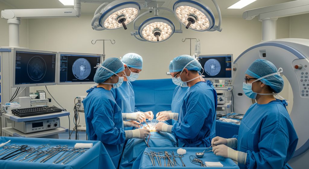
Speed is often praised as the only thing that matters in stroke care. Speed matters, but precision decides outcomes. In practice, blood clot in brain surgery is a set of disciplined choices about access, removal, protection, and repair. I will explain the main surgical options, what happens step by step, and what recovery typically involves. The aim is clear expectations, not drama.
Types of Brain Surgery for Blood Clots
Mechanical Thrombectomy for Ischaemic Stroke
When a vessel is blocked, the task is to restore flow while preserving threatened brain tissue. That is the promise of mechanical thrombectomy. I use dedicated catheters to reach the clot from the groin or wrist. Then I deploy tools that retrieve or aspirate the obstruction. The method spares healthy brain because I am inside the artery, not cutting through tissue.
Timing is critical and nuanced. As StatPearls outlines, intervention typically starts within 6 hours, and in carefully selected cases can extend to 24 hours. That wider window depends on imaging that shows salvageable tissue. I weigh age, stroke severity, clot location, and collateral flow. No single factor decides eligibility.
-
Core techniques include stent retrieval and direct aspiration.
-
Imaging guides both patient selection and device choice.
-
Antiplatelet or anticoagulant plans are tailored after the procedure.
For the avoidance of doubt, mechanical thrombectomy complements clot-dissolving drugs; it does not simply replace them. I use it when large vessel occlusion is likely, and when the anatomy allows catheter access.
Craniotomy for Brain Haemorrhage
When bleeding expands in brain tissue, the goal shifts. I must decompress the brain and control the source. A craniotomy allows direct evacuation of the haematoma and targeted haemostasis. The craniotomy procedure removes a bone flap, opens the dura, and creates a corridor to the clot. I protect vessels and white matter tracts as I work. Then I replace the bone and close in layers.
There is debate about when to operate in spontaneous intracerebral haemorrhage. Trials show mixed results for early open surgery. Yet some patients benefit, especially with deteriorating consciousness, mass effect, or superficial lobar bleeds. The judgement balances clot size, location, shift, and the patient’s physiology. I also consider whether minimally invasive methods can achieve the same effect with less disruption.
-
Indications include worsening neurology or life-threatening pressure.
-
Contraindications include small deep bleeds with stable status.
-
Image guidance and navigation reduce collateral injury.
In simple terms, brain haemorrhage surgery is not one operation. It is several approaches matched to clot depth, anatomy, and risk.
Stereotactic Aspiration for Deep Clots
For deep-seated haemorrhages, precision matters more than size. I often use stereotactic aspiration. A narrow cannula targets the clot through a small opening. I aspirate in a controlled fashion, sometimes with a gentle clot-dissolving agent through a catheter over hours. The aim is to reduce mass effect and intracranial pressure with minimal disturbance of vital tracts.
This technique suits thalamic or basal ganglia bleeds where open corridors risk more harm. Robotics and endoscopic visualisation can improve consistency. The operative footprint is small, and physiological stress is lower. Recovery pathways can then focus on neurorehabilitation rather than wound care.
-
Best for deep clots with mass effect and viable surrounding tissue.
-
Allows staged evacuation when the clot is dense.
-
Pairs well with intensive blood pressure control and ICP monitoring.
It is not a cure-all. Some clots reaccumulate, and some locations still favour open routes. But the risk-benefit profile is often favourable.
Decompressive Craniectomy for Severe Cases
When swelling threatens life, decompressive craniectomy is a rescue. I remove a larger bone section and open the dura to give the brain room. The brain needs space to swell without strangling its own blood supply. Sometimes I combine this with clot removal when pressure is extreme. The bone is stored for later cranioplasty.
This operation demands close ICU support afterwards. Helmet use protects the unshielded brain. The choice between primary decompression and delayed decompression depends on intracranial pressure trends and clinical course. The principle is straightforward. Prevent pressure-driven injury while the primary cause is treated.
-
Indicated for refractory intracranial hypertension.
-
Bone flap is replaced in a planned second procedure.
-
Outcomes vary with age, injury pattern, and timing.
Step-by-Step Surgical Process
Pre-Surgical Preparation and Anaesthesia
Preparation begins days or hours before the theatre list. I review imaging, medications, and comorbidities. The anaesthetics team assesses risks and plans airway, lines, and haemodynamic targets. We adjust anticoagulants if needed and confirm informed consent. Simple steps matter here. Fasting checks, identity checks, and site marking reduce avoidable errors.
General anaesthesia provides immobility and stable physiology for most cases. The anaesthetist titrates drugs and monitors vital signs continuously. Awake techniques are reserved for mapping eloquent cortex in select patients. I coordinate with anaesthesia to protect the brain during induction and emergence. Smooth blood pressure control reduces bleeding and secondary injury.
-
Preoperative tests confirm readiness and optimise safety.
-
Medication reconciliation prevents dangerous interactions.
-
Imaging integration aids device selection and trajectory planning.
For mechanical thrombectomy, prehospital and emergency workflows can compress door-to-puncture time. Good systems matter as much as good hands.
Opening Access to the Brain
Access must be adequate and gentle. In open cases, I plan the incision around vascular territories and venous sinuses. A tailored craniotomy exposes the target with the least tissue transgression. Keyhole variants achieve similar goals with smaller openings, given the right pathology. For posterior fossa work, retrosigmoid exposure protects cranial nerves and cerebellar veins.
For catheter-based thrombectomy, the access site is the artery. I puncture the femoral or radial artery under ultrasound guidance. Sheaths and microcatheters travel through the vasculature to the brain. Roadmap imaging and fluoroscopy guide each turn. I minimise catheter forces to protect the vessel wall.
-
Open routes prioritise safe angles to the lesion.
-
Endovascular routes prioritise atraumatic navigation.
-
Both approaches rely on meticulous planning.
Clot Removal Techniques
Technique choice follows anatomy and clot type. In endovascular work, I use a stent retriever to trap the clot and then withdraw it under aspiration. Alternatively, I place a large-bore aspiration catheter at the thrombus face and remove it with suction. I repeat passes until flow returns and perfusion is restored.
In open haemorrhage evacuation, I first decompress the cavity with controlled suction. Then I identify and secure bleeding points. I maintain gentle retraction and avoid traction on bridging veins. In stereotactic aspiration, I aspirate through a narrow cannula along a planned trajectory. Small, deliberate moves reduce collateral injury.
-
Endovascular tools focus on reperfusion and speed.
-
Open tools focus on haemostasis and decompression.
-
Stereotactic tools focus on accuracy with minimal exposure.
A brief example. A deep thalamic bleed with declining consciousness may suit stereotactic aspiration rather than a wide craniotomy. The patient avoids cortical transgression, and I achieve pressure relief. That trade-off is common in practice.
Monitoring Brain Function During Surgery
I stack three layers of protection during complex cases. First, physiological monitoring for blood pressure, gases, and temperature. Second, neurophysiology such as SSEP and MEP to track pathway integrity in real time. Third, image guidance to maintain safe corridors. If signals change, I pause, irrigate, and reassess. Small adjustments often avert larger injury.
Awake mapping is an additional safeguard when tumours or clots abut language or motor areas. The patient performs simple tasks while I stimulate cortex. This approach is not necessary for most clot evacuations. It remains valuable when millimetres matter.
-
Real-time feedback reduces postoperative deficits.
-
Collaboration between surgeon and neurophysiologist is essential.
-
Monitoring guides not only resection, but also closure timing.
The principle holds across modalities. Measure what matters and respond early. It prevents surprises that do not need to happen.
Closing the Surgical Site
Closure is not an afterthought. I restore watertight dura, replace bone if appropriate, and close muscle and skin with care. The choice of suture or staple affects infection risk and cosmesis. As PubMed reports in broader surgical data, staples can raise infection risk roughly 4 times compared with sutures. I therefore prefer sutures on cranial wounds unless circumstances dictate otherwise.
For decompressive cases, I leave the bone off and shape the dural opening to permit swelling. Drains evacuate residual blood or cerebrospinal fluid. Dressings protect the wound. I record counts, lab results, and imaging plans before handing over to ICU.
-
Watertight dural repair reduces CSF leak risk.
-
Layered closure supports healing and lowers infection rates.
-
Clear postoperative orders prevent missed steps.
Recovery After Blood Clot Surgery
Immediate Post-Operative Care in ICU
ICU care focuses on stability and early detection. Nurses and doctors track neurological status, blood pressure, and oxygenation. I set thresholds for intervention. Any decline in consciousness or new weakness prompts urgent assessment. Temperature control and glucose targets help protect the brain.
Prophylaxis against clots in the legs starts early. I use compression devices first and then introduce anticoagulation when the bleeding risk falls. Strict head positioning and ventilation strategies manage intracranial pressure. Families receive clear updates about goals for the first 24 to 48 hours.
-
Serial neurological exams detect change early.
-
Early mobilisation reduces complications from bed rest.
-
Swift imaging confirms catheter or bone flap position when needed.
Warning signs after transfer include severe headache, new confusion, or focal weakness. These merit rapid evaluation.
Managing Potential Complications
Complications fall into predictable groups. Rebleeding, swelling, and infection lead the list in the early phase. Later risks include deep vein thrombosis and pulmonary embolism. I counter these with mobility protocols, stockings, and tailored anticoagulation. The regimen depends on the index pathology and the operation performed.
Anticoagulation management is a balancing act. Restart too soon, and bleeding resumes. Restart too late, and clots form elsewhere. I use risk scores, imaging, and interdisciplinary discussion to choose timing. Communication between theatre, ICU, and ward teams prevents drift in the plan.
-
Haemostasis checks lower early rebleed risk.
-
Antibiotic stewardship targets genuine infection risk.
-
VTE prevention blends movement and medication.
A word on expectations. Perioperative dips in function can occur. Many improve with oedema resolution and therapy. Some deficits persist. I prepare patients and families for both possibilities.
Rehabilitation Timeline and Milestones
Rehabilitation starts in the ICU with simple steps. Sitting up, breathing exercises, and bedside physiotherapy come first. On the ward, occupational and speech therapists add structured tasks. The plan tracks what matters to the patient. Dressing, walking, and communication goals create momentum.
Hospital stay varies with surgery type and clinical course. As Neurosurgeons of New Jersey note, many patients stay 2 to 4 days before discharge when recovery is uncomplicated. Home recovery often spans weeks, with activity restrictions eased in steps. I schedule wound checks and imaging to confirm safe progress.
-
Week 1 to 2: wound healing and gentle mobility.
-
Week 3 to 6: strength, balance, and endurance training.
-
Week 6 to 12: targeted skills and return-to-work planning.
Two practical markers help. Tolerating a full day without new symptoms. Completing a simple cognitive task list without fatigue. Both guide return to driving and employment discussions, subject to local regulations.
Long-Term Recovery Expectations
Long-term outcomes depend on the cause of the clot, initial severity, and speed to reperfusion or decompression. Stroke related to a large vessel occlusion differs from lobar haemorrhage. Age and comorbidities matter. So does engagement with rehabilitation. Arguably, the most durable gains come from consistent therapy rather than an isolated milestone.
Secondary prevention is the other half. I address blood pressure, diabetes, atrial fibrillation, lipids, and smoking. Antiplatelet or anticoagulant therapy may be indicated. Regular follow up refines the plan. Lifestyle adjustments and medication adherence reduce the odds of a second event.
-
Expect plateaus and then bursts of improvement.
-
Fatigue and mood changes are common and manageable.
-
Cognitive therapy can push recovery beyond the early window.
One final point. Full independence is achievable for many, though not all. Realistic goals and steady work often outperform early predictions.
Conclusion
Blood clot in brain surgery is not a single operation. It is a suite of methods matched to a patient’s anatomy, timing, and goals. Mechanical thrombectomy restores flow fast when arteries are blocked. Craniotomy and stereotactic aspiration decompress and control bleeding. Decompressive craniectomy buys time when pressure rises. The steps are disciplined. Prepare, access, remove, protect, close, and then rehabilitate. Done well, this sequence gives the brain its best chance to recover.
Frequently Asked Questions
How long does blood clot brain surgery typically take?
Mechanical thrombectomy often takes 45 to 120 minutes, depending on access and clot complexity. Open evacuations vary more. A straightforward craniotomy for a superficial haematoma may take 90 to 180 minutes. Stereotactic aspiration can be shorter, though staged catheter cases run longer overall. Decompressive craniectomy adds time for dural expansion and haemostasis. I plan generous theatre time to avoid rushing critical steps.
What are the success rates of mechanical thrombectomy?
Success is judged by reperfusion quality and functional recovery. In expert centres, high reperfusion rates are routine. Functional independence improves when patients are treated promptly and appropriately selected. Results vary with clot location and collateral status. Early presentation and robust imaging increase the odds of meaningful recovery.
Can all brain blood clots be removed surgically?
No. Some clots are small, deep, or clinically silent and do not warrant surgery. Others resolve with medical management alone. Surgery is considered when there is threatened tissue, mass effect, or a reversible obstruction. I match the approach to the clinical picture, not just the scan.
What determines whether craniotomy or thrombectomy is chosen?
The type of stroke decides the route. Mechanical thrombectomy treats blocked arteries in ischaemic stroke. Craniotomy targets intracerebral bleeding with mass effect. Stereotactic aspiration bridges the gap for deep haemorrhages. Patient stability, imaging, and timing also shape the choice. The decision is multidisciplinary and time sensitive.
How soon after stroke symptoms should surgery be performed?
Sooner is generally better. For endovascular cases, many candidates benefit when treated in the first hours after onset. Select patients with favourable imaging may still benefit later. For haemorrhage, urgent intervention follows neurological decline, clot size, and pressure trends. I move swiftly once indications are met and the team is ready.