


Understanding a Brain MRI: What to Expect and Prepare For
Dr. Arunav Sharma
Most medical professionals will tell you that a brain MRI is “nothing to worry about” – just lie still for 45 minutes whilst trapped in a noisy tube. This dismissive reassurance misses the mark entirely. The experience can be genuinely challenging, and knowing exactly what happens during those 45 minutes (and how to prepare properly) transforms the whole ordeal from anxiety-inducing to entirely manageable.
What to Expect During a Brain MRI Scan
The Scanning Environment and Equipment
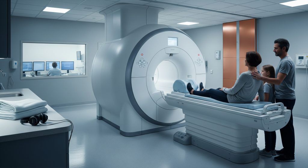
Picture walking into a room dominated by what looks like a massive white doughnut with a narrow bed sliding through its centre. That’s your MRI machine – a 7-foot-tall magnet that’s 30,000 times stronger than Earth’s magnetic field. The room itself feels oddly sterile, kept at a crisp 18-20°C to prevent the machine from overheating. You’ll notice the walls are lined with copper – that’s the Faraday cage keeping radio frequencies contained.
The technician’s booth sits behind a window, filled with monitors displaying what looks like abstract art but is actually your brain in real-time slices. They can see and hear you throughout. Always.
Most facilities now offer “wide-bore” machines with openings of 70cm instead of the traditional 60cm. It’s only 10cm difference, but when you’re lying inside, those extra centimetres feel like freedom.
Duration and Process of the Scan
Here’s what actually happens minute by minute. You’ll spend about 5-10 minutes getting positioned – the technician places padding around your head to minimise movement and fits you with headphones or earplugs. The actual scanning takes 30-60 minutes depending on what your doctor ordered. A standard brain MRI without contrast runs about 30-35 minutes. Add contrast and you’re looking at 45-60 minutes total.
The scan happens in sequences. Each sequence captures different tissue properties and takes 3-8 minutes. Between sequences, you’ll hear the technician’s voice: “This next one is four minutes, starting now.” These breaks are your chance to swallow, clear your throat, or wiggle your toes – just don’t move your head.
|
Scan Type |
Duration |
What It Shows Best |
|---|---|---|
|
T1-weighted |
5-7 minutes |
Anatomy and structure |
|
T2-weighted |
5-7 minutes |
Fluid and inflammation |
|
FLAIR |
4-6 minutes |
Lesions near ventricles |
|
DWI |
2-3 minutes |
Acute stroke |
|
Contrast sequences |
10-15 minutes |
Blood vessels and tumours |
Sounds and Sensations You’ll Experience
Nobody properly prepares you for the noise. It’s not just loud – it’s bizarre. The machine makes rhythmic banging, clicking, and buzzing sounds that reach 110 decibels (similar to a rock concert). Each sequence has its own signature sound: some like a jackhammer, others like an aggressive washing machine, and one that sounds exactly like a fire alarm going off repeatedly. The earplugs they give you knock it down to about 85 decibels. Still loud.
You might feel warmth in your head during certain sequences – that’s the radiofrequency pulses at work. Some people describe a slight tingling sensation or notice their dental fillings feeling odd (they’re perfectly safe, by the way). About 15% of patients experience mild dizziness when the table moves. Completely normal.
Communication with Medical Staff During the Scan
You’re never alone, even though it feels like you’re isolated in a sci-fi pod. The technician watches you through a camera and can hear you through a built-in microphone. They’ll hand you a squeeze ball before the scan starts – your panic button. Squeeze it and the scan stops immediately and the table slides out.
Most technicians check in every 10 minutes or so: “Doing okay in there?” A thumbs up usually suffices. If you need to speak, wait for a break between sequences. Talking during a sequence means they’ll have to repeat it. The entire 6-minute sequence. Again.
Brain MRI with Contrast: What’s Different
About halfway through your scan, the table might slide out and a nurse appears with an IV line. That’s gadolinium contrast being administered – a metal-based dye that makes certain tissues light up like a Christmas tree on the images. The injection takes about 30 seconds. You might taste metal in your mouth or feel a cool sensation travelling up your arm. Both disappear within minutes.
The contrast sequences that follow look for very specific things: active inflammation, blood vessel abnormalities, or the exact boundaries of tumours. Your body clears 90% of the gadolinium through your kidneys within 24 hours. Drink extra water afterwards – six to eight glasses over the next day helps flush it out faster.
How to Prepare for Your Brain MRI
Pre-Scan Medical History and Screening Requirements
The screening questionnaire feels endless but every single question matters. That pacemaker from 2018? Absolutely critical information. The shrapnel from your military service? Could literally be pulled through your body by the magnet. Even that tattoo you got in Bali might be relevant – some older tattoo inks contain metal particles that can heat up during scanning.
Here’s what they’re really worried about:
-
Implanted medical devices (pacemakers, insulin pumps, cochlear implants)
-
Metal fragments in your eyes (especially relevant for welders and metalworkers)
-
Surgical clips or coils from previous procedures
-
Pregnancy – especially first trimester
-
Kidney problems that affect gadolinium clearance
-
Severe claustrophobia requiring sedation
Be honest about everything. That “minor” detail you leave out could be the one that matters.
What to Wear and Items to Remove
Dress like you’re going to yoga – comfortable clothes without any metal. No underwire bras, no jeans with metal zippers or buttons, no compression clothing with metallic threads. Athletic wear usually works perfectly. You’ll be lying still for up to an hour. Comfort matters.
Everything else comes off and goes in a locker:
-
All jewellery (including wedding rings and piercings)
-
Watches and fitness trackers
-
Hair clips, bobby pins, and hair ties with metal
-
Glasses and hearing aids
-
Wallets, keys, phones, and credit cards (the magnet will wipe them)
-
Dentures and removable dental work
-
Medication patches (some contain metal backing)
Dietary Guidelines and Medication Instructions
For a standard brain MRI without contrast, eat normally. No fasting required. Actually, showing up hungry makes the experience worse – low blood sugar can trigger anxiety and make you feel lightheaded in the scanner.
With contrast, the rules change slightly. Eat a light meal 2-3 hours before. Nothing too heavy or greasy – you’ll be lying flat and nausea is a possible (though rare) contrast reaction. Take all your regular medications unless specifically told otherwise. Diabetics should check their levels before and after, as stress can spike blood sugar.
“The single most important preparation? Empty your bladder right before the scan. There’s nothing worse than needing the toilet 20 minutes into a 45-minute scan.” – Every MRI technician ever
Managing Claustrophobia and Anxiety Before the Scan
Let’s be honest – even people who aren’t normally claustrophobic can struggle with MRI machines. You’re in a narrow tube with your head immobilised whilst loud noises assault your ears. It’s not exactly relaxing. About 15% of patients experience significant anxiety and 2% can’t complete the scan without help.
What actually works? Progressive muscle relaxation starting three days before your scan. Practice lying still with your eyes closed for increasing periods. Start with 5 minutes and work up to 20. Use guided meditation apps – the same breathing techniques work brilliantly during the actual scan.
Some facilities offer “mock MRI” sessions where you can experience the machine without actually scanning. Worth asking about. If you know you’ll struggle, discuss sedation options with your doctor beforehand. A small dose of diazepam 30 minutes before can make all the difference. You’ll need someone to drive you home though.
Special Considerations for Children and Elderly Patients
Children under 6 usually need general anaesthesia – lying perfectly still for 45 minutes is beyond most young kids’ capabilities. Between ages 6-10, it depends on the child. Some manage brilliantly with preparation videos and practice sessions. Others need sedation. Many children’s hospitals have themed MRI suites – pirate ships and spaceships are popular. The scanner still makes the same noises but context helps.
Elderly patients face different challenges. Arthritis makes lying flat painful. Hearing aids must be removed, making communication harder. Confusion or dementia complicates consent and cooperation. Most facilities allow a family member to stay in the room (after screening for metal). Their familiar voice between sequences provides enormous comfort.
Brain MRI Cost and Insurance Coverage
Average Brain MRI Costs in the United States
The price variations for the same brain MRI procedure across different facilities will make your head spin (pun intended). Hospital-based imaging centres charge between £2,500-£5,000 for a brain MRI without contrast. The exact same scan at a standalone imaging centre runs £600-£1,800. Same machine, same images, same radiologist reading them. Different price.
With contrast, add another £300-£500 to those figures. Emergency department MRIs? Those can hit £7,000 because they include facility fees and emergency radiologist charges and the “convenience” of immediate scanning and often a markup just because they can.
Factors Affecting the Total Cost
Geography matters enormously. A brain MRI in Manhattan costs three times what the same scan costs in rural Kentucky. But here’s what really drives the price:
|
Cost Factor |
Impact on Price |
Typical Range |
|---|---|---|
|
Facility type |
Hospital vs standalone |
2-4x difference |
|
Time of scan |
Emergency vs scheduled |
3-5x difference |
|
Contrast needed |
Additional drug and time |
+£300-500 |
|
Radiologist reading |
In-network vs out-of-network |
+£200-800 |
|
Additional sequences |
Specialised imaging needs |
+£500-1,000 |
Insurance negotiated rates throw another wrinkle into pricing. The “retail” price might be £3,000, but your insurance company has negotiated it down to £800. If you’re paying cash? Sometimes you can negotiate an even better rate than insurance pays.
Insurance Coverage and Out-of-Pocket Expenses
Most insurance plans cover brain MRIs when deemed medically necessary. But what counts as “necessary”? Persistent headaches, seizures, suspected tumours, stroke symptoms – all typically covered. Annual screening because you’re worried? Not covered.
Your out-of-pocket costs depend on whether you’ve met your deductible. Haven’t met it yet? You’re paying the full negotiated rate until you do. Met your deductible? You’ll pay your co-insurance (typically 20-40% of the negotiated rate) until you hit your out-of-pocket maximum.
Prior authorisation is the hidden gotcha. Your doctor orders the MRI, you schedule it, you show up – then discover your insurance required pre-approval that nobody obtained. Now you’re on the hook for the full amount. Always verify authorisation 48 hours before your scan.
Cost-Saving Options and Facilities
Want to pay less? Here’s what actually works. Standalone imaging centres cost 50-80% less than hospitals for the identical scan. Pay cash upfront and many facilities offer another 20-40% discount. Some imaging centres offer payment plans at 0% interest if you pay within 12 months.
Price shopping makes a massive difference. Call five facilities within driving distance and ask for their cash price for a brain MRI with and without contrast. The variation will shock you. One facility quotes £3,200, another £750. Same scan.
Consider travelling for significant savings. That brain MRI costing £4,000 in Boston might cost £600 in New Hampshire – an hour’s drive away. Even factoring in petrol and time, you’re saving thousands. Some facilities even offer Saturday appointments to avoid missing work.
Brain MRI Side Effects and Safety Concerns
Common Temporary Side Effects
Most people walk out of a brain MRI feeling completely normal. But about 30% experience something. Headaches top the list – usually mild, lasting 2-4 hours, responding well to paracetamol. The cause? Probably the noise and stress rather than the magnetic field itself.
Dizziness affects about 10% of patients, particularly when standing up after lying flat for 45 minutes. Sit up slowly. Stand gradually. The room might spin for a few seconds. Completely normal. Some people report feeling “off” or slightly nauseated for an hour or two afterwards. Nobody knows exactly why, but it passes.
A weird one: temporary changes in taste. About 5% of patients report a metallic taste that lingers for a day or two. Harmless but annoying. Sucking on mints helps.
Gadolinium Contrast Agent Risks and Reactions
Gadolinium gets a bad reputation but serious reactions are incredibly rare – about 1 in 10,000 doses. Mild reactions (nausea, headache, dizziness) occur in about 1-2% of patients. These typically start within minutes and resolve within an hour.
The controversial bit: gadolinium retention. Tiny amounts can remain in your brain and bones years after the scan. Sounds terrifying, right? But here’s the thing – no study has shown any health effects from these deposits in people with normal kidney function. The FDA considers it safe but now requires informed consent.
Nephrogenic systemic fibrosis (NSF) – the serious gadolinium complication – only occurs in people with severe kidney disease. If your eGFR is below 30, you won’t get contrast. Between 30-60, they’ll use a different type and monitor closely. Above 60? Your kidneys clear it fine.
Long-Term Safety Considerations
Here’s what 40 years of MRI scanning has taught us: it’s remarkably safe long-term. No increased cancer risk (unlike CT scans with their radiation). No cognitive effects from repeated scans. No fertility impacts. The magnetic field doesn’t damage DNA or cells.
The biggest long-term “risk”? Incidental findings. Your brain MRI for headaches might reveal a small, harmless cyst that’s been there since birth. Now you know about it. Now you worry about it. Now you need follow-up scans to monitor something that probably never needed finding. This happens in about 3% of brain MRIs.
When to Contact Your Doctor After the Scan
Most post-scan symptoms resolve within hours. But certain things warrant a phone call. Persistent headache lasting more than 24 hours. Any rash or skin changes (possible delayed contrast reaction). Continued nausea or vomiting beyond 4-6 hours. Vision changes or confusion – though these are almost never scan-related.
If you had contrast, watch for signs of allergic reaction over the next 48 hours: widespread rash, difficulty breathing, facial swelling. Extremely rare but needs immediate attention. Your discharge papers will list everything to watch for. Actually read them.
What drives me crazy is when patients don’t report problems because they don’t want to be a bother. You’re not bothering anyone. That’s literally why the aftercare phone number exists.
Making an Informed Decision About Your Brain MRI
Brain MRIs reveal incredible detail about what’s happening inside your skull – from tiny blood vessel changes to millimetre-sized lesions that CT scans miss entirely. The technology is remarkable. But remarkable doesn’t mean comfortable or cheap or entirely risk-free.
Should you get that brain MRI your doctor recommended? Almost certainly yes. The diagnostic value usually far outweighs any temporary discomfort or minor risks. That persistent headache pattern change after 40? Worth checking. Those brief episodes where words don’t come out right? Definitely scan. The “just to be safe” annual screening your concierge doctor suggests? Maybe reconsider.
The real decision isn’t whether to have the scan – it’s how to make the experience as smooth as possible. Choose your facility based on more than just convenience. That standalone imaging centre 20 minutes further away might save you £2,000 and offer a more comfortable wide-bore machine. Ask about their specific machine model, whether they have blankets (those rooms are cold), and what music options they offer.
Don’t tough it out if you’re anxious. Mild sedation doesn’t make you weak – it makes you smart. A relaxed patient produces better images because they move less. Better images mean clearer answers. Clearer answers mean better treatment decisions.
Most importantly, remember that the weird noises and confined space and the whole surreal experience lasts less than an hour. The information gained might change everything about your treatment path. That trade-off? Worth every minute in the tube.
Frequently Asked Questions
Is a brain MRI painful or uncomfortable?
The scan itself is completely painless – you won’t feel the magnetic field or radio waves at all. The discomfort comes from lying still on a hard table for 30-60 minutes and dealing with the loud noises. Most people rate it as awkward rather than painful. The only potential pain point is if you need contrast injection, which feels like any standard IV insertion – a brief pinch.
Can I have a brain MRI if I have dental implants or fillings?
Modern dental work is almost always MRI-safe. Fillings, crowns, bridges, and dental implants made after 1990 use non-magnetic materials like titanium, ceramic, or composite resins. You might feel slight tingling or warmth in dental work during certain sequences, but it’s harmless. Braces are safe too, though they can cause image distortion around the mouth area. Always mention any dental work during screening, but it rarely prevents scanning.
How long does it take to receive brain MRI results?
The frustrating answer: it depends. In emergencies, a radiologist reads the scan within an hour. For routine outpatient scans, expect 24-72 hours for the radiologist’s report to reach your doctor. Your doctor then needs time to review and contact you – typically another 3-5 days. Total time from scan to discussion: usually a week. Some facilities offer disc copies immediately after scanning, but you’ll still need a radiologist’s interpretation.
What’s the difference between open and closed MRI machines?
Closed MRIs – the traditional tubes – have stronger magnets (1.5-3 Tesla) producing clearer, more detailed images. Open MRIs use weaker magnets (0.3-0.7 Tesla) but offer more space and less claustrophobia. The trade-off is significant: open MRI images are fuzzier and might miss subtle abnormalities. Brain scans especially benefit from the higher resolution of closed MRIs. Only choose open MRI if claustrophobia absolutely prevents you from completing a closed scan.
Can I drive myself home after a brain MRI with contrast?
Yes, unless you received sedation. Gadolinium contrast doesn’t impair driving ability or judgement. You might feel slightly dizzy or have a mild headache, but nothing that makes driving unsafe. The technician will make sure you’re steady on your feet before discharge. If you had any sedation (even mild anti-anxiety medication), you’ll need someone else to drive. No exceptions on that one.
Are brain MRIs safe during pregnancy?
MRI safety during pregnancy is complicated. No study has shown harm to developing babies from MRI scans, but we avoid them during the first trimester unless absolutely necessary. Second and third trimester scans are considered safe when medically justified. Gadolinium contrast, however, crosses the placenta and is avoided throughout pregnancy except in life-threatening situations. Each case needs individual risk-benefit analysis. Your obstetrician and radiologist will jointly decide what’s appropriate.