


Brain Cancer Stages Explained: Basics, Grading, and Diagnosis
Dr. Arunav Sharma
Most people assume brain cancer staging works just like it does for breast or lung cancer. You get a stage one through four, and that number tells you how far things have spread. Simple, right? Except when it comes to the brain, that assumption can steer you completely wrong. The grading system used for brain tumours functions on an entirely different logic. It measures how aggressive the cells look under a microscope, not how far the disease has travelled through your body. Understanding this distinction isn’t just academic. It fundamentally changes how doctors approach treatment, how prognosis gets discussed, and what questions patients should be asking their medical teams.
I’ve spent considerable time unpacking the confusion surrounding brain cancer stages, and honestly, the single most frustrating part of this topic is how often the terms “stage” and “grade” get used interchangeably when they mean very different things. Let’s clear that up and walk through what actually matters when it comes to brain tumour classification and diagnosis.
Brain Cancer Grades and Staging System
The World Health Organisation (WHO) grading system is the gold standard here. It classifies brain tumours from Grade 1 to Grade 4 based on how the cells behave and appear when examined microscopically. Think of it like assessing a building’s structural integrity. You’re not measuring where the building is located on a map; you’re measuring how likely it is to collapse based on its construction.
Grade 1: Low-Grade Tumours
These are the slow movers. Grade 1 tumours grow at a snail’s pace and typically have well-defined edges that make surgical removal more straightforward. The cells look almost normal under a microscope. Pilocytic astrocytomas fall into this category, and they’re more commonly found in children and young adults. The good news? Complete surgical removal often results in a cure. It’s basically the best-case scenario in brain tumour classification.
Patients with Grade 1 tumours frequently don’t need radiation or chemotherapy after surgery. The key is getting clean margins during the operation, which means the surgeon removes all visible tumour tissue. Follow-up imaging becomes critical to catch any potential recurrence early.
Grade 2: Low-Grade Tumours
Here’s where things get slightly more complicated. Grade 2 tumours are still considered low-grade, but they have a tendency to infiltrate surrounding brain tissue. The edges become blurrier, making complete removal more challenging. These cells show some abnormality under the microscope but aren’t dividing rapidly.
The tricky part about Grade 2 tumours? They can transform. Over time, a Grade 2 tumour can evolve into a higher grade, which is why ongoing surveillance becomes non-negotiable. Patients might live for years or even decades with a Grade 2 tumour, but that doesn’t mean the story ends there.
Grade 3: High-Grade Tumours
This is where the term “malignant” genuinely applies. Grade 3 tumours show clear signs of aggressive behaviour. The cells are actively dividing and look distinctly abnormal. Anaplastic astrocytomas and anaplastic oligodendrogliomas live in this territory. Treatment typically involves a combination approach: surgery first (to remove as much tumour as safely possible), followed by radiation and often chemotherapy.
The prognosis shifts significantly at Grade 3. We’re no longer talking about potential cures but rather about controlling disease progression and maintaining quality of life for as long as possible. That sounds grim, but advances in targeted therapies are genuinely changing outcomes for many patients.
Grade 4: High-Grade Tumours
Glioblastoma. That’s the name most people recognise and the one that generates the most fear. Grade 4 tumours are the most aggressive form of brain cancer. They grow rapidly, spread through healthy brain tissue, and are notoriously difficult to treat. The cells show extreme abnormality, with areas of dead tissue (necrosis) and new blood vessel formation feeding the tumour’s growth.
Standard treatment follows an aggressive protocol: maximal safe surgical resection, followed by concurrent radiation and temozolomide chemotherapy. But here’s the honest truth. Despite best efforts, Grade 4 tumours typically recur. Current research focuses heavily on immunotherapy and tumour-treating fields, which represent some of the more promising developments in recent years.
TNM Staging vs WHO Grading
This is the distinction that trips everyone up. The TNM system (Tumour, Nodes, Metastasis) that works brilliantly for cancers like colon or breast cancer simply doesn’t apply to brain tumours in the traditional sense. Why? Because brain tumours rarely spread through lymph nodes or metastasise to distant organs. They’re locally destructive but usually stay confined to the central nervous system.
So when someone asks about brain cancer stages, what they should really be asking about is the WHO grade. The grade tells you about cellular behaviour and aggressiveness. That’s what drives treatment decisions and prognosis predictions. Don’t even bother with TNM staging when discussing primary brain tumours. It’s essentially irrelevant.
Progression Between Grades
Here’s something that often catches patients off guard: grades aren’t static. A low-grade tumour diagnosed today can become a high-grade tumour years down the line. This transformation happens because tumour cells accumulate additional genetic mutations over time. The brain tumour that started as a manageable Grade 2 can evolve into an aggressive Grade 4.
This progression isn’t inevitable, but it’s common enough that neurologists watch for it carefully. Regular MRI scans serve as the early warning system, detecting changes in tumour behaviour before symptoms develop. When progression occurs, treatment strategies shift accordingly.
Common Brain Tumour Types and Classification
Brain tumour classification goes beyond grades. The type of cell that becomes abnormal determines what category the tumour falls into, and this classification has major implications for treatment and outcomes.
Primary Brain Tumours
Primary brain tumours originate in the brain itself or in tissues immediately surrounding it, like the meninges or pituitary gland. They develop from brain cells, glial cells, or other structures within the skull. Gliomas account for the largest share of primary brain tumours, but meningiomas and pituitary adenomas are also quite common.
The brain cancer causes behind primary tumours remain largely mysterious. Some genetic syndromes increase risk (Li-Fraumeni syndrome, neurofibromatosis), and ionising radiation exposure has clear links. But for most patients? The cause stays unknown. That uncertainty can be maddening, but it’s the reality.
Secondary Brain Tumours
Also called metastatic brain tumours, these start elsewhere in the body and spread to the brain. Lung cancer, breast cancer, melanoma and kidney cancer are the most common culprits. Secondary brain tumours are actually more common than primary ones in adults. The cancer cells travel through the bloodstream and establish new colonies in brain tissue.
Treatment for secondary brain tumours differs fundamentally from primary tumours. The approach must address both the brain lesions and the original cancer. Stereotactic radiosurgery, whole-brain radiation, and targeted therapies all play roles depending on the number and location of metastases.
Gliomas Classification
Gliomas arise from glial cells, the supportive tissue of the brain. They’re subdivided based on which type of glial cell they originate from:
-
Astrocytomas – arise from star-shaped cells called astrocytes and include the dreaded glioblastoma
-
Oligodendrogliomas – develop from cells that produce myelin, the protective coating around nerve fibres
-
Ependymomas – originate from cells lining the ventricles and central canal of the spinal cord
Recent advances in molecular testing have revolutionised glioma classification. Specific genetic markers like IDH mutation status and 1p/19q codeletion now factor into diagnosis and significantly influence prognosis. An IDH-mutant glioma, for instance, generally carries a better outlook than its IDH-wildtype counterpart.
Meningiomas Classification
Meningiomas grow from the meninges, the protective membranes covering the brain and spinal cord. They’re typically benign (Grade 1) and slow-growing. Many meningiomas get discovered accidentally during brain imaging performed for unrelated reasons.
But here’s where it gets nuanced. Not all meningiomas are harmless. Atypical meningiomas (Grade 2) and anaplastic meningiomas (Grade 3) show more aggressive features and higher recurrence rates. Location matters enormously too. A small meningioma pressing on critical structures can cause significant problems even if the cells look perfectly benign under the microscope.
Pituitary Tumours
The pituitary gland sits at the base of the brain and controls hormone production throughout the body. Tumours here, called pituitary adenomas, are almost always benign. They cause problems through two mechanisms: mass effect (pressing on nearby structures like the optic nerves) and hormonal disruption.
Some pituitary tumours actively secrete hormones, leading to conditions like acromegaly (excess growth hormone) or Cushing’s disease (excess cortisol). Others are “non-functioning” and cause symptoms only when they grow large enough to compress surrounding tissues. Treatment ranges from medication to surgery depending on tumour type and size.
Paediatric Brain Tumours
Children develop different brain tumour types than adults, and the location patterns differ too. Medulloblastomas, ependymomas, and pilocytic astrocytomas appear more frequently in paediatric populations. Many paediatric tumours arise in the posterior fossa, the area at the back of the skull containing the cerebellum and brainstem.
The developing brain adds complexity to treatment decisions. Radiation therapy, highly effective against many tumour types, carries significant long-term risks for children including cognitive impairment and endocrine dysfunction. Oncologists must balance immediate tumour control against decades of potential side effects.
Recognising Brain Tumour Symptoms
Brain tumour symptoms depend heavily on location, size, and growth rate. There’s no single warning sign that definitively points to a brain tumour, which makes early detection genuinely challenging. What drives me crazy is how often general symptoms get dismissed for months before anyone thinks to order a brain scan.
Early Warning Signs
The classic early symptoms include:
-
Persistent headaches – often worse in the morning or when lying down
-
Nausea and vomiting – particularly in the morning without clear cause
-
Vision changes – blurred vision, double vision, or loss of peripheral vision
-
Seizures – sometimes the very first symptom, especially in adults with no prior seizure history
-
Cognitive changes – difficulty concentrating, memory problems, confusion
Here’s the frustrating reality: these symptoms overlap with dozens of other conditions. Migraines, stress, eye strain, medication side effects. Most people experiencing these symptoms don’t have brain tumours. But when symptoms persist, progress, or combine in unusual ways, investigation becomes essential.
Location-Specific Symptoms
The brain has specialised regions, and tumours in different areas produce distinct symptom patterns:
|
Brain Region |
Associated Symptoms |
|---|---|
|
Frontal lobe |
Personality changes, weakness on one side, speech difficulties |
|
Temporal lobe |
Memory problems, seizures, hearing disturbances |
|
Parietal lobe |
Difficulty with spatial awareness, reading, writing |
|
Occipital lobe |
Visual disturbances, blindness in part of visual field |
|
Cerebellum |
Balance problems, coordination difficulties, dizziness |
|
Brainstem |
Double vision, swallowing problems, facial weakness |
Understanding this mapping helps explain why symptoms vary so dramatically between patients. A frontal lobe tumour might manifest primarily as personality changes that family members notice before the patient does. A cerebellar tumour might make someone look intoxicated due to balance and coordination problems.
Symptoms in Children vs Adults
Children can’t always articulate what they’re experiencing, making symptom recognition harder. Parents and caregivers often notice behavioural changes first: irritability, declining school performance, reluctance to participate in activities the child previously enjoyed.
Physical signs in children may include:
-
Head tilting to one side
-
Regression in developmental milestones
-
Increasing head circumference in infants
-
Frequent falls or clumsiness
-
Morning vomiting that improves as the day progresses
Adults tend to develop symptoms more gradually and may unconsciously compensate for subtle deficits. A person might not realise they’ve stopped using one hand as much, or that they’re having slightly more difficulty finding words. It’s often a spouse or colleague who first notices something is off.
Emergency Warning Signs
Some symptoms demand immediate medical attention:
-
Sudden severe headache – the “worst headache of your life”
-
New seizure activity – especially in someone with no seizure history
-
Sudden weakness or numbness – particularly on one side of the body
-
Sudden vision loss
-
Confusion or decreased consciousness
These can indicate rapidly growing tumours, bleeding into a tumour, or dangerous swelling around tumour tissue. They require emergency evaluation, typically starting with a CT scan followed by MRI if needed.
Brain Cancer Diagnosis Methods
Diagnosing a brain tumour involves a systematic process that moves from clinical suspicion through imaging to tissue confirmation. The journey from first symptom to definitive diagnosis typically takes weeks, though emergency presentations can compress this timeline dramatically.
Neurological Examination
Every brain tumour workup starts with a detailed neurological exam. This isn’t just about checking reflexes (though that happens too). The neurologist assesses cranial nerve function, motor strength, sensation, coordination, cognitive function, and visual fields. Each component provides clues about tumour location.
I’ve seen cases where an experienced neurologist localised a tumour with remarkable accuracy based purely on examination findings, before any imaging was performed. The physical exam remains a powerful diagnostic tool even in our imaging-heavy era.
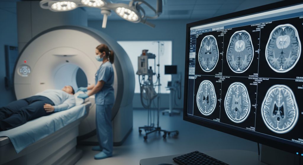
Imaging Tests
Imaging is where diagnosis becomes concrete. The key modalities include:
-
MRI (Magnetic Resonance Imaging) – the gold standard for brain tumour detection. Provides exquisite detail of brain structures and tumour characteristics. Various sequences (T1, T2, FLAIR, diffusion-weighted) reveal different tumour properties.
-
CT Scan (Computed Tomography) – faster than MRI and often the first test performed in emergency settings. Better at detecting acute bleeding or calcification within tumours.
-
MR Spectroscopy – measures chemical composition of tissue, helping distinguish tumour from other abnormalities.
-
PET Scan – shows metabolic activity and helps differentiate tumour recurrence from treatment-related changes.
The real change happened when MRI became widely available. You stopped relying purely on symptoms and started seeing exactly what was happening inside the skull. It’s a game-changer for diagnosis and treatment planning.
Biopsy Procedures
Imaging can strongly suggest a brain tumour, but definitive diagnosis requires tissue. There are two main biopsy approaches:
Stereotactic needle biopsy: Using coordinates from brain imaging, a neurosurgeon inserts a thin needle through a small skull opening to extract tissue samples. This minimally invasive approach works well for deep-seated tumours or when surgical removal isn’t planned.
Open (craniotomy) biopsy: When surgery is planned anyway, tissue diagnosis happens during the resection. The surgeon removes as much tumour as safely possible while obtaining samples for pathological analysis.
Getting tissue samples allows pathologists to examine cell appearance, perform immunohistochemistry, and send material for molecular testing. All of this information feeds into the final diagnosis and treatment plan.
Molecular Testing
Modern brain tumour diagnosis extends beyond what cells look like to how they behave at the genetic level. Key molecular markers now routinely tested include:
-
IDH mutation status – mutations in isocitrate dehydrogenase genes generally indicate better prognosis
-
1p/19q codeletion – characteristic of oligodendrogliomas and associated with better treatment response
-
MGMT promoter methylation – predicts response to temozolomide chemotherapy in glioblastomas
-
H3K27 alterations – important for classifying midline gliomas
These molecular findings are now integrated directly into the WHO classification system. Two tumours that look identical under the microscope might receive different diagnoses based on their molecular profiles, and those differences genuinely matter for treatment selection.
Staging Workup Process
Once a brain tumour is confirmed, additional testing may be needed depending on tumour type. For primary brain tumours, this typically involves spinal MRI to check for spread along the spinal cord (particularly important for certain tumour types like medulloblastoma or ependymoma).
For suspected metastatic disease, the workup reverses direction: if the brain tumour appears to be secondary, imaging of the chest, abdomen, and pelvis searches for the primary cancer. Sometimes the brain lesion is the first sign that cancer exists elsewhere in the body.
The complete diagnostic picture including tumour type, grade, location, molecular features and patient health status guides treatment recommendations. This information forms the foundation for discussions between patients, families and medical teams about next steps.
Understanding Your Brain Cancer Diagnosis
Receiving a brain tumour diagnosis ranks among the most overwhelming experiences anyone can face. The information comes fast, the terminology feels alien, and decisions seem urgent even when they’re not. Taking time to absorb what you’ve learned matters.
Most people focus immediately on prognosis numbers. But what does this actually mean for your specific situation? Survival statistics describe populations, not individuals. They’re based on historical data and may not reflect recent treatment advances. Your outcome depends on factors including tumour type and grade and location and your overall health and response to treatment. Each journey is unique.
Getting a second opinion is standard practice and expected by most neuro-oncology teams. Major treatment decisions benefit from confirmation by specialists at high-volume brain tumour centres. Don’t hesitate to seek additional perspectives, especially for complex cases.
Support resources exist at multiple levels: medical teams (oncologists, nurses, social workers), patient organisations, and peer support groups. The path ahead may be difficult, but no one needs to walk it alone. Understanding your diagnosis marks the first step toward actively participating in your care.
Frequently Asked Questions
What is the difference between brain cancer stage and grade?
Stage describes how far a cancer has spread through the body. Grade describes how abnormal and aggressive the tumour cells appear under a microscope. For brain tumours, grading (WHO Grade 1-4) matters more because brain cancers rarely spread outside the central nervous system. The grade tells doctors how the tumour is likely to behave and guides treatment decisions.
Can a low-grade brain tumour become high-grade?
Yes, this progression happens regularly. Low-grade tumours can accumulate genetic changes over time, transforming into more aggressive forms. A Grade 2 tumour might become Grade 3 or even Grade 4 years after initial diagnosis. This is why ongoing monitoring with regular MRI scans remains essential even for tumours initially classified as low-grade.
How long does brain cancer diagnosis take?
The timeline varies considerably. From first symptom to definitive diagnosis might take anywhere from days (for emergency presentations) to months (when symptoms develop gradually). The imaging itself happens quickly, but waiting for biopsy scheduling and pathology results adds time. Molecular testing can take an additional one to two weeks. Most patients have complete diagnostic information within three to four weeks of starting the formal workup.
Are all brain tumours cancerous?
No. Brain tumours can be benign (non-cancerous) or malignant (cancerous). Benign tumours like most meningiomas and pituitary adenomas don’t invade surrounding tissue aggressively. However, even benign brain tumours can cause serious problems depending on their size and location. A “benign” tumour pressing on critical brain structures still requires treatment.
What causes brain cancer to develop?
For most brain tumours, the cause remains unknown. Established risk factors include ionising radiation exposure and certain inherited genetic syndromes (like neurofibromatosis and Li-Fraumeni syndrome). Despite popular concerns, current evidence doesn’t support mobile phone use as a brain cancer cause. The honest answer is that brain cancer causes remain largely unidentified for most patients.
Can brain cancer be detected through blood tests?
Currently, no reliable blood test exists for diagnosing brain tumours. Research into “liquid biopsy” approaches that detect tumour DNA or other markers in blood is ongoing and shows promise, but these tests haven’t yet reached routine clinical practice. Brain tumour diagnosis still requires imaging and, for definitive classification, tissue biopsy.