


What You Need to Know About Oral Cancer Screening in India
Dr. Akriti Rastogi
Routine check-ups are often sold as a formality. For oral cancer screening, that thinking is costly. Early detection shifts outcomes, reduces treatment intensity, and keeps quality of life intact. I will map the practical options available in India and when each approach is useful. Then I will outline warning signs, the main oral cancer causes, programmatic access points, and what action to take next. The aim is simple. Make timely oral cancer screening feel doable and not distant.
Top Oral Cancer Screening Methods Available in India
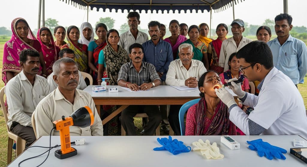
Conventional Oral Examination (COE)
I start with COE because it underpins every other method. A trained clinician inspects the lips, tongue, floor of mouth, buccal mucosa, palate, and gingiva under good light. I palpate suspicious areas gently to judge induration, borders, and tenderness. It is quick, low cost, and effective for triage in clinics and camps. As a baseline for oral cancer screening, COE helps decide who needs an adjunct test or biopsy. It also supports counselling on tobacco cessation and follow-up planning. Strengths are accessibility and immediate judgement. Limitations are subjectivity and the risk of missing subtle field changes.
-
Best use: first-line assessment and documentation.
-
What I look for: colour change, ulceration, fixation, and nerve symptoms.
-
Next step: adjunctive tool or biopsy if suspicion persists.
Toluidine Blue Staining
Toluidine Blue selectively highlights cells with increased nucleic acid content. Dysplastic and malignant epithelium often retains the dye, which helps me target the most suspicious zone. As an adjunct to COE, it improves lesion localisation for scalpel biopsy. I treat it as a decision aid, not a verdict. False positives occur with inflammation or trauma. A careful re-evaluation after local factors settle is prudent. In high-risk users of tobacco or alcohol, this method can focus attention on areas that appear borderline on routine examination. That precision can reduce sampling error and delay.
-
Best use: selecting a biopsy site within a heterogeneous patch.
-
Strength: highlights potentially dysplastic epithelium.
-
Constraint: interpret alongside clinical context and history.
Mobile Health Technology Screening
Mobile tools extend reach. That is their power. Training frontline workers to capture high-quality oral images and structured histories can lift detection at the community level. As WHO notes, digital platforms can support visual examinations, structured triage, and public education in one workflow. I have seen health workers use secure apps to tag lesion sites, track risk behaviours, and escalate urgent cases. The process shortens time to specialist review. It also creates a record for follow-up, which matters when people travel for work or miss appointments. Privacy, consent, and image quality must be handled rigorously. But the model scales in a way paper registers never could.
-
Best use: community screening and continuity of care.
-
Strength: rapid referral and audit trail of decisions.
-
Constraint: connectivity, device upkeep, and structured training.
OralScan Handheld Device
Adjunctive optics can reveal changes invisible to normal light. OralScan uses diffuse reflectance and autofluorescence principles to differentiate suspicious tissue. The device guides the examiner with real-time visual cues. Preliminary validation suggests sensitivity around 78% and specificity near 90%, as CSRBOX reports, which fits its role as a triage tool rather than a stand-alone diagnosis. In practice, I use such devices to grade concern, select biopsy sites, and document baseline images for review. Portable form factor helps in camps and district clinics. Interpretation still relies on training and a disciplined protocol.
-
Best use: triage, biopsy guidance, and longitudinal imaging.
-
Strength: objective optical contrast in real time.
-
Constraint: learning curve and need for quality control.
Brush Biopsy with Cytology
Brush cytology samples the full epithelial thickness with minimal discomfort. Results can flag atypia in lesions that look equivocal. I use it when a lesion is small, the patient is anxious about a scalpel biopsy, or when access to operative facilities is limited. A positive or atypical result strengthens the case for an incisional biopsy. A negative result reduces immediate concern but does not override a strong clinical suspicion. It is an adjunct for oral cancer screening, not a replacement for histopathology. Good lab support and clear reporting templates improve value.
-
Best use: low-to-moderate suspicion lesions where reassurance or escalation is needed.
-
Strength: minimally invasive sampling with quick turnaround.
-
Constraint: confirmatory scalpel biopsy is still required for diagnosis.
Key Warning Signs and Oral Cancer Symptoms to Recognise
Persistent Sores and Ulcers
An ulcer that does not heal within a short, defined period deserves evaluation. I inspect the base, margins, and any induration. Non-healing, contact bleeding, and rolled edges raise concern. In the presence of risk behaviours, I escalate promptly. These patterns are among the classic oral cancer symptoms encountered in clinics.
White or Red Patches
Leukoplakia and erythroplakia demand attention. Homogeneous white patches may be low grade, yet mixed or speckled areas carry higher risk. Red patches with a velvety look and easy bleeding are particularly concerning. I document size, texture, and site to support timely review.
Lumps and Swellings
Any new mass in the oral cavity or neck requires systematic assessment. Fixation to deeper planes and irregular borders increase suspicion. I palpate regional nodes and consider imaging when clinically justified. Early referral is wise when features cluster unfavourably.
Numbness and Pain
Altered sensation or persistent pain without obvious cause can signal neural involvement. Trigeminal or lingual nerve changes deserve a careful mapping of affected areas. I am cautious when pain is disproportionate to visible findings. That mismatch often points to deeper disease.
Difficulty Swallowing and Jaw Problems
Dysphagia, trismus, or bite changes attract attention. I check inter-incisal opening and masticatory tenderness. Ulcers near the retromolar trigone or floor of mouth may impair function early. These signs, alongside other oral cancer symptoms, support urgent review and structured staging.
Understanding Oral Cancer Causes and Risk Factors in India
Tobacco and Areca Nut Use
Smoked and smokeless tobacco drive risk. Areca nut, with or without tobacco, contributes to oral submucous fibrosis and malignant change. Habit type, frequency, and placement site matter. I ask about quid composition and duration. Behavioural support to quit is integral to care. These exposures sit at the centre of oral cancer causes observed across clinics.
Alcohol Consumption Patterns
Alcohol acts synergistically with tobacco. Heavy intake compromises mucosal barriers and alters metabolism. I assess weekly units, binge patterns, and co-use with tobacco. Brief counselling helps, though sustained change needs follow-up. Risk falls after cessation, but not immediately.
HPV Infection Risk
High-risk HPV subtypes are implicated in oropharyngeal disease and, to a limited extent, oral sites. Sexual practices and immune status shape risk. I do not overstate this factor for typical buccal or tongue lesions in India, yet I keep it in mind. Vaccination policies will shape future trends.
Age and Gender Factors
Risk rises with age due to cumulative exposure and field cancerisation. Male predominance reflects habit profiles, though women with areca nut use also present. I adjust screening advice for those with long-standing habits. Biology explains part of the pattern. Access and stigma explain the rest.
Government Screening Programmes and Access Points
Health and Wellness Centres
Health and Wellness Centres under Ayushman Bharat are designed to deliver community-based prevention and screening. Trained multipurpose workers can perform visual checks and refer. When integrated with oral cancer screening workflows, these centres bring services closer to rural households. The model works best with standard forms, risk scoring, and clear referral pathways.
-
Walk-in visual assessment and counselling.
-
Referral to dental or ENT clinics for escalation.
-
Local follow-up for habit cessation and wound checks.
NPCDCS Implementation Status
The National Programme for Prevention and Control of Cancer, Diabetes, Cardiovascular Diseases and Stroke supports opportunistic screening. Implementation varies by district, which affects coverage and follow-up. Some districts run regular camps and digital registries. Others rely on ad hoc activities with paper records. I advise clinicians to align with district NCD cells to standardise referral and data capture. It reduces loss to follow-up and duplication.
Cost-Effectiveness of Screening Approaches
Cost-effectiveness hinges on three variables. Reach, triage accuracy, and time to treatment. COE remains the lowest cost first step. Adjuncts add value when they shorten the path to definitive biopsy, not when they delay it. Community models that use trained workers and tele-triage tend to reduce travel costs and lost wages. The right blend is context specific. Urban outreach may favour clinic-based optics. Remote blocks may favour mobile imaging with rapid referral.
Challenges in Programme Delivery
Programme gaps are predictable. Staff turnover, inconsistent supplies, and limited supervision. Data fragmentation still hampers continuity across facilities. I see improvement when teams standardise forms and commit to monthly audit. Another point is public awareness. Screening uptake improves when people know what it involves and where to go. Technology helps, but leadership and simple routines decide pace.
Taking Action for Early Detection
Here is the practical workflow I recommend. It is straightforward and scalable.
-
Risk classify in minutes. Record tobacco, areca nut, and alcohol use. Note age and comorbidities.
-
Perform a structured COE. Document sites, colour, borders, and palpation notes with photographs.
-
Use an adjunct tool when clinical doubt remains. Choose toluidine blue, optical imaging, or brush cytology.
-
Escalate to biopsy without delay when red flags cluster. Fixation, contact bleeding, or persistent ulceration.
-
Book follow-up at the time of the visit. Do not leave it open ended.
-
Provide cessation support for habits driving risk. It improves outcomes more than any gadget.
This is oral cancer screening used well. Focused, documented, and connected to treatment. Small steps compound when the system is consistent.
Frequently Asked Questions
How often should high-risk individuals undergo oral cancer screening?
For people with daily tobacco or areca nut use, I advise a structured check every six months. If a suspicious lesion is under observation, I shorten the interval. Once healing is confirmed or risk behaviours change, the schedule can widen. Oral cancer screening is a process, not a single event. The cadence should reflect risk and clinical findings.
What are the survival rates for early-stage oral cancer in India?
Survival improves markedly when disease is caught early. Stage I and II disease often allow curative treatment with better function preserved. Figures vary by centre and follow-up rigour, and results depend on margins and nodal status. The reliable takeaway is simple. Earlier oral cancer screening increases the chance of organ-sparing treatment and durable control.
Is oral cancer screening covered under Ayushman Bharat?
Health and Wellness Centres support preventive services, including visual checks and referral. Under Ayushman Bharat – PMJAY, hospital procedures are covered for eligible beneficiaries. Screening per se happens in primary care. I suggest confirming local enrolment and referral processes. This reduces delays if a biopsy or surgery becomes necessary.
Can I perform self-examination for oral cancer at home?
Self-checks can help you notice change. Use a mirror and good light to inspect the tongue, cheeks, and floor of mouth. Look for ulcers, colour change, or lumps. If a lesion persists or hurts, seek a professional opinion promptly. Self-checks are not a substitute for oral cancer screening by a trained clinician. They complement it.
Which oral cancer stage requires immediate medical attention?
Any suspected malignancy warrants immediate attention regardless of stage. That said, symptoms pointing to advanced local invasion or nodal disease demand urgent escalation. Difficulty swallowing, trismus, nerve numbness, or a fixed neck node are examples. Oral cancer stages guide treatment planning, but the first task is prompt biopsy and staging workup.
Earlier, I emphasised community pathways and documentation. One more point. Digital tools only matter when they shorten time to diagnosis. As MoHFW highlights for primary care, mobile systems can support screening and follow-up when embedded into routine practice. That is worth building into local protocols.
To close, a concise reminder. Use COE as the foundation. Add adjuncts thoughtfully. Refer early when red flags persist. This is disciplined oral cancer screening. It protects lives and preserves function.