


What Is Uveitis Treatment? Symptoms, Causes and Relief Options
Dr. Krishna Vaitheeswaran
Standard advice says any red, painful eye can wait for a routine check. That counsel is risky. With uveitis, delay invites damage, and swift, precise uveitis treatment preserves vision. I will set out the options, the likely course, and how to avoid common pitfalls. It is basically a practical guide for patients and clinicians who want clarity without drama.
Uveitis Treatment Options and Medications
Corticosteroid Eye Drops for Anterior Uveitis
For anterior disease, I start with topical corticosteroids as first line. They reduce intraocular inflammation quickly when dosed intensively, then tapered in step with clinical response. As Mayo Clinic notes, stronger formulations such as prednisolone acetate 1 percent penetrate better than 0.1 percent, and prolonged use may raise intraocular pressure or accelerate cataract formation, so I schedule pressure checks.
-
Indications: new anterior flare, unilateral or bilateral.
-
Dosing: frequent in the first 24 to 72 hours, then gradual taper.
-
Monitoring: ocular pressure, lens clarity, symptomatic response.
Combination drops with an antibiotic and steroid can be useful when surface infection risks exist, though I separate infection control from inflammation control in most cases. The principle holds. Use the least invasive route that controls inflammation, keep taper steady, and avoid abrupt stops.
When discussing uveitis medications with patients, I emphasise technique. A good instillation routine improves bioavailability and outcomes. A poor one leads to undertreatment and avoidable clinic returns.
Oral and Injectable Steroids for Severe Cases
Systemic corticosteroids enter when the disease is bilateral, sight threatening, or posterior. In practice, I choose oral prednisolone for broad control and add periocular or intravitreal steroid when a local boost is needed. Intravenous dosing is reserved for fulminant inflammation or when oral therapy cannot wait to act. Tapering follows activity, not the calendar.
-
Systemic benefits: rapid suppression of widespread inflammation.
-
Systemic risks: hyperglycaemia, hypertension, mood change, bone loss.
-
Mitigation: proton pump inhibitor when indicated, calcium and vitamin D, and a clear taper plan.
This step is often the bridge within an overall uveitis treatment plan. It buys time while I organise steroid-sparing therapy, or it rescues vision during a dangerous flare. But still, prolonged systemic steroids without a plan usually create more problems than they solve.
Immunosuppressive Medications for Chronic Uveitis
When flares recur or steroid burden climbs, I introduce immunosuppressive medications. Methotrexate, mycophenolate mofetil, azathioprine, and ciclosporin are familiar steroid-sparing tools. They reduce relapse frequency and allow a lower maintenance steroid dose. Multidisciplinary coordination with rheumatology is standard, especially when systemic disease is present.
-
Selection factors: uveitis type, comorbidities, pregnancy plans, adherence.
-
Baseline checks: full blood count, liver enzymes, renal function, viral status where relevant.
-
Follow up: scheduled labs and structured symptom review.
The aims are simple. Reduce cumulative steroid exposure, stabilise inflammation, and protect vision. Costs and access can shape choices in some settings, so I adapt the regimen to what is available without compromising safety. This is uveitis treatment as a long game, not a sprint.
Mydriatic Eye Drops to Prevent Complications
Mydriatic and cycloplegic drops support comfort and prevent posterior synechiae. I use short acting agents for mild cases and longer acting agents when the iris is sticky or very inflamed. As Mayo Clinic records, tropicamide often acts within 25 to 30 minutes and can last 4 to 8 hours, while some individuals experience longer effects, so I advise on light sensitivity and driving.
-
Benefits: pain relief, photophobia reduction, synechiae prevention.
-
Typical issues: blurred vision, glare, near focus difficulty while active.
In a severe anterior flare, I may step up to a longer acting option for several days. The goal is mechanical prevention of iris adhesions while the anti inflammatory therapy takes effect. It is routine and quietly effective.
Biologic Therapy for Refractory Cases
When conventional agents fail or are intolerable, I escalate to biologics. These agents target cytokines and pathways central to autoimmune activity. They offer a decisive steroid-sparing effect for many patients with non infectious uveitis.
In a small refractory cohort, adalimumab accounted for **61 percent** of use and infliximab for **22 percent**, and **16 of 18** patients showed visual improvement after therapy, as the published study reports. The signal is consistent with broader clinical experience. Response is often brisk, though monitoring for infection risk and rare adverse events is essential.
I agree clear entry criteria upfront: inadequate control on immunosuppressants, unacceptable steroid dose, or sight threatening relapse. Then I define objective milestones. Reduced cell grade, fewer flares, and stable imaging. No guesswork.
Surgical Interventions When Medical Treatment Fails
Surgery enters the picture for complications or when opaque media hide pathology. I consider cataract extraction, glaucoma procedures, or vitrectomy depending on the problem. The inflammation should be controlled for several months before elective surgery where possible. That reduces postoperative flare and improves outcomes.
-
Cataract from steroids or chronic inflammation.
-
Secondary glaucoma from steroid response or angle damage.
-
Persistent vitreous haze or traction needing vitrectomy.
Combined perioperative steroid and immunosuppression strategies help. The surgical plan must reflect uveitis aetiology, macular status, and systemic factors. Precision matters more than speed.
Recognising Uveitis Symptoms and Getting Diagnosed
Early Warning Signs of Eye Inflammation
I look for pain, redness around the limbus, photophobia, and sudden blur. Floaters suggest vitreous involvement. Pressure sensation can point to secondary ocular hypertension. These are classic uveitis symptoms, but they overlap with other eye disorders, so slit lamp confirmation is non negotiable.
-
Pain that worsens with light exposure.
-
New floaters or hazy vision developing over days.
-
Perilimbal redness and tender eye on palpation.
Swift triage matters. A same day review is prudent when pain and photophobia combine with blur.
Differences Between Anterior, Intermediate and Posterior Symptoms
Anterior disease skews towards pain and light sensitivity. Intermediate disease brings more floaters and variable blur. Posterior uveitis can be deceptively painless yet dangerous, with distortion or scotomas leading the complaint. Panuveitis mixes features across segments.
Here is why this distinction matters. The segment guides the uveitis treatment route of delivery and the urgency of systemic work up. It also shapes the follow up cadence and imaging plan.
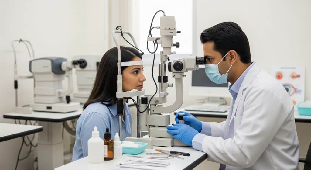
Diagnostic Tests and Eye Examinations
Diagnosis remains clinical at the slit lamp and indirect ophthalmoscope. I grade cells and flare, assess intraocular pressure, and examine the fundus. Imaging supports management rather than replaces the examination. Optical coherence tomography clarifies macular status. Fundus photography tracks change over time.
-
Anterior assessment: cell grade, keratic precipitates, synechiae.
-
Posterior assessment: vitreous haze, vasculitis, choroiditis, macular oedema.
-
Laboratory tests: targeted to suspected uveitis causes, not a fishing expedition.
I order tests to answer a question, not to fill a panel. That restraint reduces false positives and needless referrals.
When to Seek Emergency Eye Care
Immediate care is appropriate when vision drops quickly, pain is severe, or the eye is very red with nausea. New central distortion, a black curtain, or flashes with a shower of floaters warrant urgent assessment. Steroid naive patients with a painful red eye must be reviewed before any drop is used.
Speed prevents damage. Delay risks synechiae, macular oedema, or elevated pressure that spiral beyond simple fixes.
Understanding Uveitis Causes and Risk Factors
Autoimmune Conditions Linked to Uveitis
Several systemic autoimmune diseases associate with intraocular inflammation. I commonly see links with HLA-B27 spondyloarthropathy, sarcoidosis, juvenile idiopathic arthritis, and inflammatory bowel disease. These conditions can set tempo and recurrence. They also determine which systemic partner specialties I involve early.
This is where a joined plan matters. Shared protocols limit duplication of tests and streamline therapy.
Infectious Causes of Eye Inflammation
Infective aetiologies must stay on the table, especially in posterior disease. Herpetic disease, toxoplasmosis, tuberculosis, and syphilis are the usual suspects. I rule these out before starting heavy immunosuppression. Treatment changes direction if infection is present. Antimicrobials first, then cautious anti inflammatory support.
Misclassification leads to harm. An antiviral or antibiotic can reverse the course when chosen correctly.
Genetic and Environmental Risk Factors
Genetic predisposition, such as HLA markers, raises risk in some uveitides. Environmental exposures, including pathogens and smoking, shape incidence and severity. The combination often decides relapse patterns more than any single factor.
Roughly speaking, risk is multifactorial. That is why a checklist approach outperforms a single hunch.
Age-Related Patterns in Different Types
Children are more prone to silent anterior disease in the context of juvenile idiopathic arthritis. Adults present across the spectrum, with anterior flares being the most frequent clinic encounter. Older adults bring more posterior pathology and comorbidity. Age guides screening intensity and communication strategies with families.
Different ages, different risks. The therapeutic principles remain stable.
Managing Complications and Long-Term Care
Common Uveitis Complications to Monitor
Secondary glaucoma, cataract, macular oedema, band keratopathy, and epiretinal membrane are the usual complications. I check pressure at every visit, review the macula on OCT when blur lingers, and watch the lens carefully during steroid courses. These uveitis complications are predictable. Vigilance keeps them manageable.
-
Pressure rise during steroid therapy.
-
Macular swelling after a sudden taper.
-
Capsular fibrosis in chronic inflammation.
Preventing Vision Loss Through Regular Follow-ups
Follow up cadence matches risk. Early on, I compress intervals to catch a rebound. As stability persists, I widen visits with clear return precautions. Structured monitoring converts a volatile disease into a controlled one.
In practice, I use a simple rule. Taper the medicine and stretch the appointments only when the eye stays quiet.
Lifestyle Adjustments for Better Management
Lifestyle does not replace treatment, but it supports it. I counsel on smoking cessation, medication adherence, and sick day rules. For those on systemic therapy, vaccination planning and infection avoidance matter. Blue light filters do not treat inflammation. Regular sleep and stress control help symptom tolerance.
-
Keep a flare diary with date, trigger, and therapy response.
-
Carry a current medication list for emergency care.
Paediatric Considerations for Children with Uveitis
Children need gentle examinations and strong coordination with paediatrics and rheumatology. Asymptomatic inflammation is common, so screening is scheduled, not symptom driven. Growth, schooling, and psychosocial support sit alongside therapy. I adjust dosing forms and teach families the instillation routine carefully.
Parents manage most doses. Make it simple, consistent, and documented.
Taking Control of Your Uveitis Treatment Journey
Good outcomes come from early diagnosis, a tailored plan, and disciplined follow up. That is the spine of effective uveitis treatment. I recommend agreeing a written plan that lists the current drops, systemic agents, taper steps, and triggers for urgent review. Small details matter, such as one pharmacy, one calendar, and one point of contact.
-
Clarify the type of uveitis and what that implies for relapse risk.
-
Set objective targets: quiet anterior chamber, OCT baseline, pressure stability.
-
Define boundaries: how far to taper, what to do if pain returns.
A brief example helps. A patient with HLA-B27 anterior flares uses a fixed steroid taper and a short mydriatic course, with a follow up at two weeks and six weeks. A text line is available for early warnings. Flares halve, and vision stabilises. Simple, documented, enforced.
All of this sits within a wider conversation about uveitis causes, the expected horizon, and the rare need for surgery. The intent is steady control and preserved sight, not firefighting. That is achievable.
Frequently Asked Questions
How long does uveitis treatment typically last?
Duration varies by type and severity. Anterior flares may need weeks of drops with a measured taper. Chronic or posterior disease often requires months of systemic therapy and planned maintenance to prevent relapse.
Can uveitis be cured permanently or will it return?
Some cases are single events, but many are relapsing. With precise uveitis treatment, relapses become less frequent and less severe. Long remission is common, though permanent cure cannot be guaranteed.
Are there natural remedies that complement medical uveitis treatment?
Supportive measures like rest and smoking cessation help comfort. They do not suppress intraocular inflammation. Evidence based therapy remains essential. Discuss supplements with your clinician to avoid interactions with uveitis medications.
What happens if uveitis is left untreated?
Risks include synechiae, elevated pressure, macular oedema, cataract, and vision loss. Some damage becomes irreversible. Early assessment and targeted uveitis treatment prevent most complications.
Can children develop uveitis and how is their treatment different?
Yes. Children, especially with juvenile idiopathic arthritis, can have silent inflammation. Screening is scheduled, not symptom led. Dosing, monitoring, and family education are adapted to growth and school life.
Is uveitis treatment covered by health insurance in India?
Coverage varies across insurers and policies. Outpatient drops are sometimes out of pocket, while hospital therapies and biologics may be covered. Verify preauthorisation rules and keep detailed prescriptions and reports.