


What Is the Cataract Surgery Procedure? From Start to Finish
Dr. Krishna Vaitheeswaran
Conventional wisdom says cataract surgery is simple because it is quick. The truth is more disciplined. The Cataract Surgery Procedure is a sequence of tightly controlled steps, each designed to protect the cornea, stabilise the lens capsule, and deliver predictable vision. This guide explains what happens, why it matters, and how your choices affect outcomes. You will see the full pathway from preparation to follow-up, with practical detail you can use in real consultations.
Step-by-Step Breakdown of the Cataract Surgery Procedure
Pre-operative Eye Preparation and Anaesthesia
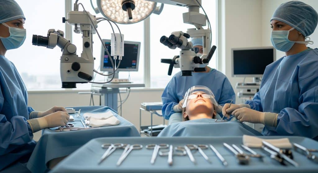
Before the Cataract Surgery Procedure begins, your eye is measured, cleaned, and numbed. Nurses instil antibiotic and dilating drops to reduce microbial load and widen the pupil. Anaesthesia is usually topical with lidocaine gel, though a sub-Tenon block may be chosen for added comfort. You must remain still. A sterile drape and lid speculum maintain a clean field. The aim is straightforward. Minimise risk and ensure you feel pressure only, not pain. This preparation sets the tone for the next precise steps.
-
Topical or sub-Tenon anaesthesia based on case complexity.
-
Pupil dilation for safe access to the lens capsule.
-
Antisepsis with povidone-iodine to lower infection risk.
Creating the Corneal Incision
Your surgeon begins the Cataract Surgery Procedure with a clear corneal incision of roughly 2 to 2.8 mm. The tunnel architecture is trapezoidal and self-sealing, which preserves globe integrity. A secondary paracentesis is made for instrument access. Balanced salt solution deepens the anterior chamber. The location is chosen to avoid astigmatism induction or to neutralise it strategically. Precision here pays off later, because a stable chamber improves control during lens work.
Performing Capsulorhexis to Access the Lens
The continuous curvilinear capsulorhexis is the disciplined centrepiece of the Cataract Surgery Procedure. Using a cystotome or forceps, the surgeon creates a round opening of about **5** to **5.5** mm in the anterior capsule. The circle must be centrically overlapped by the intraocular lens optic for long-term stability. A well-sized rhexis facilitates nuclear manipulation and reduces posterior capsular opacification to an extent. If the pupil is small, mechanical dilation or viscomydriasis is used.
Breaking Up the Cataract with Phacoemulsification
Phacoemulsification uses ultrasound to segment and liquefy the nucleus. In the Cataract Surgery Procedure, this step determines efficiency and corneal clarity after surgery. The phaco tip emulsifies the lens while the sleeve cools and protects the incision. Energy settings are tailored to lens hardness, which varies with age and comorbidity. Lower effective phaco time usually correlates with faster recovery. Technique choice here links directly to astigmatism control and endothelial safety.
Lower ultrasound energy and stable fluidics often mean crisper corneas on day one.
Removing Lens Fragments Through Aspiration
Once the nucleus is divided, fragments are aspirated safely. The Cataract Surgery Procedure then advances to cortical clean-up. Bimanual or coaxial irrigation-aspiration removes residual cortex from the capsule equator. The posterior capsule is polished judiciously. Overly aggressive polishing risks tears. Too little leaves lens epithelial cells behind. The goal is a clear bag with an intact capsule that will hold the intraocular lens implant centrally and securely for years.
-
Steady foot pedal control to maintain chamber depth.
-
Gentle equatorial stripping for full cortical removal.
Inserting the Intraocular Lens Implant
After cohesive viscoelastic opens the capsular bag, the intraocular lens implant is folded and delivered via an injector. In the Cataract Surgery Procedure, optic capture and haptic positioning matter. The lens unfolds in the bag, aligns with the visual axis, and sits behind the iris. If a toric lens is used, alignment to the planned meridian is essential. Centration affects quality of vision, halos, and contrast. Care is taken to remove viscoelastic to prevent pressure spikes.
|
Step |
Purpose |
|---|---|
|
Lens delivery |
Secure optic within the capsular bag |
|
Rotation/centring |
Optimise refractive outcome and stability |
|
Viscoelastic removal |
Reduce postoperative pressure rise |
Self-Sealing Incision Closure
The Cataract Surgery Procedure concludes with chamber pressurisation and incision testing. Hydration of the stromal tissue seals the tunnel without sutures in most cases. If the wound is not fully watertight, a single suture may be placed. Antibiotic drops are instilled. A clear shield protects the eye as you leave theatre. Stability at this point reduces foreign body sensation and lowers infection risk. Small actions here prevent large problems later.
Types of Intraocular Lens Implants Available
Monofocal IOLs for Single Distance Vision
Monofocal lenses provide one focal point, typically distance. In the Cataract Surgery Procedure, they remain the standard for predictable clarity and contrast sensitivity. You may still need reading glasses. Monofocal optics offer reduced photic phenomena compared with complex designs. They suit individuals who value crisp distance performance and simplicity. If you have ocular surface disease, monofocals often provide the most stable quality of vision across lighting conditions.
-
Clear distance vision for driving or television.
-
Reading glasses required for near tasks.
Multifocal IOLs for Multiple Distance Correction
Multifocal lenses split light for distance, intermediate, and near. When chosen during the Cataract Surgery Procedure, they can reduce spectacle dependence. Trade-offs include halos and reduced contrast, particularly at night. Careful selection and counselling are critical. If you frequently drive at night, consider whether visual phenomena would be acceptable. The best candidates have healthy maculae and controlled dry eye. Expectations must align with optical physics, not wishful thinking.
Toric IOLs for Astigmatism Correction
Toric lenses neutralise corneal astigmatism and sharpen the image. The Cataract Surgery Procedure includes preoperative keratometry and axis planning to position the toric optic accurately. Rotation after implantation degrades performance, so surgeons aim for rotational stability within a few degrees. Toric optics can be combined with monofocal or multifocal designs. The result is cleaner edges and finer detail, especially for distance tasks like road signs or sport.
Extended Depth of Focus (EDOF) Lenses
EDOF lenses stretch the focal range to enhance intermediate vision while maintaining distance clarity. In the Cataract Surgery Procedure, EDOF options often suit computer-heavy work and active lifestyles. Night halos are typically milder than multifocals, though not absent. Reading glasses for very small print may still be needed. For many patients, this is a sensible compromise between range and visual comfort across varying light conditions.
Light Adjustable Lenses for Post-Surgery Customisation
Light Adjustable Lenses allow postoperative refractive tuning with targeted ultraviolet treatments. When incorporated into the Cataract Surgery Procedure, they create a safety valve for small prediction errors. You wear UV-blocking glasses until the lock-in treatment. This approach helps achieve precise outcomes in prior refractive surgery eyes, where measurements are tricky. It is a premium path. Yet for complex optics, customisation after healing is a persuasive advantage.
Phacoemulsification Techniques and Surgical Approaches
Divide and Conquer Technique
Divide and conquer sculpts deep grooves, then cracks the nucleus into quadrants. In the Cataract Surgery Procedure, it balances control and visibility. The grooves reduce stress on the capsule during subsequent emulsification. Energy is delivered into the nuclear core, away from the endothelium. For moderately dense lenses, it is dependable and reproducible. It may take slightly longer than chop methods, which is an acceptable exchange for control.
Stop-and-Chop Method
Stop-and-chop starts with a central trench, stops to crack the nucleus, then proceeds with chopping. The Cataract Surgery Procedure benefits from this hybrid because it combines predictable initial control with efficient fragmentation. It suits a range of densities. Surgeons often favour it when the red reflex is dull, because the trench provides depth orientation. Fewer large manipulations reduce zonular stress, which protects the capsular bag.
Direct Chop Technique
Direct chop engages the nucleus and splits it immediately without trenching. Within the Cataract Surgery Procedure, it can reduce ultrasound time and fluid usage. The technique relies on firm nuclear purchase and stable fluidics. It excels in very hard lenses, where quick segmentation saves energy. The trade-off is a steeper learning curve and less margin for error. In experienced hands, results are excellent and efficient.
Femtosecond Laser-Assisted Cataract Surgery
Laser assistance automates corneal incisions, capsulotomy, and nucleus pre-fragmentation. When integrated into the Cataract Surgery Procedure, it offers geometric precision and potentially less phaco energy. Docking, suction, and imaging add steps and time. Outcomes are comparable in many series, though centration and rhexis regularity can be superb. The value case is strongest for premium IOLs and dense nuclei. Cost and access remain practical considerations in routine care.
Manual Small Incision Cataract Surgery
Manual small incision cataract surgery uses a self-sealing scleral tunnel and manual nucleus delivery. In the Cataract Surgery Procedure, this approach is useful where phaco technology is limited, or in very dense lenses. It reduces ultrasound exposure entirely. The incision is larger than micro-incision phaco, which may induce more astigmatism. With careful tunnel design and suturing strategy, outcomes are consistently good. It remains a vital technique globally.
Cataract Surgery Recovery Timeline and Expectations
First 24-48 Hours Post-Surgery Care
Expect mild irritation, watering, and light sensitivity. Your drops schedule begins immediately. For most patients, the cataract surgery recovery time starts with clear improvement in the first **24** hours. You should wear the shield at night and avoid eye rubbing. Gentle walking is acceptable. Heavy lifting, dusty environments, and swimming are deferred. If pain is significant or vision worsens suddenly, contact the clinic promptly. Early vigilance prevents most complications from escalating.
-
Shield at night for **1** week.
-
Topical antibiotics and steroids as prescribed.
-
No eye makeup for several days.
Week 1: Initial Healing Phase
By week one, vision is typically functional for daily tasks. During this stage of the Cataract Surgery Procedure pathway, inflammation settles and the cornea clears. You can read, cook, and manage screens, provided comfort allows. Avoid high-impact exercise. Use artificial tears if dryness appears. The first follow-up confirms pressure, wound integrity, and IOL centration. Most patients feel confident by day **7**. Minor glare at night is common and often fades.
Weeks 2-4: Vision Stabilisation Period
Between weeks two and four, refraction stabilises. You will discuss final spectacle needs, especially if monofocal lenses were selected. The Cataract Surgery Procedure aims to minimise dependence on glasses, but small adjustments can sharpen acuity. If you chose toric or multifocal optics, neuroadaptation continues through this period. Maintain drop tapering as instructed. Reading fine print may still improve incrementally as contrast recovers and the tear film normalises.
Full Recovery at 4-8 Weeks
Most eyes reach stable clarity by **4** to **8** weeks. At this milestone of the Cataract Surgery Procedure, the capsule has settled and the ocular surface is steady. Any residual refractive error can be corrected with glasses or, selectively, laser enhancement. If posterior capsular opacification develops later, a quick YAG laser capsulotomy usually restores brightness. Long-term stability is excellent for the majority of patients, though regular checks remain sensible.
Long-Term Visual Outcomes and Follow-up
Expect sustained clarity and improved contrast, assuming healthy retinas and controlled comorbidities. The Cataract Surgery Procedure is also a form of lens replacement surgery, so presbyopia management is part of the discussion. Annual optometry checks are advisable. If you notice new floaters, flashes, or sudden blur, seek review swiftly. Small behavioural choices help maintain results. Good lid hygiene and dry eye management support consistent quality of vision.
Conclusion
The Cataract Surgery Procedure is not a single moment. It is a carefully choreographed sequence that begins with preparation, proceeds through precision lens work, and ends with disciplined aftercare. Technique selection influences energy usage and corneal clarity. IOL choice determines how you see across distances and in varied light. Recovery hinges on adherence to drops, activity restraint, and timely review. With the right plan, you gain clarity and confidence. With the right expectations, you keep both.
Frequently Asked Questions
How long does the actual cataract surgery procedure take?
The operating time for a single eye is typically **10** to **20** minutes. Allow longer for preparation, checks, and recovery room monitoring.
Is cataract surgery painful during or after the procedure?
Anaesthetic drops prevent pain, though you may feel pressure or cool fluid. Mild soreness later responds to prescribed drops and simple analgesia.
When can I resume driving after cataract surgery?
Drive once legal vision standards are met and glare is manageable. This is often within a few days, subject to individual healing and clinician advice.
What are the potential risks and complications of cataract surgery?
Risks include infection, inflammation, pressure rise, cystoid macular oedema, and capsule tear. Serious complications are uncommon, but prompt reporting improves outcomes.
Will I still need glasses after cataract surgery with lens replacement?
Possibly. Monofocals usually need readers. Multifocal or EDOF lenses reduce dependence, yet small spectacles may refine night driving or fine print.
How soon can I return to work following cataract surgery?
Desk work often resumes in **2** to **3** days. Physically demanding or dusty roles may require **1** to **2** weeks, pending surgeon guidance.
Can both eyes undergo cataract surgery at the same time?
Most surgeons stage eyes days or weeks apart to reduce compounded risk. Same-day bilateral surgery is selective and protocol driven in certain settings.
A final note for clarity: throughout the Cataract Surgery Procedure, choices on phacoemulsification settings, the intraocular lens implant, and perioperative care shape your result and your cataract surgery recovery time. Treat each decision as part of one continuous pathway.