


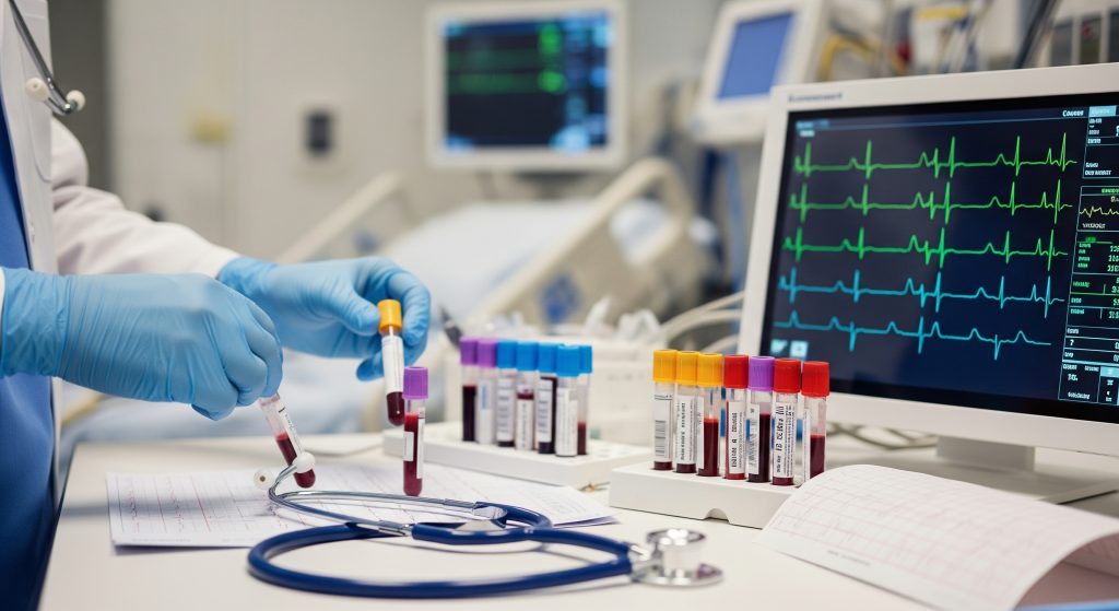


What Cardiac Markers Reveal About a Heart Attack
Dr. Hriday Kumar Chopra
Disclaimer: The content shared here is for informational purposes only. Always consult a specialist doctor before attempting any treatment, procedure, or taking any medication independently.
Most advice still treats a heart attack as a single moment. In practice, it is a moving target. Cardiac Markers help you catch that movement in real time, quantify the damage, and separate genuine emergencies from lookalikes. This guide clarifies which tests matter, how their levels change, and how to interpret results without guesswork.
Key Cardiac Markers That Diagnose Heart Attacks
1. Troponin I and T: Gold Standard for Heart Attack Detection
Troponin I and T are the frontline Cardiac Markers for diagnosing acute myocardial infarction. They are specific to cardiac muscle and rise when heart cells are injured. You rely on the 99th percentile upper reference limit as the decision boundary, not a fixed universal number. A rising or falling pattern with at least one value above that limit signals acute injury.
-
High clinical specificity for heart muscle injury.
-
Detectable rise typically within several hours of symptom onset.
-
Remain elevated long enough to confirm late presenters.
A single result rarely tells the full story. Serial sampling does. It shows dynamics, not a snapshot.
2. CK-MB Test: Monitoring Heart Muscle Damage Timeline
The ck-mb test sits behind troponin today, yet it retains value for timing. CK-MB rises and falls faster than troponin, which helps you estimate the window of injury. It is less specific, so you interpret it alongside symptoms, ECG changes, and troponin. It supports pattern analysis rather than standing alone.
-
Useful for assessing reinfarction when troponin remains elevated.
-
Faster normalisation allows rough timing of the event.
-
Particularly informative when paired with serial troponin testing.
3. Myoglobin: Early Detection Within First Hours
Myoglobin appears very early after muscle injury. That speed is attractive. Specificity is poor because skeletal muscle also releases it. Use it as an early signal, then confirm with troponin. If your department has high-sensitivity assays, myoglobin adds little. If not, it can flag risk in the first hours.
-
Very early rise supports rapid triage.
-
Low specificity limits stand-alone diagnostic value.
-
Best used as a rule-in prompt pending troponin results.
4. Natriuretic Peptides: BNP and NT-proBNP for Heart Stress
Natriuretic peptides reflect ventricular wall stress rather than direct necrosis. They help distinguish cardiac from non-cardiac breathlessness and quantify heart failure severity. As Biomarkers in Routine Heart Failure Clinical Care – PMC explains, BNP and NT-proBNP function as the practical gold standard markers for diagnosing heart failure and for monitoring its course.
Evidence is broad based. As BNP and NT-proBNP as Diagnostic Biomarkers for Cardiac … reports, both peptides indicate cardiac dysfunction risk and support early diagnostic decisions. Complementary research emphasises prognostic value. As Role of B-type natriuretic peptide (BNP) and NT-proBNP in … notes, levels correlate with wall stress and stratify risk, which is critical during acute presentations that may mimic infarction.
Cut-offs are not one size fits all. As Guideline for the Use of Natriuretic Peptides in the Early … highlights, thresholds depend on age, renal function, and clinical context. This nuance matters for accuracy. Recent bioinformatic work widens the lens. As Integrative bioinformatic analysis of prognostic biomarkers … underscores, these peptides mirror pathophysiology and support both diagnosis and prognosis in heart failure.
Practical takeaway. Use natriuretic peptides to measure stress load and heart failure probability. Use troponin to confirm myocardial injury.
5. High-Sensitivity Troponin: Enhanced Early Detection Capabilities
High-sensitivity troponin assays detect very low concentrations with high precision. This upgrade improves early rule-out and rule-in pathways. You can safely discharge low-risk patients using validated algorithms at 0 and 1 hour or 0 and 2 hours. Precision at the low end enables fast decisions with fewer admissions.
-
Detects smaller injury earlier compared with conventional assays.
-
Reduces uncertainty in the grey zone around the 99th percentile.
-
Supports accelerated diagnostic protocols when paired with ECG and risk scores.
How Cardiac Markers Change During Heart Attack
Timeline of Cardiac Enzyme Release Patterns
Cardiac Markers follow predictable kinetics after myocardial injury. The sequence helps you time the insult and plan care. Roughly speaking:
-
Myoglobin rises earliest within a few hours, then declines quickly.
-
CK-MB rises shortly after, peaks within a day, and normalises earlier than troponin.
-
Troponin rises later than myoglobin, peaks within one to two days, and remains elevated longer.
These patterns provide temporal clues. They do not replace clinical context.
Peak Levels and Duration for Each Biomarker
|
Biomarker |
Typical Kinetics After Infarction |
|---|---|
|
Myoglobin |
Early rise within hours. Peaks quickly. Returns to baseline within roughly **24-36** hours. |
|
CK-MB |
Rises within several hours. Peaks by about **24** hours. Normalises within **2-3** days. |
|
Troponin I or T |
Rises within several hours. Peaks at **24-48** hours. Elevated for **5-14** days depending on assay. |
|
BNP or NT-proBNP |
Reflects stress load. Not used to time infarction. Tracks haemodynamic status over days to weeks. |
Assay methods vary, so timing estimates shift by several hours. Methodology matters.
Serial Testing Protocols for Accurate Diagnosis
Diagnosis relies on change over time. A cardiac enzyme test strategy with serial sampling captures that change. High-sensitivity pathways commonly test at presentation and at **1-2** hours. Conventional assays may need longer intervals.
-
Obtain baseline troponin at arrival with ECG and clinical review.
-
Repeat troponin at a validated interval to detect rise or fall.
-
Consider CK-MB to time reinfarction if troponin remains elevated from a recent event.
Document the delta. A significant absolute or relative change carries more weight than a single value just above the threshold.
Interpreting Rising and Falling Marker Trends
Look for a coherent pattern. A rising troponin with symptoms and ECG changes supports infarction. A falling pattern after revascularisation suggests stabilisation and reperfusion success. Flat, low values across serial tests help you rule out myocardial infarction with confidence.
-
Rising trend + clinical features = probable acute injury.
-
Falling trend = evolving or resolving injury.
-
No change = consider non-cardiac causes or chronic elevations.
The interpretation hinges on pre-test probability. Numbers do not stand alone.
Understanding Normal vs Elevated Cardiac Marker Ranges
Reference Ranges for Troponin I and T
Troponin uses the 99th percentile upper reference limit from a healthy population. Each assay sets its own value. You should avoid comparing numbers across platforms. Focus on whether the result exceeds that limit and whether there is a dynamic rise or fall.
|
Principle |
Decision boundary is the 99th percentile for the specific assay. |
|
Reporting |
Use whole numbers or ng/L as reported by the laboratory method. |
|
Interpretation |
Elevated plus change over time suggests acute injury; static elevation may be chronic. |
In practice, reference documentation from your laboratory is decisive. Assay heterogeneity is considerable.
CK-MB Normal Values and Heart Attack Thresholds
CK-MB reference ranges vary by laboratory. Many labs report a small upper limit at rest, then a higher alert threshold for suspected infarction. The pattern over time remains central. A marked rise and fall supports a diagnosis when aligned with symptoms and ECG findings.
-
Use the ck-mb test to clarify reinfarction timing.
-
Avoid cross comparing absolute values from different labs.
-
Confirm with troponin where possible, as it is more specific.
BNP and NT-proBNP Cut-off Points
BNP and NT-proBNP cut-offs depend on age, renal function, and clinical setting. Thresholds for acute presentations differ from chronic care. As Guideline for the Use of Natriuretic Peptides in the Early … indicates, clinicians should adapt cut-off points to demographic and clinical context for best accuracy.
|
Acute dyspnoea setting |
Use lower rule-out thresholds and higher rule-in thresholds to balance sensitivity and specificity. |
|
Chronic monitoring |
Track personal baselines and trends rather than a single universal number. |
|
Renal impairment |
Expect higher baseline levels; interpret with clinical judgement. |
|
Advanced age |
Cut-offs often shift upward; age-adjusted values are advisable. |
These peptides measure haemodynamic stress, not infarct size. Align interpretation with the clinical picture.
Factors Affecting Cardiac Enzyme Test Results
Several factors influence Cardiac Markers and their interpretation. Some increase baseline levels, others change clearance or assay performance.
-
Renal dysfunction can elevate troponin and natriuretic peptides chronically.
-
Sepsis and myocarditis can raise troponin without plaque rupture.
-
Strenuous exercise and tachyarrhythmias may cause small transient troponin rises.
-
Macrocomplexes, heterophile antibodies, or biotin can interfere with assays.
-
Timing of symptom onset affects detectability and peak assessment.
If results contradict the presentation, repeat testing and review assay notes. Then decide.
Clinical Applications Beyond Heart Attack Detection
Cardiac Biomarkers in Heart Failure Diagnosis
Cardiac biomarkers inform heart failure assessment at both diagnosis and follow up. Troponin elevations signal ongoing myocardial injury, even at low levels. BNP and NT-proBNP quantify wall stress and correlate with symptom burden. The combination guides decisions on diuretics, afterload reduction, and escalation to advanced therapies.
-
Use natriuretic peptides to gauge congestion and response to treatment.
-
Use troponin to detect concurrent injury in decompensated states.
-
Track trends to judge therapy effectiveness and patient stability.
Risk Stratification for Future Cardiac Events
Persistent low level troponin elevation associates with higher risk of future events. The signal is modest per test, yet meaningful at population scale. Combining Cardiac Markers with clinical scores refines risk assessment. You move from a hunch to a quantified prediction.
-
High-sensitivity troponin helps identify subclinical injury.
-
Natriuretic peptides reflect haemodynamic stress burden.
-
Together they inform preventive therapy and follow up intensity.
Monitoring Treatment Response and Prognosis
Trends matter more than single values. Falling natriuretic peptide levels during therapy generally align with improved outcomes. Stable or rising levels invite a review of adherence, dosing, and comorbidities. After revascularisation, a declining troponin confirms progress.
|
Rising troponin post procedure |
Consider procedural injury versus reinfarction; interpret with symptoms and ECG. |
|
Falling BNP or NT-proBNP |
Suggests improved decongestion and haemodynamics. |
|
Stable high values |
Reassess therapy, renal function, and adherence. |
A clear trend can simplify complex decisions. Sometimes that is the difference between waiting and acting.
Non-Cardiac Conditions That Elevate Markers
Several non-cardiac states raise Cardiac Markers. This does not negate their utility. It demands careful interpretation.
-
Renal failure increases baseline troponin and natriuretic peptides.
-
Pulmonary embolism can elevate troponin through right ventricular strain.
-
Severe anaemia or sepsis may cause supply demand mismatch injury.
-
Intense exercise can cause small troponin rises in healthy individuals.
-
Stroke, burns, and critical illness can also alter levels to an extent.
Anchor your decision to the clinical story and ECG. Numbers assist. They do not define the diagnosis alone.
Conclusion
Cardiac Markers turn a chaotic clinical scene into something measurable. Troponin confirms myocardial injury. CK-MB refines timing in specific scenarios. Myoglobin flags very early risk when sensitivity is scarce. BNP and NT-proBNP quantify stress and support heart failure decisions. Used together with ECG and clinical judgement, these tests reduce uncertainty and improve outcomes.
Adopt serial testing, respect assay limits, and watch the trend. That approach is rigorous and humane. It gets the right patient to the right treatment at the right time.
Frequently Asked Questions
How quickly do cardiac markers rise after chest pain starts?
Myoglobin can rise within a few hours. CK-MB follows soon after. Troponin usually rises within several hours as well, with high-sensitivity assays detecting smaller changes earlier. Timelines vary by assay and patient factors, so serial sampling remains essential.
Can cardiac markers be elevated without a heart attack?
Yes. Cardiac biomarkers can rise in renal failure, pulmonary embolism, myocarditis, sepsis, and after strenuous exercise. These elevations reflect injury or stress, not always plaque rupture. You should interpret results with symptoms, ECG, and imaging where needed.
What is the difference between troponin I and troponin T tests?
Both measure cardiac specific proteins released during injury. Troponin I and T are equally useful in practice, though assays differ by manufacturer. Values are not interchangeable across platforms. Focus on your laboratory reference limits and on serial change.
How long do cardiac enzymes remain elevated after a heart attack?
CK-MB usually normalises within about two to three days. Troponin can remain elevated for five to fourteen days, depending on the assay and infarct size. Myoglobin returns to baseline within roughly one to two days. These figures are approximate, yet clinically helpful.
Are cardiac marker tests accurate for ruling out heart attacks?
High-sensitivity troponin protocols provide reliable early rule-out when combined with ECG and clinical assessment. A single negative test is not sufficient if symptoms are recent. Serial measurements at validated intervals deliver dependable safety.