


Week 10 Ultrasound Explained: What to Expect and Why It Matters
Dr. Beenish Khan


Standard advice says to wait for the 12-week scan before paying close attention. That sounds sensible. It also overlooks the practical value of a week 10 ultrasound for dating, reassurance, and early planning. I treat this appointment as an information-rich checkpoint. It helps me confirm what matters now and prepares me for the next steps with fewer unknowns.
What Happens During a Week 10 Ultrasound
Transvaginal vs Abdominal Ultrasound at 10 Weeks
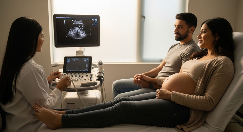
At ten weeks, the sonographer will choose the approach that best balances clarity and comfort. A transabdominal scan uses gel on the abdomen with a handheld probe. A transvaginal scan uses a slim probe in the vagina for closer views of the uterus. For many patients, a transabdominal approach suffices. Yet early pregnancy sometimes benefits from the higher resolution of the internal method.
As Mayo Clinic explains, transvaginal imaging often delivers clearer pictures in early gestation, which can help confirm viability and visualise detail. I consider both methods routine. The sonographer will discuss preferences, explain the rationale, and confirm consent. Comfort matters, but image quality does too. If I need precise measurements, I am happy to opt for the clearer view.
-
Transabdominal: broader field, generally more comfortable at this stage.
-
Transvaginal: closer proximity to the embryo, sharper detail for early checks.
-
Both are safe and use the same non-ionising sound waves.
Duration and Process of the Scan
The appointment is structured and efficient. I am checked in, asked brief medical questions, and taken to the scanning room. The sonographer reviews the plan, applies gel for an abdominal scan or prepares the transvaginal probe, and begins image capture. The core sequence focuses on viability, dating, and number of embryos.
In practice, a week 10 ultrasound usually takes **15 to 30 minutes**, depending on position and visibility. As the Fetal Medicine Centre notes, the choice between abdominal and transvaginal is guided by stage and clarity needs. If the baby’s position is not ideal, I may be asked to adjust posture or cough. Small adjustments can improve alignment for measurements.
-
Initial orientation: identify the uterus and gestational structures.
-
Check for heartbeat and movement.
-
Measure crown-rump length for gestational age.
-
Assess yolk sac and surrounding anatomy as needed.
Measurements Taken During the 10 Week Pregnancy Scan
Key metrics guide clinical decisions. I expect these standard measurements and observations:
|
Measurement |
Purpose |
|---|---|
|
Crown-rump length (CRL) |
Primary metric for dating at this stage. |
|
Heartbeat presence and rate |
Confirms viability; the rate naturally varies. |
|
Yolk sac size and shape |
Supporting sign of a healthy early environment. |
|
Gestational sac and location |
Confirms intrauterine pregnancy. |
|
Number of embryos |
Screens for twins or higher-order multiples. |
These data points feed directly into the report. They also underpin the due date and any early recommendations.
Checking Heartbeat and Movement
By ten weeks, cardiac activity is usually visible on screen. The sonographer will show the flicker and may play the sound briefly. Motion is often subtle but present. I find this reassuring. It is a practical confirmation, not just a sentimental moment.
The week 10 ultrasound does not aim for a full cardiac assessment. It confirms the heartbeat and notes general rhythm. Detailed cardiac anatomy comes later. Still, this early check is valuable. It signals the pregnancy is on track, at least from current imaging.
Dating the Pregnancy Accurately
CRL-based dating at this stage is robust. The embryo’s growth is relatively uniform in early weeks, which supports consistent estimates. The week 10 ultrasound reduces uncertainty from recall of last menstrual period, cycle length, or implantation timing. The sonographer will measure several times and take the best average.
Why this matters is straightforward. Accurate dating aligns testing windows and planned care. It also reduces later confusion when fundal height or anatomy timelines are reviewed. Small differences at this stage can correct weeks of unnecessary worry later.
Screening for Multiple Pregnancies
The scan checks for more than one embryo and records chorionicity if twins are present. Early identification changes monitoring plans and follow-up scheduling. The week 10 ultrasound is well placed to clarify these points while growth patterns remain distinct.
If multiples are seen, the sonographer will flag the finding and suggest appropriate follow-up. I expect tighter surveillance in those cases, with clear instructions on the next appointments.
What Can Be Seen on a 10 Week Pregnancy Scan
Baby’s Size and Development at 10 Weeks
At this stage, the embryo is transitioning into a fetus. The body is recognisably human in outline. With a week 10 ultrasound, the crown-rump length anchors the estimate of gestational age. Limb buds are more defined, and the head appears proportionally larger. It looks top heavy. That is normal in early development.
-
CRL is the headline metric for growth and dating.
-
Basic limb movement can sometimes be seen.
-
The neural tube has closed by now in most pregnancies.
Visible Body Parts and Features
On a clear view, I may see head, torso, limb buds, and the beginnings of hands and feet. The face has early outlines. Details are limited, so I avoid over-interpreting shadows. The week 10 ultrasound establishes presence and pattern, not full anatomy.
If the angle is favourable, the sonographer can capture a profile. That often makes the images easier to interpret later. It also helps me orient the position on screen.
Placenta and Umbilical Cord Formation
The placenta is forming and the cord is in place. Blood flow is not the focus at ten weeks, though Doppler may be used sparingly if indicated. The week 10 ultrasound will note the placental location in broad terms. Final placental position is usually assessed later. Migration is common as the uterus expands.
Amniotic Sac and Fluid Levels
A normal amniotic sac surrounds the fetus with adequate fluid. At ten weeks, the qualitative view is sufficient. Quantitative fluid assessment takes priority later in gestation. The sac’s shape and borders are reassuring when smooth and regular. The week 10 ultrasound captures those impressions.
10 Week Ultrasound 3D Images vs 2D
Some centres can generate 3D previews at this stage. The effect is interesting, but diagnostic value remains with 2D. A week 10 ultrasound in 2D delivers the crisp measurements. A 10 week ultrasound 3d image can be a helpful keepsake, though results vary with fetal position and maternal anatomy.
-
2D: best for CRL, heartbeat, and viability checks.
-
3D: optional visualisation, variable clarity this early.
Limitations of Week 10 Ultrasound
There are natural limits to what can be concluded. Many structural anomalies are simply not visible yet. In its guidance on early scans, London Pregnancy Clinic notes that genetic testing usually happens later and that visible abnormalities are harder to assess this early. Accuracy improves as the pregnancy progresses. Early reassurance does not replace mid-trimester checks.
The week 10 ultrasound is powerful for dating and viability. It is not the final word on anatomy. Both truths can sit together without contradiction.
Preparing for Your 10 Week Ultrasound
Bladder Preparation Requirements
If the scan is transabdominal, a moderately full bladder can lift the uterus for a clearer line of sight. I aim to drink water 30 to 60 minutes before the appointment. For a transvaginal scan, an empty bladder is usually preferred. The clinic will confirm their protocol in the appointment letter.
This is a minor step. It improves the week 10 ultrasound quality more than most realise.
What to Wear to Your Appointment
Comfortable, two-piece clothing helps. A top with trousers or a skirt is practical. It allows easy access to the lower abdomen or, if needed, for a transvaginal scan. Avoid heavy belts and one-piece outfits. They slow the process without adding comfort.
Documents and Information to Bring
-
Photo identification and any referral letters.
-
Prenatal records or previous scan reports.
-
Medication list and known allergies.
-
Estimated last menstrual period, if relevant.
-
Insurance documents or payment method.
Having these at hand keeps the week 10 ultrasound efficient and accurate.
Questions to Ask Your Sonographer
-
Is the dating based on CRL, and what margin of error applies?
-
Is the heartbeat rate appropriate for this stage?
-
Is the pregnancy single or multiple, and what does that imply?
-
Are any follow-up scans recommended, and when?
-
Will I receive printed images or a digital link?
Specific questions clarify the report. They also anchor expectations for the next visits.
Partner and Family Attendance Guidelines
Policies vary by clinic and local guidance. Many centres allow one partner to attend. Larger groups are often discouraged because the room is tight and the focus is clinical. I plan ahead and confirm the rules at booking. The week 10 ultrasound is a clinical appointment first, with moments of joy built in.
Understanding Your 10 Week Ultrasound Pictures and Results
Reading 10 Week Ultrasound Pictures
Images can look abstract at first glance. Labels and markers help. I expect to see CRL measurement lines, the date, and the gestational age estimate. Contrast is high, and shallow angles can distort shape. This is normal for ultrasound. The sonographer’s annotations point to the head and rump so I can orient myself.
If I requested 10 week ultrasound pictures, I keep both the printed and digital versions. Later comparisons are easier with the same views.
Normal Measurements and Ranges
Ranges vary with growth curves and equipment. Roughly speaking, CRL at ten weeks sits within a predictable band. The precise millimetres matter less than the overall fit to expected development. The sonographer will note whether measurements align with dates. If there is a mismatch, they may adjust the estimated due date.
|
Parameter |
Typical Finding at 10 Weeks |
|---|---|
|
CRL |
Used to date the pregnancy; a tight predictor at this stage. |
|
Cardiac activity |
Present and regular; exact rate varies by individual. |
|
Gestational sac |
Intrauterine, with smooth borders. |
|
Yolk sac |
Visualised and appropriately sized. |
If I want specifics, I request the numeric readout and ask how it maps to the expected chart.
Crown-Rump Length Significance
CRL is the most reliable early dating metric. It is less affected by individual variation than later biometric measures. A precise CRL during a week 10 ultrasound can refine the due date window. That helps schedule screening tests accurately. It also reduces later confusion when anatomy scans report different sizes.
CRL at ten weeks gives the due date its backbone. The rest of the plan follows from that estimate.
Nuchal Translucency Screening Basics
Nuchal translucency (NT) is generally measured between 11 and 13 weeks plus six days. The week 10 ultrasound is slightly early for a formal NT measurement. Still, I can use this appointment to plan the NT and any combined screening. Getting the dates correct now ensures the NT window is not missed.
Some centres offer a combined first-trimester screen with bloods and NT. Others offer cell-free DNA later. A clear week 10 ultrasound helps choose the right path.
When Results Indicate Concerns
Occasionally, the report raises questions. Examples include an absent heartbeat, a CRL not aligning with dates, or concerning sac features. These findings do not automatically predict outcomes. They do guide next steps. The sonographer or clinician will explain the concern and outline options.
-
Repeat scan after a short interval to assess progression.
-
Targeted blood tests or hormone levels, if indicated.
-
Referral to an early pregnancy unit for closer review.
The week 10 ultrasound opens the discussion early. That timeline helps decision-making and support.
Follow-up Scans and Next Steps
Follow-up depends on findings and local protocols. For uncomplicated results, the next milestone is the NT window. If dates were adjusted, I log the new due date and confirm the booking accordingly. If I am carrying twins, I expect more frequent monitoring. The week 10 ultrasound sets that cadence with fewer surprises.
Documentation matters. I keep the report, images, and any revised schedule in one place. Simple organisation reduces future friction.
Making the Most of Your Week 10 Ultrasound Experience
Approach this scan with a clear plan. The aim is to obtain accurate data and leave with fewer uncertainties. Here is how I make the most of the appointment:
-
Arrive with a short list of priorities: dating, viability, number of embryos.
-
Ask for numeric outputs where helpful, especially CRL.
-
Confirm the next time-sensitive appointment, such as NT.
-
Request both printed and digital images for reference.
-
Note any advice on supplements, symptoms, or early warning signs.
In product terms, this is a checkpoint. In clinical terms, it is a foundation for the first trimester plan. The week 10 ultrasound provides enough certainty to move ahead with confidence. It is basically the pivot between early questions and structured care.
Frequently Asked Questions
Can you determine gender at a 10 week ultrasound?
Sex determination by ultrasound at ten weeks is not reliable. External genitalia have not developed enough for confident visual assessment. Some blood tests can indicate fetal sex from about ten weeks, depending on availability and policy. The week 10 ultrasound focuses on dating and viability, not sex.
Is it normal not to see much on a 10 week scan?
Yes, detail is limited at this stage. The sonographer looks for the big markers: CRL, heartbeat, sac, and basic structure. A week 10 ultrasound is not intended to replace the mid-trimester anatomy scan. It is normal to leave with essential data rather than fine-grained imagery.
How accurate is dating at the 10 week ultrasound?
CRL-based dating is considered reliable in early pregnancy. Variability is low compared with later biometric measures. The week 10 ultrasound often refines or confirms the due date, which improves scheduling for screening and later scans. Minor day-level adjustments are common and useful.
What if no heartbeat is detected at 10 weeks?
The clinician will review dates, measurements, and technique. Sometimes a repeat scan is scheduled after a short interval to confirm findings. The week 10 ultrasound creates a clear reference. Subsequent imaging clarifies whether development progresses or not. Emotional support and clear next steps are essential in that scenario.
Can I get 10 week ultrasound pictures to take home?
Most centres provide printed images or a secure digital link. I request both if possible. 10 week ultrasound pictures are helpful for personal records and comparisons. Policies and fees vary by clinic, so I ask in advance.
Is the 10 week scan covered by insurance in India?
Coverage varies by insurer, plan type, and whether the scan is prescribed. Some policies cover the scan when medically indicated within prenatal care. I check the policy wording and confirm with the clinic. The week 10 ultrasound is often billed as an obstetric ultrasound with a standard code.
Stepping back, the week 10 ultrasound does one job very well. It establishes a trustworthy baseline. The data inform timelines, tests, and expectations. And yet, it does not need to answer everything. That balance is the strength of an early, well-executed scan.