


Understanding Lung Cancer Stages: Early Signs, Diagnosis, and More
Dr. Kunal Luthra
Stage labels are often treated as destiny. They are not. Understanding lung cancer stages clarifies treatment options, prognosis, and when to escalate care. I focus on what the labels mean in practice, how staging is established, and which early clues warrant investigation. I also outline key tests so patients and clinicians can share a clear map. The aim is straightforward comprehension, not oversimplification of a complex disease.
Lung Cancer Stages Explained
I use lung cancer stages to describe how far disease has progressed and where it has spread. This guides surgery, radiotherapy, systemic therapy, and clinical trial choices. Staging is built from imaging, pathology, and the TNM system. It is a framework, not a sentence.
Stage 0 (Carcinoma in Situ)
Abnormal cells are confined to the innermost lining of the airway. No invasion is seen. Treatment may include endobronchial therapy or limited resection. Surveillance is essential because progression risk exists, though it varies by pathology and site.
Stage I (Early-Stage Disease)
The tumour is localised within the lung without nodal spread. Surgical resection is usually preferred if fitness allows. I often consider lobectomy with lymph node sampling. Adjuvant therapy may be discussed when risk features are present. The intent is curative.
Stage II (Locally Advanced)
The tumour is larger or involves nearby structures or hilar nodes. Surgery remains possible in many cases. Treatment planning often includes adjuvant chemotherapy. Multidisciplinary evaluation matters here because margins, nodal status, and recovery capacity guide decisions.
Stage III (Regional Spread)
Regional lymph nodes are involved, often mediastinal. Management is complex and team based. I typically weigh concurrent chemoradiation, consolidation therapy, or surgery in select IIIA scenarios. Goals can still be curative to an extent, but toxicity and sequencing require precision.
Stage IV (Metastatic Cancer)
Disease has spread beyond the chest to distant organs. Systemic therapy leads: targeted agents, immunotherapy, or chemotherapy depending on biomarkers. Symptom control is central. Long control is possible for some molecular subsets, though not universally.
TNM Classification System
TNM translates anatomy into a reproducible stage. I summarise the core definitions below for quick reference.
|
Term |
Definition |
|
T (Tumour) |
Size and local invasion, from Tis to T4, based on diameter and extension. |
|
N (Nodes) |
Regional lymph node involvement, from N0 to N3, defining laterality and stations. |
|
M (Metastasis) |
Presence and extent of distant spread, M0 or M1 subcategories. |
Combining T, N, and M yields the final stage group. Small details shift categories. And outcomes.
Symptoms vary and often overlap with benign disease. I watch for persistence, escalation, and combinations. Those patterns raise suspicion for early signs of lung cancer.
Persistent Cough and Blood
A cough lasting beyond eight weeks warrants assessment. Any haemoptysis, even streaks, needs prompt review. Chronic smokers are not exempt from new causes. Neither are never smokers.
Breathing Difficulties
New or worsening breathlessness, wheeze, or unexplained hoarseness can reflect airway obstruction. I correlate with exertion capacity and oxygen saturation. Recurrent infections in one lobe suggest an endobronchial lesion.
Chest Pain and Discomfort
Pleuritic pain, shoulder ache, or chest wall tenderness can indicate local invasion. The distribution offers clues. So does pain that disturbs sleep or fails to resolve with standard measures.
Unexplained Weight Loss
Involuntary weight loss, fatigue, or appetite change may signal systemic activity. I review thyroid, infection, and gastrointestinal causes. But sustained loss often prompts imaging and labs.
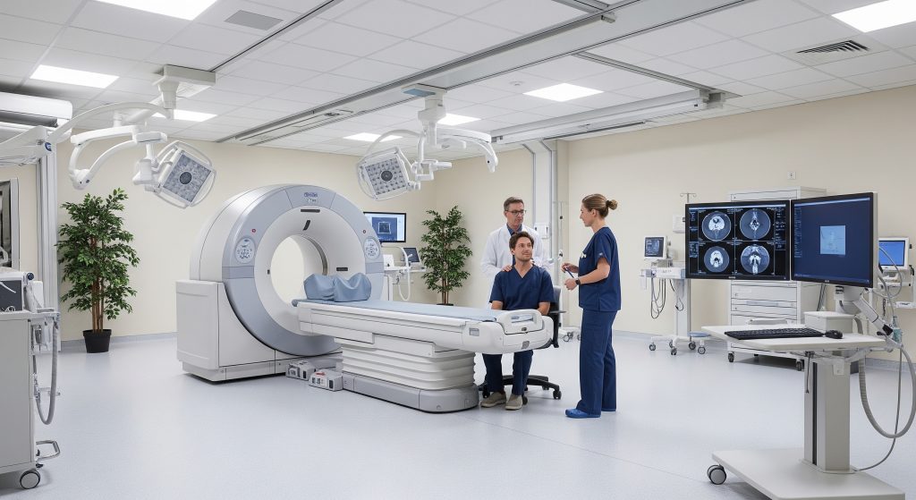
Diagnostic Tests and Scans
Imaging builds the staging picture. The usual sequence is pragmatic, not rigid.
- Chest X ray for a first look when symptoms persist.
- Contrast CT chest and upper abdomen for anatomy and nodes.
- PET CT to assess metabolic activity and detect occult spread.
- Brain MRI if neurological signs or advanced stage is suspected.
- Pulmonary function tests to assess surgical fitness.
Biopsy Procedures
Tissue is essential because therapy depends on histology and biomarkers. Options include CT guided needle biopsy, bronchoscopy with EBUS, or surgical biopsy. I prioritise the safest route that yields adequate material for full molecular testing.
Moving Forward with Lung Cancer Knowledge
Clear understanding of lung cancer stages helps align decisions with goals and values. It also reduces uncertainty during a stressful period. I encourage early presentation when symptoms persist, especially the early signs of lung cancer mentioned above. Screening with low dose CT fits high risk groups and catches disease earlier. One note of caution. Screening does not replace clinical judgement or follow up for new symptoms.
Use the stage to inform choices and to time second opinions, biomarker testing, and trial exploration. The map is valuable. The journey still requires skill.
What are the survival rates for different lung cancer stages?
Survival varies by stage, histology, biomarkers, and treatment access. Early stages generally have higher cure rates after resection. Advanced stages rely on systemic therapy and long control is possible for biomarker matched disease.
Can lung cancer be detected before symptoms appear?
Yes, in high risk individuals through low dose CT screening. That approach detects nodules earlier and supports curative treatment. It also reduces unnecessary delays from symptom driven pathways.
How quickly does lung cancer progress through stages?
Progression speed differs by subtype and biology. Some tumours grow slowly for months. Others accelerate within weeks. As current data suggests, growth kinetics vary with genomics and microenvironment.
What imaging tests best detect early-stage lung cancer?
Low dose CT detects small nodules better than chest X ray. Contrast CT refines anatomy for staging. PET CT helps distinguish active disease from scar, especially when planning treatment.
Are there differences between small cell and non-small cell staging?
Yes. Non small cell uses the full TNM system and stage groups. Small cell traditionally uses limited versus extensive stage, though TNM can also apply. Treatment pathways differ substantially.