


Understanding ERCP Stents: Uses, Types, and Recovery
Dr. Prajwal S
The most repeated advice about stents is to pick the strongest device and be done. That view oversimplifies a nuanced clinical decision. In practice, the right ercp stent depends on anatomy, diagnosis, and anticipated complications. I will set out the choices, the process, and the recovery, so an informed decision is achievable without guesswork.
Types of ERCP Stents and Their Primary Applications
Plastic Biliary Stents
Plastic devices remain the workhorse for short to medium term drainage. I use them when inflammation or stones are the primary issue, and when planned ercp stent removal is expected within weeks. They are simple to exchange, cost effective, and compatible with narrow ducts. The trade off is a higher occlusion rate as biofilm and sludge build up.
-
Typical dwell time: 4 to 12 weeks.
-
Best for benign obstruction, post stone clearance, and temporary decompression.
-
Lower upfront cost, higher likelihood of early replacement.
When drainage is urgently needed, a plastic biliary stent often achieves immediate relief with minimal device complexity. It is basically a reliable first step while the underlying cause is addressed.
Self-Expanding Metal Stents
Self expanding metal stents offer larger diameter and longer patency. I prioritise them for malignant obstruction where a single durable intervention is desirable. The expansion provides better flow, which reduces the risk of recurrent jaundice or cholangitis. Placement requires precise positioning and a clear strategy for future access.
-
Typical dwell time: months, often until disease progression.
-
Best for malignant distal or hilar strictures, when long term drainage is needed.
-
Higher upfront cost, fewer early exchanges.
Metal options can be life extending to an extent, because stable drainage supports systemic therapy and nutrition. But they demand careful selection, since removal may not be feasible in some configurations.
Covered vs Uncovered Stents
The membrane on a covered device resists tissue ingrowth and can improve patency. As Experimental and Therapeutic Medicine notes, the decision often hinges on expected risks such as migration or occlusion. Covered stents help when tumour or reactive mucosa tends to grow into the mesh.
Economics matters too. In a model of clinical trade offs, Cost-Effectiveness Decision Analyses Comparing Covered reported that covered stents may be more cost effective in selected settings by reducing re interventions over time. The counterpoint is a higher migration tendency in some anatomies. Uncovered designs anchor well and permit side branch drainage but can clog with ingrowth.
-
Covered: better resistance to tissue ingrowth, higher migration risk.
-
Uncovered: better fixation and side branch patency, harder to remove later.
I align the choice with duct geometry, stricture length, and the plan for ercp stent removal or exchange. A small shift in anatomy can change the calculus entirely.
Pancreatic Duct Stents
Pancreatic stents are smaller calibre devices used for prophylaxis and therapy. I place them to reduce post ERCP pancreatitis risk in high risk cannulations, or to bypass a tight pancreatic stricture. They are usually short, with flanges or pigtails that limit migration into the duct.
-
Prophylactic stents tend to pass spontaneously within days.
-
Therapeutic stents need planned removal or exchange.
-
Placement requires delicate wire control due to the duct’s angulation.
In practice, a correctly sized pancreatic device can prevent a serious complication. Small detail, large effect.
Temporary vs Permanent Stents
Temporary placement aligns with benign disease, stone disease, and post operative leaks. Permanent, or at least long term, placement corresponds to malignant obstruction and unresectable strictures. I counsel patients that the term permanent is contextual. It means indefinite support rather than literal permanence.
|
Scenario |
Preferred approach |
|---|---|
|
Benign stricture |
Sequential plastic stents with planned ercp stent removal |
|
Malignant distal bile duct obstruction |
Self expanding metal stent with covered or uncovered design as indicated |
|
Post cholecystectomy bile leak |
Short term plastic biliary stent |
|
High risk for pancreatitis |
Temporary pancreatic prophylactic stent |
The end goal is uninterrupted drainage with minimal interventions. Everything else is a means to that end.
ERCP Procedure Steps and Stent Placement Process
Pre-Procedure Preparation
I review anticoagulation, allergy profile, and infection risk in detail. Antibiotics are considered for suspected cholangitis or incomplete drainage risk. Patients fast for several hours, and consent covers the benefits, ercp procedure steps, and alternatives.
-
Laboratory review: full blood count, clotting, renal function, and bilirubin pattern.
-
Imaging: ultrasound or MRCP to map obstruction level and length.
-
Optimisation: correct coagulopathy and manage sepsis before the ercp stent is placed.
Anaesthesia support is arranged for airway protection, especially in high aspiration risk cases. Preparation reduces surprises. It also improves outcomes.
Endoscopic Retrograde Cholangiopancreatography Technique
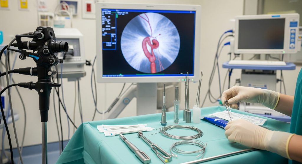
The endoscopic retrograde cholangiopancreatography sequence is systematic. I pass a side viewing duodenoscope to the second part of the duodenum. Cannulation of the papilla follows, with gentle wire access into the target duct. Contrast is used sparingly to calibrate the stricture and the downstream drainage path.
-
Scope to duodenum and identify the papilla.
-
Wire guided cannulation of bile or pancreatic duct.
-
Diagnostic cholangiogram or pancreatogram as needed.
-
Sphincterotomy if required for access or stone extraction.
-
Preparation and deployment of the selected ercp stent.
I maintain wire access throughout, which preserves control if repositioning is required. Small corrections prevent large problems.
Stent Selection Criteria
Selection blends anatomy, disease, and patient goals. I consider duct diameter, length and location of stricture, expected survival, and plans for systemic therapy. A biliary stent for a short distal stricture is not the same device as one for a complex hilar lesion.
-
Benign vs malignant aetiology guides plastic vs metal choice.
-
Covered vs uncovered follows the risk of ingrowth and migration.
-
Need for future access argues for removable or revisable designs.
I aim for the simplest ercp stent that achieves reliable drainage. Simplicity correlates with fewer complications.
Placement Methods and Positioning
Positioning is the decisive step. I align the distal end just across the papilla or above it, depending on the plan for future access. In hilar disease, bilateral or side by side metal stents might be appropriate, though not without exceptions.
-
Deploy under fluoroscopy with clear markers on the wire and catheter.
-
Check for free flow of contrast and bile after deployment.
-
Confirm final position with a limited cholangiogram.
For pancreatic stents, I advance gently over a soft wire and avoid deep instrumentation. Less trauma means lower pancreatitis risk.
Immediate Post-Procedure Monitoring
Observation targets pain, fever, and early bleeding. I check vitals, abdominal tenderness, and the trend in liver enzymes. If the patient had cholangitis, clinical improvement should follow within 24 to 48 hours.
-
Pain control with careful titration, avoiding excess opioids when possible.
-
Hydration and early nutrition as tolerated.
-
Clear instructions on ercp stent symptoms that warrant urgent review.
Most patients leave the same day or next morning. Those with severe infection or complex anatomy remain for closer monitoring.
Recovery After ERCP Stent Placement
First 24 Hours Post-Procedure
The first day sets the tone. I advise rest, oral hydration, and light meals. Mild throat soreness and bloating are common after scope passage. Transient nausea can occur from anaesthesia or contrast.
-
Keep a simple pain log to identify escalation early.
-
Measure temperature twice on the first evening.
-
Avoid alcohol and heavy meals until appetite is normal.
If pain localises to the upper abdomen and intensifies, contact a clinician. Early alerts prevent serious events.
Managing Common Side Effects
Mild abdominal discomfort, low grade fever, or dark urine may occur. As the biliary system clears, stool colour often normalises. I use simple analgesia and hydration first, then escalate if symptoms persist.
-
Gas related bloating usually resolves within 24 hours.
-
Sore throat responds to lozenges and warm fluids.
-
Itching from cholestasis eases as bile flow improves through the ercp stent.
Side effects should move in the right direction. If they do not, reassessment is prudent.
Diet and Activity Restrictions
I recommend a light diet for the first day, then normal meals as tolerated. Avoid high fat feasts that challenge early biliary drainage. Gentle walking is safe the same day, while strenuous activity can wait for 48 hours.
-
Hydrate well to support bile flow and prevent constipation.
-
Resume prescribed medications, with anticoagulation per the plan.
-
No driving for 24 hours after sedation.
In practical terms, stability is the goal. Energy returns as bilirubin falls and infection settles.
Warning Signs to Watch For
Clear red flags merit urgent care. I instruct patients to seek help if severe pain, persistent vomiting, fever over 38.5 C, or jaundice worsens. Bleeding, black stools, or uncontrolled chills also deserve immediate evaluation.
-
Sudden severe epigastric pain could indicate pancreatitis.
-
High fever and rigors suggest cholangitis or stent blockage.
-
New confusion in older patients may signal sepsis.
When in doubt, attend an emergency department. Delay turns manageable issues into crises.
Follow-up Schedule
Follow up is tailored to stent type and diagnosis. For plastic devices, review at 4 to 6 weeks is typical, with planned exchange or ercp stent removal soon after. For metal devices, clinic review at 2 to 4 weeks confirms symptom relief and biochemical response.
-
Benign disease: set an exchange calendar before discharge.
-
Malignant disease: integrate oncology timelines and nutrition support.
-
Pancreatic prophylaxis: confirm spontaneous passage or arrange early removal.
Structured follow up prevents silent occlusions. It also provides accountability for the care plan.
ERCP Stent Removal and Long-term Management
When Stent Removal Is Necessary
Removal is planned when the underlying problem is resolved or when device exchange is due. Plastic devices in benign disease should not overstay, since occlusion risk climbs with time. Pancreatic prophylactic stents are intentionally short term.
-
Resolution of stone disease or leak permits timely extraction.
-
Benign strictures may require staged exchanges before final removal.
-
Suspected infection or blockage can prompt early intervention.
The principle is simple. The ercp stent supports healing, it is not the destination.
Removal Procedure Overview
Removal mirrors placement but is usually faster. I perform an endoscopic retrograde cholangiopancreatography with wire access as needed. Graspers or a snare extract the device under fluoroscopy. Covered metal designs may be removed if placed with that intent, though operator judgement is crucial.
-
Confirm position and plan with a brief cholangiogram.
-
Secure the stent with appropriate retrieval tools.
-
Withdraw under vision, maintaining wire access for exchange.
Analgesia and sedation match patient factors. Recovery tends to be straightforward.
Stent Migration and Occlusion
Migration and occlusion are the two common device problems. Distal migration can be asymptomatic or cause obstruction. Proximal migration complicates retrieval and may require advanced techniques.
-
Occlusion presents with fever, rising bilirubin, or recurrent pruritus.
-
Biofilm formation is common in plastic devices after several weeks.
-
Covered metal stents resist ingrowth but may move in smooth ducts.
When I suspect blockage, I arrange urgent imaging and plan for cleaning, exchange, or upsizing. Speed matters because cholangitis escalates quickly.
Replacement Schedules
Replacement is not guesswork. Plastic devices in the bile duct are usually exchanged every 8 to 12 weeks. Pancreatic stents for therapy follow a shorter timeline. Metal devices are replaced only if dysfunctional or if the treatment plan changes.
|
Device |
Typical replacement plan |
|---|---|
|
Plastic biliary stent |
Exchange at 8 to 12 weeks or earlier if symptomatic |
|
Covered metal stent |
Observe and replace only if obstructed or migrated |
|
Uncovered metal stent |
Generally not removed; manage dysfunction with additional stenting |
|
Pancreatic stent |
Early confirmation of passage; remove if retained |
An explicit schedule avoids uncertainty for both the team and the patient. It reduces emergency calls and hospital returns.
Living with Permanent Stents
Some patients will live with an indwelling device for the long term. I advise on infection vigilance, hydration, and timely reporting of any cholestatic symptoms. Routine life is achievable, including travel, with a clear emergency plan and contact points.
-
Carry a procedure note summarising the device type and date.
-
Keep an updated medication list, including antibiotics that previously worked.
-
Schedule periodic liver tests to detect early dysfunction.
Quality of life improves when expectations are aligned. Clear information reduces anxiety and delays.
Making Informed Decisions About ERCP Stenting
Decision quality improves with structured thinking. I frame decisions around goals of care, anatomy, and timing. If the objective is durable relief in malignant disease, a metal choice often prevails. If the target is short term support while stones are cleared, a plastic device is sufficient.
Right patient, right device, right timing. That triad prevents most complications.
I also emphasise coordination. Gastroenterology, radiology, surgery, and oncology should align on intent and sequence. It sounds obvious. Coordination still fails without explicit ownership and dates. Earlier, I noted that covered devices can reduce re interventions in selected cases. That matters when scheduling chemotherapy or major surgery that cannot be repeatedly delayed.
One final point. The ercp stent is a means to restore flow and reduce infection risk. It is not a substitute for addressing the cause, whether that is a stone, a stricture, or a tumour.
Frequently Asked Questions
How long can an ERCP stent stay in place?
It depends on the device and diagnosis. Plastic devices usually remain for 4 to 12 weeks, then require exchange or ercp stent removal. Metal devices can remain for months and often until they fail or the plan changes.
What are the risks of ERCP stent placement?
Risks include pancreatitis, bleeding, infection, perforation, and device migration or occlusion. The overall risk is low but non zero. I mitigate risk with careful technique, antibiotic cover when needed, and clear follow up.
Can I resume normal activities after getting a biliary stent?
Light activity is acceptable within 24 to 48 hours for most patients. Strenuous exercise can resume when pain is absent and appetite is normal. Travel is possible with a summary letter and an emergency contact plan.
How do I know if my ERCP stent is blocked?
Watch for fever, dark urine, pale stools, recurrent itching, or rising jaundice. Abdominal pain with chills is concerning. If any occur, seek urgent assessment for possible cleaning or exchange of the ercp stent.
Is ERCP stent removal painful?
Removal is done with sedation or anaesthesia. Discomfort is usually mild and short lived. Most patients go home the same day with minimal restrictions.
What happens if a stent migrates from its original position?
Migration can cause symptoms or remain silent. Distal migration may pass spontaneously, but evaluation is required. If it causes blockage or pain, I plan retrieval or a new ercp stent based on imaging and symptoms.