


Understanding Duplex Ultrasound for Vascular Disease Diagnosis
Dr. Hriday Kumar Chopra
Disclaimer: The content shared here is for informational purposes only. Always consult a specialist doctor before attempting any treatment, procedure, or taking any medication independently.
Standard advice suggests most vascular problems need advanced scans or invasive angiography. That approach is often unnecessary and sometimes slows care. In practice, I use duplex ultrasound as the first-line test for most suspected vascular disease. It offers real-time anatomy and blood flow in one sitting, without needles or radiation. It is basically a precise extension of the physical examination that I can repeat, compare, and act on with confidence.
Types of Vascular Conditions Diagnosed with Duplex Ultrasound
Peripheral Artery Disease
For suspected lower limb ischaemia, I start with duplex ultrasound. It maps vessel anatomy and captures flow velocities that reflect stenosis severity. I can localise a focal narrowing, grade it, and assess the downstream effect. Claudication patterns and rest pain correlate well with waveform changes. When I see damped monophasic waveforms beyond a lesion, critical disease is likely. Short segment lesions are often suitable for angioplasty. Diffuse calcific disease points towards medical optimisation and targeted revascularisation.
-
Identifies inflow versus outflow disease quickly.
-
Guides whether endovascular or surgical planning is sensible.
-
Monitors response to exercise therapy and medication.
Deep Vein Thrombosis
Compression ultrasound with Doppler is the practical standard for DVT screening. I evaluate vein compressibility, intraluminal echoes, and flow augmentation with calf squeeze. Non-compressible segments or absent phasicity suggest acute thrombus. Proximal extension risk changes management urgency. In pelvic segments that are less accessible, I combine targeted duplex findings with clinical probability scores. Safety matters here. A prompt negative study can limit unnecessary anticoagulation.
Chronic Venous Insufficiency
Chronic venous insufficiency presents with oedema, skin changes, and heaviness. Duplex ultrasound shows reflux duration, vein diameter, and the reflux pathway. I map the great and small saphenous systems, tributaries, and perforators. Standing or reverse Trendelenburg positions help reveal true reflux. The report becomes a procedural roadmap for ablation, foam, or phlebectomy. Better maps yield cleaner results.
Varicose Veins Detection
Varicose veins reflect valve failure and segmental venous dilation. Population burden is high. As CARDIOVASCULAR UPDATE reports, roughly 27% of the US population is affected, and non-invasive duplex is widely used for detection. When I assess ultrasound for varicose veins, I focus on reflux origin, axial competency, and perforator status. That level of detail drives targeted, durable treatment choices.
-
Clinical exam defines symptoms. Duplex ultrasound confirms reflux and maps anatomy.
-
Reflux timing in seconds matters for grading and for procedural planning.
-
Clear vein marking on the skin simplifies theatre execution.
Carotid Artery Disease
Carotid duplex blends structural imaging with flow dynamics, which is valuable for stroke prevention. In symptomatic disease, speed matters. As Duplex ultrasound for diagnosing symptomatic carotid… notes, duplex reliably identifies clinically significant stenosis and helps triage candidates for timely revascularisation. I evaluate peak systolic velocity, end-diastolic velocity, and plaque morphology. Ulceration features, though nuanced, can be inferred when image quality is optimal.
-
Triages between medical therapy and carotid intervention.
-
Enables serial follow-up for moderate stenosis.
-
Detects contralateral disease that may change perioperative risk.
Aneurysm Detection
Abdominal aortic aneurysm surveillance fits duplex ultrasound well. I measure outer-to-outer diameter, mural thrombus, and iliac involvement. Growth rate influences surveillance intervals. Rapid expansion prompts vascular referral. For peripheral aneurysms, particularly popliteal, duplex adds thrombus pattern and runoff assessment. These details shape bypass or endovascular choices and limb risk mitigation.
How Duplex Ultrasound Works for Vascular Assessment
B-mode Imaging Component
B-mode supplies the structural picture. I visualise vessel walls, plaque, thrombus, and relative diameters. Echogenicity hints at plaque stability. Calcified caps cast acoustic shadows that sometimes obscure the lumen. I adjust gain, depth, and focus to optimise edges. Good B-mode frames anchor the entire study. Without a clean anatomical baseline, velocity data can mislead.
Duplex ultrasound marries B-mode anatomy with flow data. Structure and function in one examination.
Doppler Velocity Measurements
Pulsed Doppler quantifies haemodynamics. I align the sample volume with the vessel axis and maintain a consistent angle. Velocity rises as luminal area narrows. Ratios between stenotic and pre-stenotic segments improve grading confidence. In veins, prolonged reversed flow indicates reflux. In arteries, tardus-parvus beyond a lesion signals a proximal stenosis. It is precise when technique is disciplined.
-
Angle correction typically standardised around 60 degrees.
-
Velocity ratios reduce inter-operator variability.
-
Multiple samples across a lesion define the jet profile.
Colour Flow Mapping
Colour overlays speed and direction onto the grey-scale frame. I use it to locate jets, aliasing, and flow separation. Colour priority and scale need careful setting. Too high, and slow flow disappears. Too low, and everything looks turbulent. In venous work, colour reflux during manoeuvres clarifies incompetent segments. Subtle but decisive findings.
Spectral Waveform Analysis
Spectral traces translate complex flow into measurable curves. Triphasic waveforms imply normal peripheral arterial tone. Biphasic can be acceptable, depending on age and vessel segment. Monophasic waveforms suggest significant proximal disease. In the carotids, I assess systolic peaks, diastolic tail, and spectral broadening. As Carotid artery duplex scanning describes, parameters like peak systolic velocity and spectral waveform support classification of stenosis across the 0 to 100% range. Those numbers guide critical decisions.
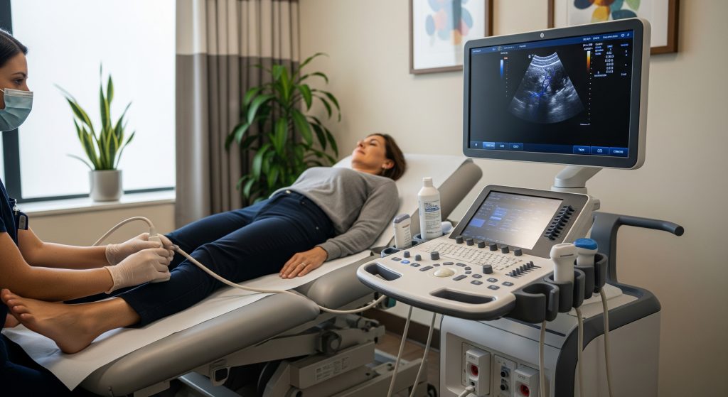
Procedure and Patient Experience
Pre-examination Preparation
Preparation is simple for most studies. I advise comfortable clothing and easy limb access. For abdominal aorta studies, fasting helps reduce bowel gas. Hydration supports venous filling. Medication schedules usually remain unchanged. A concise referral question improves focus and efficiency. Precision begins with clarity.
Examination Process Steps
-
Brief clinical review and consent.
-
Positioning and exposure of the relevant anatomy.
-
Application of gel to ensure acoustic coupling.
-
B-mode survey to define landmarks and pathology.
-
Colour flow to identify jets and reflux paths.
-
Pulsed Doppler sampling with measured angle correction.
-
Provocation manoeuvres where needed, such as calf squeeze or Valsalva.
-
Labelling, measurements, and image capture for the report.
Duration and Comfort
Most focused vascular ultrasound studies take 20 to 45 minutes. Comprehensive mapping may take longer. The process is painless, with brief pressure when I compress veins or hold a probe to steady an angle. No needles, no contrast, no radiation. That matters for repeat assessments over time.
Post-examination Protocol
Results are explained promptly, then compiled into a structured report. I include key measurements, velocity data, and a clear impression. For urgent findings, I contact the referrer immediately. For elective cases, I add specific next steps. That might be compression therapy, referral for intervention, or a follow-up interval. Clarity lowers anxiety and prevents delays.
Clinical Benefits and Diagnostic Advantages
Non-invasive Assessment Method
Duplex ultrasound is non-invasive and repeatable. I can reassess progress without exposing patients to radiation or contrast. This is valuable for chronic disease where longitudinal data matters. The safety profile enables proactive care.
Real-time Blood Flow Visualisation
Real-time imaging captures dynamic physiology. I can provoke reflux or assess vasoreactivity in the same session. Changes are immediate on screen. It shortens the feedback loop between assessment and action.
Treatment Planning Applications
Accurate maps reduce procedural uncertainty. For varicose veins, precise reflux routes define ablation targets. For PAD, lesion length and morphology shape endovascular strategy. Cleaner plans mean fewer surprises and better outcomes.
Post-treatment Monitoring Uses
After intervention, I verify patency, residual stenosis, and flow normalisation. For venous ablation, I confirm successful closure and check for EHIT. For stents, I track velocities at edges where restenosis may start. Early detection preserves results.
Comparison with Other Imaging
|
Modality |
Typical Use and Considerations |
|---|---|
|
Duplex ultrasound |
First-line for most vascular beds. No radiation. Portable. Operator dependent yet highly informative. |
|
CT angiography |
Excellent anatomy and 3D planning. Uses radiation and iodinated contrast. Limited in severe calcification. |
|
MR angiography |
Detailed vessels without radiation. Gadolinium considerations in renal disease. Availability varies. |
|
Catheter angiography |
Interventional gold standard. Invasive with contrast and radiation. Best when treatment is anticipated. |
Accuracy and Reliability Metrics
Diagnostic performance depends on technique, protocols, and equipment. Arterial grading correlates well with angiography when angle correction and velocity ratios are applied. Venous reflux timing is reproducible with standard manoeuvres. Carotid velocity criteria provide consistent classification across centres. The result is reliable, though not infallible.
Making Informed Decisions About Vascular Ultrasound
Choosing the right test starts with the clinical question. I consider symptom pattern, risk profile, and the decision the result will drive. If a test does not change management, I defer it. Duplex ultrasound usually meets the threshold of relevance and safety. It also integrates smoothly with follow-up planning. That continuity supports better care to an extent.
-
Use duplex ultrasound to confirm a working diagnosis and quantify severity.
-
Escalate to CTA or MRA when anatomical ambiguity persists or intervention needs detailed mapping.
-
Reserve catheter angiography for cases where treatment will likely proceed.
There is a contrary view that advanced imaging is always superior. The position is understandable, but it overlooks cost, contrast risk, and time. Duplex often answers the question with fewer trade-offs.
Frequently Asked Questions
What is the difference between duplex ultrasound and regular ultrasound?
Regular ultrasound shows structures in grey scale. Duplex ultrasound combines that view with Doppler measurements to display and quantify blood flow. It reveals both anatomy and haemodynamics in a single examination. This dual view is decisive in vascular disease.
How much does duplex ultrasound cost in India?
Fees vary by city, facility level, and study scope. A focused limb study usually costs less than comprehensive mapping. Hospital-based services may charge more than standalone centres. It is sensible to confirm inclusions such as a formal report and images.
Can duplex ultrasound detect all types of varicose veins?
It detects reflux in superficial and deep systems and maps perforators. Small reticular veins or cosmetic spider veins may be visible yet clinically minor. For treatment planning, duplex is the preferred ultrasound for varicose veins in routine practice.
How long does a complete vascular ultrasound examination take?
Most focused exams take 20 to 45 minutes. Complex mapping can extend beyond an hour, depending on anatomy and the clinical question. Preparation quality and patient comfort also influence duration.
Is duplex ultrasound safe for repeated examinations?
Yes. It uses sound waves, not radiation. There is no contrast load. I schedule repeat studies for surveillance or post-procedure checks without safety concerns. Repeatability is a major advantage.
When should I consider getting a duplex ultrasound for vascular problems?
Consider it for limb pain on exertion, non-healing ulcers, limb swelling, suspected DVT, neck bruit, or follow-up after treatment. New neurological symptoms suggest urgent carotid assessment. Early testing often clarifies the next step.
Supplementary context: I integrate doppler ultrasound techniques within every comprehensive vascular ultrasound session when appropriate and justified by the clinical indication.