


How an Early Pregnancy Scan Confirms Pregnancy Week by Week
Dr. Manju Hotchandani
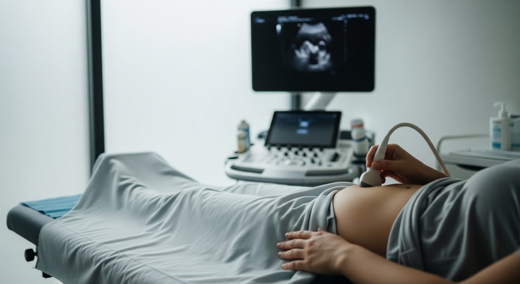
Conventional advice says the first scan should wait until the end of the first trimester. That guideline helps in many contexts. It also delays clarity that parents and clinicians value in the earliest weeks. I use early pregnancy ultrasound to establish what is present, where it is, how far along it likely is, and whether development matches expectations. Step by step. Week by week. It is basically a structured way to turn uncertainty into a practical plan.
What Early Pregnancy Ultrasound Reveals Week by Week
4-5 Weeks: Gestational Sac Detection
At this stage I focus on location and confirmation. An early pregnancy ultrasound can reveal a small gestational sac within the uterine cavity. That single finding helps exclude ectopic pregnancy to an extent, while not eliminating risk entirely if symptoms evolve. The sac is usually round or oval with smooth margins.
What I look for is consistency with the claimed dates. A gestational sac may measure only a few millimetres. A yolk sac might not yet be visible. That is acceptable this early. If the sac is irregular or too small for the presumed dates, I recommend repeat scanning after 7 to 10 days. Timed follow up is vital.
-
Goal: confirm intrauterine pregnancy and baseline measurements.
-
Typical view: gestational sac only, sometimes a yolk sac near week 5.
-
Action: schedule a follow up if structure is present but too early for a heartbeat.
In practice, an early pregnancy scan at this point provides orientation. Not closure. I prefer clarity over false certainty.
6 Week Pregnancy Ultrasound: Heartbeat and Foetal Pole
By six weeks, a 6 week pregnancy ultrasound often shows a foetal pole and a heartbeat. On an early pregnancy ultrasound, cardiac activity appears as a flicker within the embryo. Visibility depends on dating accuracy and scan method. Transvaginal imaging typically provides clearer visualisation now.
If a heartbeat is not yet visible, I reassess three variables. First, are the dates exact or approximate. Second, is the image quality sufficient for a definitive call. Third, does the crown-rump length suggest a younger gestation. A short interval repeat scan usually answers the question without drama.
-
Expected: foetal pole, yolk sac, and cardiac flicker.
-
If absent: repeat scan after 5 to 7 days is reasonable.
-
Method: transvaginal if appearances are borderline.
The key is restraint. Early reassurance matters, but so does diagnostic accuracy.
7-8 Weeks: Crown-Rump Length Measurement
From seven to eight weeks I move into measurement and precision. On early pregnancy ultrasound, the crown-rump length becomes the anchor metric for gestational age. CRL dating is highly reliable in this window and usually more accurate than memory of the last menstrual period.
At this stage, I expect a well defined embryo, a clear yolk sac, and a regular heartbeat. I also review sac position, uterine shape, and any peri-gestational collection. Small discrepancies are normal. Large discrepancies prompt a careful review of biology and timing. It is a clinical judgement, not a rush to label.
-
Primary metric: crown-rump length for gestational age estimation.
-
Secondary checks: yolk sac size, sac shape, and chorionicity if twins appear.
-
Plan: note baseline for comparison at the next visit.
Precision improves as structures become unambiguous. That is the advantage of this week.
9-10 Weeks: Limb Buds and Movement
By weeks nine and ten, anatomical detail increases. An early pregnancy ultrasound now shows limb buds, head-body differentiation, and early spontaneous movement. Cardiac activity is stable. The embryo usually fills much of the gestational sac.
This is a good time to confirm single or multiple pregnancy, check chorionicity in twins, and refine dates if needed. I also assess the yolk sac. A very large or irregular yolk sac may warrant closer observation. It is a clue, not a verdict.
-
Expected features: limb buds, head contour, and subtle movement.
-
Clinical focus: viability, multiplicity, and alignment with expected age.
-
Next step: plan the first trimester ultrasound screening timetable.
Development accelerates rapidly here. So do parental questions. I keep guidance clear and calm.
11-12 Weeks: Nuchal Translucency and Dating Accuracy
At eleven to twelve weeks, I can combine dating and screening. On early pregnancy ultrasound, the nuchal translucency can be measured if the position is optimal. CRL at this point still offers strong dating accuracy. The combination supports well timed screening decisions.
Position matters for NT measurement. If foetal posture is unfavourable, I do not force the issue. A short pause, a gentle position change, or a repeat appointment may be appropriate. Quality over haste. Screening depends on technique and timing.
-
Key measures: nuchal translucency and CRL.
-
Secondary observations: nasal bone presence and basic anatomy overview.
-
Outcome: integrated risk assessment when paired with bloods, if chosen.
This is where data and context meet. Numbers inform decisions, but they do not dictate them.
13-14 Weeks: Gender Determination Possibilities
From thirteen weeks, external genitalia may occasionally suggest sex, but confidence varies. I present this as tentative if asked. The purpose of an early pregnancy ultrasound here remains growth, positioning, and general wellbeing. Definitive sex determination is more reliable later.
-
Primary aim: confirm ongoing normal development and refine dates if needed.
-
Secondary note: sex prediction is possible but not guaranteed at this stage.
-
Advice: treat early sex indicators as provisional.
Curiosity is understandable. Accuracy takes precedence.
Types of Early Pregnancy Scans and Their Purposes
Transvaginal vs Transabdominal Ultrasound
Choice of approach is a technical decision. For very early assessment, a transvaginal scan usually provides superior resolution. It allows earlier detection of a gestational sac and cardiac activity on an early pregnancy ultrasound. Transabdominal scanning becomes more informative as the pregnancy progresses and the uterus rises.
|
Approach |
Primary use |
|---|---|
|
Transvaginal |
Optimal visualisation at 4 to 8 weeks, early heartbeat, precise CRL |
|
Transabdominal |
Useful from around 8 to 10 weeks onward, broader overview |
Comfort, privacy, and image quality guide the final choice. I discuss options and proceed with consent.
Dating Scan Accuracy and Timing
Dating accuracy is highest when using CRL in early gestation. An early pregnancy ultrasound between 7 and 12 weeks aligns gestational age estimates closely with true embryonic age. Accuracy decreases when based on sac size alone, particularly before the foetal pole emerges.
-
Best window for dating: 7 to 10 weeks using CRL.
-
Acceptable but less precise: 5 to 6 weeks using sac metrics.
-
If discrepancy occurs: prioritise CRL over recollected cycle length.
Reliable dating underpins every subsequent interpretation. It is the first building block.
Viability Scan at Different Gestational Ages
A viability scan answers a narrow question. Is the pregnancy developing in line with expectations today. On an early pregnancy ultrasound, viability is inferred from structural progression and cardiac activity. The earlier the scan, the more cautious the inference should be.
-
Very early viability: presence of sac and yolk sac, to be confirmed later.
-
Definitive viability: foetal pole with visible heartbeat.
-
Progress checks: serial scans a few days apart when appearances are borderline.
One scan gives a snapshot. Serial scans define a trend. That distinction matters.
First Trimester Ultrasound Screening Tests
Screening in the first trimester combines measurements with maternal blood markers. The first trimester ultrasound provides CRL and nuchal translucency. When paired with biochemistry, it yields a risk estimate for chromosomal conditions. It is screening, not diagnosis.
-
Components: NT, CRL, and maternal serum markers.
-
Outcome: risk estimation that may guide further testing.
-
Timing: typically 11 to 13+6 weeks for a complete assessment.
Results inform choices. They do not oblige any action. Autonomy remains central.
Understanding Early Pregnancy Scan Measurements and Results
Gestational Sac Size and Growth Rates
In the earliest window, I may use mean sac diameter as a provisional sign of progress. On early pregnancy ultrasound, the sac should enlarge steadily over days. A visible yolk sac generally appears when the mean sac diameter reaches a modest threshold.
-
Healthy trend: consistent sac enlargement on serial scans.
-
Concerning patterns: irregular sac shape or absent yolk sac over time.
-
Action: align findings with dates and repeat for confirmation when borderline.
Growth trends trump single measurements. Pattern over points.
Foetal Heart Rate Normal Ranges by Week
Heart rate rises in the early weeks, then stabilises. On early pregnancy ultrasound, I expect lower rates near week six and higher by week eight. Variation exists, and posture or technical factors can influence readings.
|
Gestational period |
Typical heart rate pattern |
|---|---|
|
6 weeks |
Lower normal limits, early cardiac flicker visible |
|
7 to 8 weeks |
Rising rates with regular rhythm |
|
9 to 10 weeks |
Stable pattern, consistent visibility across views |
Outliers warrant context and follow up, not instant conclusions. Caution avoids needless alarm.
Crown-Rump Length Charts and Accuracy
CRL is the most robust single measure for dating in early gestation. On early pregnancy ultrasound, the correct plane is essential: midline, neutral flexion, tips clearly defined. Repeating the measurement improves confidence and reduces operator bias.
-
Best practice: obtain multiple CRL measurements and use an average.
-
Check: avoid hyperflexion or hyperextension when freezing the image.
-
Use: anchor estimated due date on CRL when available.
CRL reduces debate about dates. That clarity streamlines every later assessment.
Signs of Healthy vs Concerning Findings
Healthy early scans show a round sac, appropriate yolk sac, clear foetal pole, and a regular heartbeat. On an early pregnancy ultrasound, mismatch between dates and structures may simply reflect late ovulation. But it can also signal a developmental issue. The solution is paced reassessment.
-
Reassuring: steady growth and stepwise appearance of expected features.
-
Ambiguous: features present but not aligned with reported dates.
-
Concerning: deteriorating appearances on serial scans.
Good news is common. Bad news, when it occurs, should be delivered with clarity and support.
Preparing for Your Early Pregnancy Scan
When to Schedule Your First Scan
Timing depends on intent. If the goal is simple confirmation, a visit around five to six weeks can suffice. For strong dating accuracy, I suggest seven to eight weeks. The early pregnancy ultrasound at that point provides both reassurance and reliable measurements.
-
Confirmation only: book at 5 to 6 weeks.
-
Dating focus: book at 7 to 8 weeks.
-
Screening plan: align with first trimester timetable after dating is set.
A short delay often avoids repeat attendance. That is respectful of time and wellbeing.
Full Bladder Requirements for Different Scan Types
A moderately full bladder helps with transabdominal views in early gestation. It displaces bowel gas and lifts the uterus into the acoustic field. For transvaginal imaging, an empty bladder usually produces better detail on an early pregnancy ultrasound.
|
Scan type |
Bladder guidance |
|---|---|
|
Transabdominal |
Arrive with a comfortably full bladder |
|
Transvaginal |
Empty bladder unless advised otherwise |
Comfort is a legitimate clinical factor. If discomfort arises, I pause and adjust.
Questions to Ask Your Sonographer
Preparation improves the value of the visit. I encourage concise, practical questions that target clarity.
-
What is visible today, and how does it align with the reported dates.
-
Is a heartbeat present, and what follow up is advised if not yet visible.
-
What is the estimated gestational age from CRL, and how precise is that estimate.
-
Do the findings suggest any specific next steps or repeat scanning.
The aim is shared understanding. Everyone leaves with the same plan.
Understanding Scan Limitations Before 6 Weeks
Before six weeks, an early pregnancy ultrasound offers limited inference. Structures are tiny. Cardiac activity may not be visible even when development is normal. A non-diagnostic early scan is not a negative result. It is simply too soon.
-
Risk of false reassurance or undue worry is higher very early.
-
Short interval follow up is often the best solution.
-
Transvaginal imaging may improve clarity but cannot accelerate biology.
Biology sets the pace. The scanning strategy respects that pace.
Making Informed Decisions About Early Pregnancy Scanning
Early scanning has clear benefits and a few drawbacks. Benefits include accurate dating, early localisation of the pregnancy, and timely detection of concerning patterns. Drawbacks include the possibility of ambiguous results that require repeat visits. I keep the balance explicit to support informed consent.
Pros
-
Clarifies intrauterine location early.
-
Provides reliable dating from CRL at the right window.
-
Identifies multiple pregnancy and chorionicity sooner.
Cons
-
Very early scans may be non-diagnostic and prompt repeat visits.
-
Incidental findings can create anxiety without altering management.
Here is how I approach decisions in practice:
-
Define the objective. Confirmation, viability check, dating, or screening alignment.
-
Choose the optimal window. Earlier is not always better for decision quality.
-
Select the method. Transvaginal or transabdominal based on timing and image need.
-
Plan contingencies. If results are equivocal, agree a repeat interval now.
I also set expectations. An early pregnancy ultrasound gives answers, but sometimes it gives a timeline. That is still useful. It turns vague waiting into a clear next step.
On language, I use two simple terms for clarity. CAC means clinical assessment checkpoint, a defined review point when results are borderline. SOP means standard operating plan, the agreed sequence of scan, repeat, and referral if criteria are met. A little structure reduces stress when uncertainty appears.
Early certainty is ideal. Early clarity is achievable. When certainty must wait, clarity carries the day.
One final point. A first trimester ultrasound later in the window remains essential even after an early check. The early study does not replace screening or anatomical review. It complements them and improves scheduling.
Frequently Asked Questions
Can an early pregnancy scan detect twins at 6 weeks?
Yes, twins can be identified at this stage, especially with transvaginal imaging. An early pregnancy ultrasound may show two gestational sacs, two yolk sacs, or two foetal poles. Chorionicity assessment begins early, but confirmation improves as the pregnancy advances. If the view is uncertain, I schedule a repeat to ensure accuracy.
Why might heartbeat not be visible at 6 week ultrasound?
Timing and technique are the usual reasons. Ovulation may have occurred later than assumed, so the pregnancy is simply earlier than expected. On an early pregnancy ultrasound, image quality and foetal position also influence visibility. I recommend a repeat scan within a week before drawing conclusions, provided symptoms are stable.
How accurate is gestational age determination from early scans?
CRL-based dating between seven and ten weeks is highly reliable. It is often more precise than calculations from the last menstrual period. An early pregnancy ultrasound before the foetal pole appears is less precise, because sac measurements are proxies. Once CRL is measurable, I use it to anchor the due date.
Is transvaginal ultrasound safe in early pregnancy?
Yes, it is considered safe when performed by trained professionals. It offers better resolution in the early weeks and improves the diagnostic yield of an early pregnancy ultrasound. If there is any discomfort, I pause and adjust. Consent and communication remain central throughout.
What if my dates don’t match the ultrasound measurements?
This is common. Cycle variability and ovulation timing explain most discrepancies. When CRL is available, I prioritise the scan-derived date. If the difference is substantial, I propose a short interval recheck. That approach respects biology and avoids premature conclusions while using early pregnancy ultrasound to refine timing.
Can early pregnancy scan detect chromosomal abnormalities?
Ultrasound alone provides risk indicators, not a diagnosis. Features such as nuchal translucency contribute to a screening estimate when combined with blood tests. An early pregnancy ultrasound supports this process by securing accurate dating and suitable timing. Diagnostic testing, if chosen, follows standard pathways.