


Haemorrhage Explained: Everything You Need to Know
Dr. Rajesh Kumar Mishra
Stop thinking of bleeding as a single problem. It is a set of distinct failure states that challenge oxygen delivery, organ perfusion, and clot stability. If you understand the mechanics, you act faster and with fewer errors. This guide gives you a clear structure for spotting, prioritising, and managing Haemorrhage so you protect function, not just survival.
Types of Haemorrhage
External Haemorrhage
External Haemorrhage is bleeding you can see. You identify the source, apply targeted pressure, and control it early. Arterial bleeding is typically brisk and pulsatile. Venous bleeding is darker and flows steadily. Capillary bleeding looks diffuse and oozing.
Here is why the basics matter. Blood loss reduces preload, cardiac output, and tissue oxygen delivery. That cascade worsens quickly. Early control stops the slide into shock.
-
Use direct pressure with a clean cloth or dressing.
-
Apply a tourniquet for life-threatening limb bleeding if pressure fails.
-
Elevate the limb if no fracture is suspected.
-
Do not remove embedded objects; pack around and stabilise them.
Haemorrhage control is not complicated. It is basically prompt, firm, and sustained pressure in the right place.
Internal Bleeding Classifications
Internal bleeding occurs within body cavities or tissues. You cannot see it directly, but you can recognise patterns. Classifications often reflect location and mechanism.
-
Intrathoracic: chest injuries, ruptured vessels, or lung lacerations.
-
Intra-abdominal: liver, spleen, or mesenteric injury causing hidden loss.
-
Retroperitoneal: pancreatic or renal sources that bleed silently for hours.
-
Intramuscular and soft tissue: large thigh or pelvic haematomas after trauma.
Haemorrhage inside a confined space can be substantial. A single femur fracture can bleed significantly. Pelvic injuries can bleed even more. Watch for pallor, tachycardia, and cool extremities. These subtle signs often precede collapse.
Brain Haemorrhage Types
Intracranial bleeding is a different problem. The skull is rigid, so even small volumes increase pressure and threaten tissue. The major types include intracerebral, subdural, epidural, and intraventricular bleeding.
-
Intracerebral: bleeding within brain tissue, often stroke or trauma related.
-
Subdural: venous bleeding between dura and arachnoid, typically gradual onset.
-
Epidural: arterial bleeding between skull and dura, classically a lucid interval then rapid decline.
-
Intraventricular: bleeding into cerebrospinal fluid spaces, raising intracranial pressure.
With any brain haemorrhage, timing is everything. Deterioration can be sudden. You cannot afford delays.
Subarachnoid Haemorrhage
Subarachnoid Haemorrhage involves bleeding into the cerebrospinal fluid around the brain. Many cases follow ruptured aneurysms. The hallmark is a thunderclap headache. Patients may report the worst headache of their life. Neck stiffness, photophobia, and vomiting are common.
Complications include vasospasm, hydrocephalus, and rebleeding. You manage the airway, control blood pressure, and arrange definitive neurovascular care. Haemorrhage in this space is unforgiving, though good outcomes are possible with rapid stabilisation.
Gastrointestinal Haemorrhage
Gastrointestinal Haemorrhage is bleeding anywhere from oesophagus to rectum. Upper sources include peptic ulcers and varices. Lower sources include diverticula, angiodysplasia, or colorectal lesions. Presentation varies from haematemesis to melena or haematochezia.
Risk stratification guides next steps. You assess shock, haemoglobin trend, and comorbid disease. Endoscopy provides diagnosis and control. Haemorrhage here often recurs if the driver remains untreated, such as uncontrolled portal hypertension.
Post-Partum Haemorrhage
Post-Partum Haemorrhage remains a leading cause of maternal morbidity. Primary cases occur within 24 hours of delivery. Common causes include uterine atony, retained placenta, lacerations, and coagulopathy.
-
Uterine massage and uterotonics are first-line measures.
-
Repair lacerations and remove retained tissue promptly.
-
Escalate to balloon tamponade or surgical options if bleeding continues.
Haemorrhage in obstetrics demands rehearsal and checklists. Teams that practice manage stress better and act faster.
Causes and Risk Factors
Trauma-Related Causes
Trauma drives many cases of severe bleeding. High-energy impacts produce multi-cavity injury. Low-energy mechanisms, like falls in the elderly, still cause substantial Haemorrhage given fragile vessels and medications.
-
Penetrating injuries can sever major vessels and cause rapid shock.
-
Blunt trauma can shear organs and rupture splenic or hepatic tissue.
-
Fractures, especially pelvis and femur, conceal large blood volumes.
In practice, assume internal bleeding until proven otherwise after significant trauma. That mindset prevents under-triage.
Medical Conditions Leading to Haemorrhage
Several conditions increase the likelihood of Haemorrhage. Platelet disorders, liver disease, and vascular malformations are frequent culprits. Hypertension contributes to intracerebral events. Infections can trigger disseminated intravascular coagulation.
-
Liver cirrhosis reduces clotting factors and increases portal pressures.
-
Aneurysms and arteriovenous malformations predispose to rupture.
-
Haematological disorders impair platelet number or function.
The context matters. An identical injury can bleed minimally in one patient and extensively in another.
Medications That Increase Risk
Common drugs increase bleeding risk. Knowing them helps you act decisively.
|
Medication |
How it raises bleeding risk |
|---|---|
|
Warfarin |
Reduces vitamin K dependent factors; elevated |
|
Direct oral anticoagulants |
Factor Xa or thrombin inhibition; limits thrombin burst and clot stability. |
|
Antiplatelets |
Impairs platelet activation and aggregation at the injury site. |
|
NSAIDs |
Reversible platelet dysfunction and gastric mucosal injury. |
|
SSRIs |
Reduced platelet serotonin storage; modestly higher bleeding tendency. |
Haemorrhage is more likely when multiple agents interact. A careful medication history changes management.
Age and Lifestyle Factors
Bleeding risk rises at age extremes. Very young patients compensate poorly. Older adults often take anticoagulants and have comorbid disease. Lifestyle also plays a role.
-
Excess alcohol damages liver and disrupts clotting.
-
Poor nutrition reduces platelet function and vessel integrity.
-
High blood pressure increases risk of intracranial events.
Modify what you can. Control blood pressure and limit alcohol. Small changes reduce the odds of catastrophic Haemorrhage.
Recognition and Emergency Response
Warning Signs of Severe Bleeding
Severe bleeding shows a consistent pattern. You will see pallor, rapid pulse, cold clammy skin, and sometimes confusion. Breathlessness and thirst are frequent. Capillary refill prolongs as perfusion falls.
Haemorrhage is often underestimated in early stages. Look for swelling under clothing, expanding bruises, and persistent oozing. These clues point to ongoing loss.
-
Monitor mental status changes, even small ones.
-
Track pulse rate and quality. A rising rate is a warning.
-
Watch for falling urine output if you can measure it.
One more sign matters. Silence. A patient who stops talking may be losing cerebral perfusion.
Symptoms of Internal Bleeding
Internal bleeding can be subtle. Abdominal pain, shoulder tip pain, dizziness, or fainting may occur. Chest pain or shortness of breath suggests thoracic involvement. Back pain can indicate retroperitoneal bleeding.
With brain haemorrhage, note sudden severe headache, weakness, slurred speech, or seizures. These symptoms require urgent evaluation. Do not wait to see if they settle.
Haemorrhage that you cannot see still counts. Treat the physiology you observe, not only the visible wound.
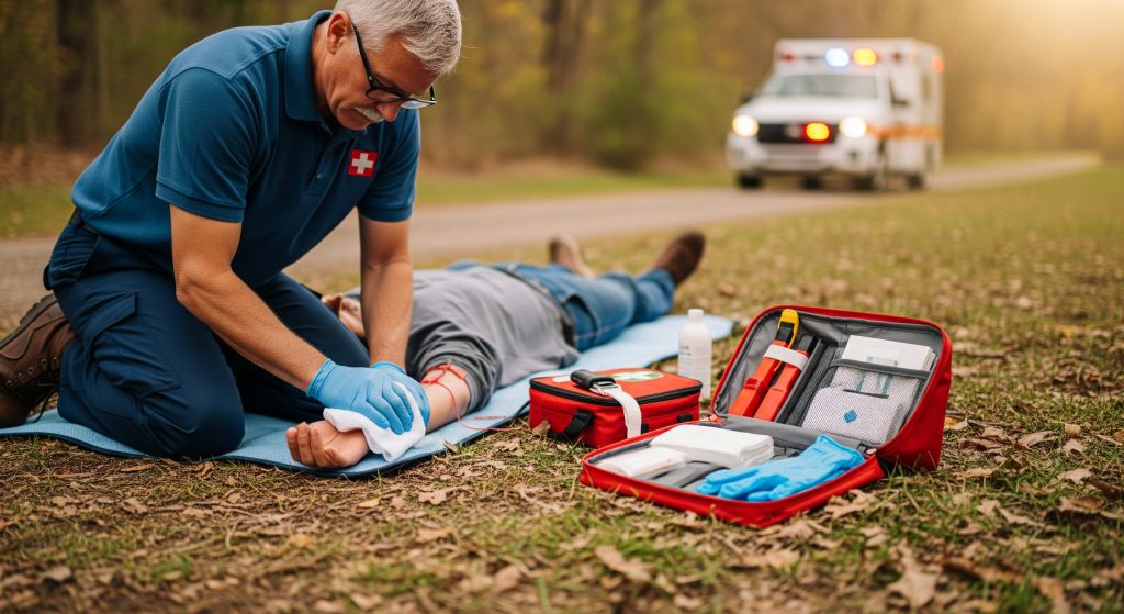
First Aid for External Haemorrhage
Control bleeding quickly and systematically. The steps are simple and effective.
-
Call for help. Alert emergency services early.
-
Expose the wound. Remove or cut clothing to see the source.
-
Apply direct pressure with a clean dressing. Maintain firm pressure.
-
If bleeding continues, add more dressings. Do not remove the first layer.
-
Use a tourniquet for limb bleeding that does not stop. Note the time applied.
-
Immobilise the area and keep the patient warm.
These steps buy time. Haemorrhage control at the scene reduces downstream complications.
When to Call Emergency Services
Call immediately if you see spurting blood, soaking dressings, or signs of shock. Call if the person becomes drowsy, confused, or short of breath. Call if internal bleeding is suspected.
With any subarachnoid haemorrhage symptoms, seek care without delay. The same applies to large gastrointestinal bleeds, such as repeated vomiting of blood or black stools. Time is the variable you can influence.
What Not to Do During Haemorrhage
Some actions worsen outcomes. Avoid them.
-
Do not remove impaled objects. Stabilise them in place.
-
Do not apply tourniquets loosely. Tighten adequately or do not use them.
-
Do not pack the chest or abdomen in the field. Focus on transport.
-
Do not give food or drink before assessment. Aspiration risk is real.
-
Do not delay escalation while trying multiple dressings repeatedly.
Haemorrhage responds to decisive action. Caution with momentum beats hesitant improvisation.
Medical Treatment and Recovery
Emergency Medical Interventions
In hospital, priorities follow a clear sequence. Airway, breathing, and circulation come first. Massive transfusion protocols address shock and coagulopathy together.
-
Tranexamic acid may be used early in trauma-related bleeding.
-
Point-of-care coagulation tests guide reversal and factor replacement.
-
Targeted blood pressure management balances perfusion with bleeding control.
Haemorrhage is ultimately a perfusion problem. You restore circulating volume and ensure oxygen delivery to vital organs.
Surgical Options for Haemorrhage
Procedures aim to expose, control, and repair the source. In trauma, damage control surgery limits time in theatre. Definitive repair follows when physiology stabilises.
-
Ligation or repair of bleeding vessels.
-
Packing of cavities with planned relook after resuscitation.
-
Endovascular embolisation or stenting where feasible.
-
Neurosurgical decompression for expanding intracranial bleeds.
Surgical judgement matters. Sometimes less is safer in the first pass. Then more, once physiology is steady.
Blood Transfusion Protocols
Modern transfusion strategy emphasises balanced products. A ratio-based approach provides red cells, plasma, and platelets in concert. This limits dilutional coagulopathy.
|
Goal |
Restore oxygen-carrying capacity and clotting ability together. |
|
Monitoring |
Use |
|
Adjuncts |
Calcium, fibrinogen concentrate, and cryoprecipitate as indicated. |
|
Temperature |
Warm all products. Hypothermia worsens coagulopathy. |
Haemorrhage control improves quickly when you correct all elements of the lethal triad. That is acidosis, hypothermia, and coagulopathy.
Recovery Timeline and Rehabilitation
Recovery is variable. Roughly speaking, soft tissue injuries settle in days to weeks. Major abdominal or orthopaedic injuries need months. Intracranial bleeds may require extended rehabilitation.
-
Physiotherapy rebuilds strength and mobility.
-
Occupational therapy adapts tasks to residual deficits.
-
Neuropsychology supports cognition after brain injury.
Measure progress with clear metrics. Six-minute walk distance, grip strength, and independence in daily activities are practical. Haemorrhage is an acute crisis. Rehabilitation converts survival into quality of life.
Understanding Haemorrhage for Better Health Outcomes
Think in systems. Source control, volume replacement, and clot optimisation move together. That mindset reduces errors and shortens time to stability. It also improves outcomes for internal bleeding where delays are common.
Adopt a simple operating picture. Identify the source. Control it. Refill the tank. Rebuild the clot. Protect temperature. Then reassess every few minutes. Use team briefs for clarity and speed.
Two practical habits pay off. Carry a dedicated tourniquet in first aid kits. Learn the basics of pressure application properly. Small skills change the trajectory of Haemorrhage before an ambulance arrives.
For organisations, drill the Massively Bleeding protocol. Track time to first unit, time to theatre, and proportion of balanced products. Use a dashboard with a few critical metrics. You live and die by your MAP, lactate, and base deficit in the first hours. MAP is mean arterial pressure. These numbers tell you if your strategy is working.
There is a counterpoint. Some bleeds stop with rest and observation. Over-intervention can harm. But waiting when physiology is sliding is worse. The art is deciding which case you have.
Frequently Asked Questions
What is the difference between haemorrhage and normal bleeding?
Normal bleeding is a small, self-limited loss that stops with simple measures. Haemorrhage is uncontrolled or large-volume loss that threatens perfusion, organ function, or both. In practice, severe bleeding that soaks dressings or causes dizziness and pallor meets the threshold for escalation.
Consider the mechanism and the patient. Anticoagulated patients cross into Haemorrhage earlier. Even modest loss can be significant in small children.
How quickly can someone die from severe haemorrhage?
Death can occur within minutes from exsanguination, especially from major arterial injury. Timeframes vary widely, depending on the source and access to care. Internal bleeding may present a slower decline and still be fatal without intervention.
Treat every minute as material. Early pressure, early transport, and early transfusion make the difference between recovery and collapse.
Can haemorrhage occur without visible bleeding?
Yes. Internal bleeding is common in trauma and in conditions like ruptured ectopic pregnancy or gastrointestinal ulcers. The absence of external blood does not exclude significant loss. Watch for abdominal distension, pain, dizziness, or sudden weakness.
If you suspect Haemorrhage, seek urgent assessment. Bedside ultrasound and rapid blood tests often identify the problem early.
What are the long-term effects of brain haemorrhage?
Outcomes vary. Some people recover fully, particularly after small bleeds with swift care. Others have lasting deficits, including motor weakness, speech problems, memory issues, or seizures. Fatigue and mood changes are frequent, even when scans normalise.
Structured rehabilitation matters. Regular follow-up with neurology and therapy teams improves the odds of meaningful recovery after any brain haemorrhage.
Is haemorrhage preventable in most cases?
Many cases are preventable to some extent. Control blood pressure, moderate alcohol, and manage anticoagulants carefully. Use protective equipment during high-risk activities. Treat ulcers and liver disease where possible.
Not everything is avoidable. Aneurysm rupture or unexpected trauma can still occur. Risk reduction remains worthwhile and effective.
What should I keep in a first aid kit for haemorrhage emergencies?
-
A quality tourniquet with clear instructions.
-
Large sterile dressings and haemostatic gauze.
-
Compression bandage to maintain pressure hands-free.
-
Gloves, trauma shears, and a foil blanket for warmth.
-
A concise instruction card for bleeding control steps.
If you support a team or workplace, add a second tourniquet and more gauze. Practice application on a training limb once a year. Haemorrhage control is a skill. Keep it fresh.