


Bone Scan Test Explained: Procedure, Results and Safety
Dr. Beenish Khan


Routine X-rays miss early metabolic changes in bone. That is where a bone scan test earns its place. I will explain how each scan type works, what to expect on the day, how results are interpreted, and how to weigh benefits against risks. Clear, formal, and practical. That is the brief.
Types of Bone Scans and Their Medical Uses
Under the umbrella of a bone scan test sit several techniques with distinct roles. The core idea is simple. Use a tracer or X-ray beam to capture bone turnover or density so clinicians can act early. Different questions demand different tools.
Bone Scintigraphy for Cancer Detection
Bone scintigraphy maps osteoblastic activity across the skeleton. In practice, that means it lights up areas where bone is actively remodelling in response to injury, inflammation, or tumour activity. For oncology, this functional view is valuable because skeletal metastases often trigger repair processes before structural change is obvious on plain X-ray.
Clinically, I consider bone scintigraphy a broad survey. It helps stage many cancers, triage unexplained bone pain, and direct targeted imaging. There is an important caveat. Uptake is not specific to malignancy. Arthritic joints, healing fractures, and infections can also appear active. A multimodal approach is therefore common. In breast cancer, for instance, FDG PET/CT may outperform scintigraphy in picking up certain metastatic lesions, a point highlighted in Journal of Nuclear Medicine Technology.
-
Strength: Whole skeleton coverage in a single session.
-
Limitation: Activity highlights a process, not a diagnosis in isolation.
-
Best used for: Staging, surveillance, and problem solving when symptoms outpace X-ray findings.
Here is the practical takeaway. If the clinical question is widespread involvement or a search for occult sites, a bone scan test based on scintigraphy often provides the quickest map.
DEXA Scan for Osteoporosis
A DEXA scan is a bone density test. It measures bone mineral density at key sites to assess fracture risk and diagnose osteoporosis. Unlike scintigraphy, it does not look at biological activity. It measures density with high precision and low radiation. The output is numerical and comparable over time, which suits monitoring.
For workflow planning, DEXA is efficient. As RadiologyInfo notes, most exams take about 10 to 15 minutes and results are reported using T-scores and Z-scores. In primary care and endocrinology, that speed and reproducibility matter. They help clinicians decide on calcium, vitamin D, exercise prescriptions, and when to escalate to antiresorptive or anabolic therapies.
-
Strength: Quantitative, reproducible measurement for longitudinal follow up.
-
Limitation: Focused on density, not pain generators or active lesions.
-
Best used for: Baseline risk, therapy decisions, and treatment response in osteoporosis.
Think of DEXA as the right tool when the question is risk over the next years. It is not built to locate an acute lesion today.
Three Phase Bone Scan
The three phase bone scan is a targeted form of bone scintigraphy. It sequences images in three phases to evaluate both blood flow and bone turnover. The phases are performed rapidly at injection, shortly after for blood pool, and later for delayed skeletal uptake. When infection or complex regional pain syndrome is suspected, this sequencing can sharpen interpretation.
-
Phase 1: Flow – assesses regional perfusion.
-
Phase 2: Blood pool – shows soft tissue hyperaemia.
-
Phase 3: Delayed – demonstrates osteoblastic activity within bone.
This structure helps distinguish vascular and inflammatory patterns from purely osseous processes. In practice, it reduces ambiguity when conventional imaging is equivocal.
SPECT Bone Scan Technology
SPECT adds depth to standard scintigraphy. The reconstruction produces three dimensional images that allow precise localisation of uptake in complex anatomy such as the spine or foot. That extra localisation can shift management because it answers where the activity sits within bone, joint, or adjacent structures.
Clinical studies have shown that SPECT or SPECT/CT can improve characterisation of spinal lesions compared with planar scans, supporting decisions when MRI or CT findings are non-specific. In difficult axial pain, that incremental clarity often tips the balance toward the correct intervention. A considered reading protocol is crucial here.
-
Strength: 3D localisation for complex regions and overlapping structures.
-
Limitation: Availability and slightly longer acquisition compared with planar scans.
-
Best used for: Focal questions in the spine, pelvis, or foot where overlap obscures planar images.
As a rule of thumb, I escalate to SPECT when the question is more about exact location than simply presence or absence of disease.
Common Conditions Diagnosed
A bone scan test can support diagnosis or management across several areas:
-
Oncology: staging and surveillance of skeletal metastases; problem solving for suspected primary bone tumours.
-
Infection: evaluation of osteomyelitis patterns and differentiation from sterile inflammation.
-
Trauma and sports: detection of stress reactions and occult fractures where X-ray is normal.
-
Arthritis: assessment of activity in degenerative or inflammatory arthropathies to correlate with symptoms.
-
Post-operative: distinguishing loosening from infection in prosthetic joints with appropriate protocols.
Different tests answer different questions. I match the tool to the decision at hand.
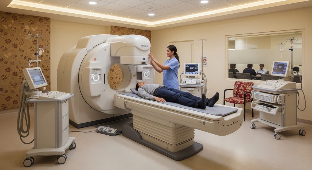
Complete Bone Scan Test Procedure
Despite the technical language, the process is straightforward. The team prepares, injects, waits for tracer distribution, then acquires images. A bone scan test follows a predictable rhythm, with variations for SPECT, three phase, or focused areas.
Pre-Test Preparation Requirements
Preparation is usually light. I advise ordinary hydration unless restricted for another reason. Metal objects should be removed to prevent artefacts during imaging. For DEXA, calcium supplements are typically paused the day before to avoid spurious readings, and clothing without metal fasteners is preferred. If there is any chance of pregnancy, disclosure is essential so the team can advise appropriately.
-
Bring prior imaging and reports for comparison.
-
Share all medications, especially recent radionuclide or contrast studies.
-
Inform the team about implants, previous surgeries, or prostheses.
Simple preparation prevents avoidable repeats and keeps image quality high.
Radiotracer Injection Process
For scintigraphy, a small amount of radiotracer is injected into a peripheral vein. The dose is selected for adequate image quality while maintaining low exposure. The injection is typically brief. I recommend keeping the limb warm and gently mobile, which can help reduce local pooling and minor discomfort at the site.
For a three phase bone scan, initial imaging starts immediately after injection to capture the flow and blood pool phases. For a standard whole skeleton study, the delayed phase occurs after the tracer binds to bone mineral.
Waiting Period Activities
After injection, distribution time is required. Light walking and good hydration can support tracer circulation and renal clearance of background activity. Most centres provide a waiting area and suggest returning at a set time. It is reasonable to use the interval for a light snack, normal fluids, and a brief walk unless advised otherwise.
This period is not wasted time. It enhances image contrast between active bone and soft tissue background.
Imaging Process Steps
Imaging is conducted with a gamma camera for planar scintigraphy or with a rotating detector for SPECT. The technologist will position the table and guide the sequence. For DEXA, the table and arm move in a defined path across the lumbar spine and hips while the patient remains still. For a bone scan test using scintigraphy, whole body sweeps and spot views are common, followed by SPECT if localisation is needed.
-
Identity and safety checks.
-
Positioning and initial scout images as needed.
-
Acquisition of whole body or regional planar images.
-
Optional SPECT or SPECT/CT for targeted regions.
-
Quality review to ensure complete coverage and clarity.
I encourage questions before the camera moves. A clear plan reduces anxiety and movement artefacts.
Duration and Positioning
Planar whole body imaging is typically efficient. SPECT adds time for rotation and reconstruction. Most of the appointment is still and quiet. The technologist will ask for absolute stillness during each acquisition to avoid blur. Blankets, cushions, and gentle prompts help maintain position without strain. If discomfort is likely, flag this early so positioning can be adjusted.
For patients with back pain or recent surgery, careful padding and short breaks between sequences can be arranged. Precision matters. Comfort supports precision.
Post-Procedure Instructions
Normal activities can usually be resumed immediately after a bone scan test. I advise increased fluids for the rest of the day to help clear residual tracer via the kidneys. The injection site may be tender briefly. If redness or swelling develops, notify the imaging department.
-
Continue prescribed medicines unless instructed otherwise.
-
Delay close, prolonged contact with infants or pregnant individuals for the remainder of the day as a conservative precaution.
-
Keep the appointment summary for reference should another imaging test be scheduled soon.
Image interpretation is performed by a nuclear medicine physician or radiologist. The report is then shared with the referring clinician for action.
Interpreting Bone Scan Results
Interpretation combines patterns, intensity, distribution, and clinical context. A bone scan test shows biology in motion. That makes the discussion nuanced and, at times, deceptively simple on paper.
Normal vs Abnormal Findings
A normal study shows expected uptake in joints, growth plates in younger patients, and consistent distribution in axial and appendicular skeleton. Abnormal findings vary. Focal intense uptake may indicate a fracture, metastasis, or infection depending on location and pattern. Diffuse uptake can reflect metabolic bone disease or systemic factors.
I look for symmetry, correlation with pain, and the match or mismatch with other imaging. A single hot focus in a rib of a coughing patient suggests a stress fracture. Multiple asymmetric foci in the pelvis and spine in a patient with known carcinoma shifts suspicion toward metastases.
Hot Spots and Cold Spots
Hot spots represent increased tracer uptake. They reflect osteoblastic activity. Cold spots represent reduced uptake and can indicate lytic lesions, infarcts, or vascular compromise where bone turnover is suppressed. Both patterns can be clinically significant.
-
Hot: stress injury, arthritis flare, osteomyelitis, or healing fracture.
-
Cold: aggressive lytic metastasis, avascular necrosis, or certain benign lesions.
Context rules. I pair the pattern with labs, symptoms, and targeted imaging before drawing conclusions.
Bone Density Test Scores
DEXA reports provide T-scores and Z-scores. T-scores compare bone density with a young healthy reference. Z-scores compare with age matched peers. Thresholds delineate normal, low bone mass, and osteoporosis. These cut offs guide treatment choices and follow up intervals. Repeatability allows monitoring of therapy effect over time and across sites.
In reports, I review site specific values, artefacts from degenerative changes, and any discordance between hip and spine. Artefact recognition is vital to avoid over or under treatment.
False Positive Results
False positives occur when non malignant or non infectious processes cause increased uptake. Healing fractures, degenerative joints, post operative change, or benign tumours can mimic serious disease. The three phase bone scan can help narrow infection versus sterile inflammation. SPECT can localise activity to a facet joint rather than a vertebral body, changing the interpretation completely.
When the stakes are high, correlation with MRI, CT, or targeted ultrasound is prudent. It reduces diagnostic drift from pattern recognition alone.
Follow-up Testing Requirements
Follow up depends on the question asked. For suspected metastasis with indeterminate foci, I may request SPECT/CT or PET/CT for clarification. For osteoporosis, repeat DEXA at appropriate intervals tracks response and adherence. For infection, laboratory trends and, in certain cases, a tagged white cell scan form a robust pair.
Clear next steps protect against over investigation and delays. A short, focused plan beats open ended uncertainty.
Safety Considerations and Special Precautions
Safety with a bone scan test is shaped by three factors. Radiation exposure, physiological status, and medication context. The absolute risk is generally low for most adults, but careful screening and clear instructions keep it that way.
Radiation Exposure Levels
Scintigraphy involves a small radiation dose from the injected tracer. DEXA involves an even lower dose from an X-ray source. In routine clinical use, both are considered low exposure tests. The decision threshold remains clinical benefit versus minimal risk. This balance usually favours scanning when there is a clear diagnostic question that will change management.
Dose varies with protocol and body habitus. As far as current data suggests, optimised protocols continue to reduce exposure without compromising image quality.
Pregnancy and Breastfeeding Guidelines
For pregnancy, scintigraphy is generally deferred unless benefits clearly outweigh risks and alternatives are inadequate. If scanning is essential, protocols are adapted and specialist consent is required. For breastfeeding, some tracers may warrant temporary interruption with expressed milk management. Department specific guidance should be followed because tracer pharmacokinetics differ.
Disclosure at booking protects both patient and baby. No surprises on the day.
Allergic Reactions Risk
True allergic reactions to radiotracers are rare. Local injection site irritation is more common and usually minor. Prior contrast reactions do not automatically predict tracer reactions because the compounds differ. Nonetheless, I record all prior reactions and ensure observation capacity on site.
When anxiety is high, a brief pre procedure discussion often reduces perceived risk and improves cooperation during imaging.
Paediatric Safety Measures
Paediatric studies use weight adjusted dosing and child friendly positioning. Distraction techniques, parental presence, and shortened sequences where feasible can reduce motion and repeat imaging. In selected cases, child life specialists assist with preparation and coping strategies.
The principle is ALARA (as low as reasonably achievable). It guides dosing, timing, and the choice between modalities.
Medication Interactions
Most medicines do not interfere significantly with a bone scan test. However, recent high dose steroids, bisphosphonates, or denosumab are relevant for interpretation in osteoporosis follow up. Recent radionuclide therapies or iodinated contrast can also influence scheduling. For DEXA, calcium supplements on the day can cause artefacts, hence the common pause beforehand.
Provide a full medication list. It saves time and improves report quality.
Making Informed Decisions About Bone Scan Tests
Decision making is contextual. I approach it by matching the decision need with the smallest test that answers it.
|
Clinical question |
Preferred test |
|---|---|
|
Is there widespread skeletal involvement |
Planar scintigraphy as a first pass; escalate to SPECT for localisation |
|
Where exactly is the pain generator in the spine |
SPECT or SPECT/CT for 3D localisation |
|
What is the long term fracture risk |
DEXA bone density test with T-score and Z-score interpretation |
|
Is this infection or sterile inflammation |
Three phase bone scan, plus correlation with labs and targeted imaging |
|
Is a prosthesis loose or infected |
Appropriate nuclear protocol with comparison imaging and clinical markers |
One more consideration is escalation logic. If a bone scan test shows equivocal foci in a cancer patient and treatment depends on certainty, I escalate to a higher resolution or different modality rather than guess. In oncological pathways, this disciplined escalation prevents both under treatment and unnecessary toxicity.
Earlier, I noted that FDG PET/CT may surpass planar scintigraphy in certain cancers. That observation matters because it explains why protocols often layer modalities rather than choose a single test for every scenario.
The point is simple. Start with the question. Choose the fastest, safest test that answers it with confidence. Then act.
Frequently Asked Questions
How painful is a bone scan test?
The procedure involves a brief intravenous injection. Most patients describe this as a standard needle stick with minor discomfort. Imaging itself is painless. The main challenge is keeping still while the camera acquires images. If lying flat is difficult, inform the team so they can optimise positioning.
Can I drive home after a bone scintigraphy?
Yes, in most cases it is safe to drive after bone scintigraphy. The tracer does not impair alertness. If sedatives were not used and no other procedure occurred, routine driving is acceptable. I still suggest a short rest if the appointment was lengthy or if back pain is flared by lying still.
How long does radiotracer remain in the body?
Radiotracers used for a bone scan test clear gradually through the kidneys. A substantial proportion is excreted over the first day. Hydration and normal activity support clearance. Residual activity falls with physical decay and excretion, so levels drop quickly with time.
What is the difference between bone scan and bone density test?
They answer different questions. A bone scan test using scintigraphy shows active bone turnover and is used for cancer staging, infection evaluation, and occult fractures. A bone density test measures mineral density to estimate fracture risk and diagnose osteoporosis. One is functional. The other is quantitative structural assessment.
How often should bone scans be repeated?
Repetition depends on the clinical indication. For cancer surveillance, intervals are driven by disease biology and treatment plans. For suspected stress injury, repeat imaging is guided by symptoms and return to activity. For DEXA, repeat intervals are usually set to track meaningful change, not trivial variation. The referring clinician will time follow up to decision points.
Are bone scans covered by health insurance in India?
Coverage typically depends on the policy and the medical indication. Insurers often require a clinician referral and evidence that the test will change management. Hospital billing teams can confirm pre authorisation requirements and any co payment. It is advisable to check benefits before scheduling if cost predictability is important.