


What Is the EUS Procedure and When Is It Needed?
Dr. Urvashi Gupta
Popularity alone is not a reason to choose a test. Some patients are sent for scans that answer the wrong question. I use the EUS procedure when tissue detail and proximity matter, not because it is fashionable. Here is why this test earns its place in a careful workup, and when it changes management decisively.
Understanding the EUS Procedure
What Is Endoscopic Ultrasound
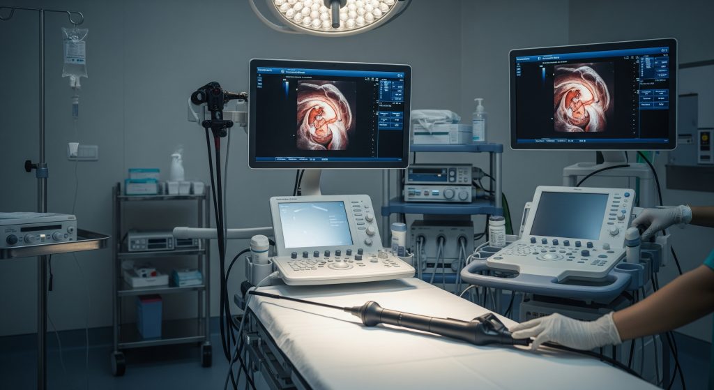
I describe endoscopic ultrasound as a hybrid test. It combines an endoscope with a high frequency ultrasound probe to visualise the digestive tract and nearby organs from within. The EUS procedure places the ultrasound source a few millimetres from the area of interest. That proximity produces high resolution images that external scans cannot match at similar depth. In practice, I use it for the pancreas, bile duct, and submucosal lesions in the gut.
-
Endoscope: a flexible tube with a camera and working channel.
-
Ultrasound transducer: generates detailed cross sectional images in real time.
-
Fine needle system: enables sampling when tissue is required.
This configuration allows both diagnosis and intervention in one sitting. It is efficient and precise.
How EUS Works
The EUS procedure relies on physics that are simple in principle. Ultrasound waves reflect differently from soft tissue, vessels, and cysts. The probe translates these echoes into a live image that guides decisions moment by moment. I navigate to the target segment under direct endoscopic vision. Then I rotate the ultrasound array to map structures in layers. It is basically a high resolution ultrasound from the inside out.
For example, to assess a suspected pancreatic mass, I position the probe in the stomach or duodenum. This provides a short acoustic path to the pancreas. The image reveals margins, vascular contact, and any suspicious nodes. If sampling is needed, I pass a needle through the endoscope under continuous ultrasound guidance.
Types of EUS Procedures
I group the EUS procedure into three broad types based on intent:
-
Diagnostic EUS: imaging alone to characterise lesions, stages, or structural changes.
-
EUS with tissue acquisition: fine needle aspiration or biopsy for cytology or histology.
-
Therapeutic EUS: interventions such as cyst drainage, celiac plexus block, or biliary access in selected cases.
Choice depends on clinical questions, safety profile, and available expertise. Not every centre offers therapeutic EUS, so planning matters.
EUS vs Traditional Endoscopy
Standard endoscopy shows mucosal surfaces in high detail. The EUS procedure extends this view beneath the mucosa into the wall and adjacent organs. The difference is decisive when pathology is subepithelial or extrinsic. An ordinary endoscopy may see a bulge. EUS defines whether that bulge is a lipoma, a GIST, or compression from outside. Same patient, different outcome.
In clinical terms, endoscopy answers the question on the surface. EUS answers the question below the surface. Both have value. They solve different problems.
Equipment and Technology Used
An EUS suite includes echoendoscopes, processors, ultrasound consoles, and needles in various gauges. Linear array scopes enable needle guidance. Radial array scopes create 360 degree views that are helpful for orientation. I choose the platform based on the task at hand. Needle size is matched to the target. A cyst with thick fluid may need a larger gauge. A small lymph node often does not.
Ancillary technologies improve yield. Suction techniques, stylet control, and rapid on site evaluation of cytology samples (ROSE) can raise diagnostic accuracy. These details sound minor. They are not. Execution quality drives outcomes.
When Is EUS Procedure Needed
1. EUS for Pancreatic Cancer
I reserve the EUS procedure for questions that imaging alone cannot resolve. For suspected pancreatic cancer, EUS for pancreatic cancer serves two key purposes. It refines visual assessment of the lesion and it enables tissue diagnosis with fine needle aspiration. I also evaluate vascular involvement and nodal disease to guide staging. A small lesion invisible on CT may be visible on EUS. That can move a case from uncertainty to a plan.
-
Characterise solid vs cystic lesions accurately.
-
Assess proximity to arteries and veins that determine resectability.
-
Obtain cytology to confirm malignancy before systemic therapy.
There is a counterpoint. Not every pancreatic abnormality needs immediate sampling. In high risk cysts with clear surgical features, I may proceed to definitive management first. Judgement matters.
2. Digestive System Tumours
For oesophageal, gastric, rectal, and duodenal tumours, the EUS procedure assesses depth of invasion and nodal status. This information shapes neoadjuvant therapy and surgical approach. I use EUS to clarify T stage in early cancers where organ preserving strategies may be viable. It helps avoid both under treatment and overtreatment.
3. Chronic Pancreatitis Evaluation
When standard imaging is equivocal, the EUS procedure can reveal subtle parenchymal changes in chronic pancreatitis. I look for lobularity, hyperechoic foci, and duct irregularity. EUS also assists in identifying complications such as pseudocysts or biliary compression. In carefully selected cases, celiac plexus block under EUS guidance can relieve pain to some extent.
4. Biliary Tract Disorders
In suspected choledocholithiasis or indeterminate biliary strictures, the EUS procedure offers high sensitivity. I can detect small distal stones and assess the ampulla, distal bile duct, and pancreas head. When ERCP findings are unclear, EUS adds clarity and may reduce unnecessary interventions. It can also direct tissue acquisition in malignant strictures.
5. Staging of Cancers
Regional staging informs prognosis and therapy. The EUS procedure contributes to T and N staging in oesophageal, gastric, rectal, and pancreatic cancers. I identify suspicious nodes, assess wall layers, and determine extramural spread. This complements cross sectional imaging. In practice, a combined approach provides the best map before treatment starts.
6. Unexplained Abdominal Pain
Persistent upper abdominal pain without explanation demands a careful approach. The EUS procedure can uncover small pancreatic lesions, biliary microlithiasis, or subtle ductal changes not seen on ultrasound or CT. I use it when symptoms persist and the pre test probability of a structural cause is meaningful. It is not a first test for nonspecific pain. It is a precise tool for a focused question.
7. Lung and Mediastinal Masses
With an echoendoscope placed in the oesophagus, I can assess posterior mediastinal structures. The EUS procedure helps sample mediastinal lymph nodes and masses adjacent to the oesophagus. It complements EBUS performed via the airway. Together, EUS and EBUS provide comprehensive access to thoracic nodes without surgical incisions.
The EUS Procedure Process
Pre-Procedure Preparation
I begin with an assessment of indications, co morbidities, and medications. Anticoagulants and antiplatelets are managed according to bleeding risk and local protocols. Patients fast to reduce aspiration risk and to clear the stomach. I obtain informed consent covering diagnostic and therapeutic possibilities. Expectations are set plainly. Clarity reduces anxiety.
-
Review recent imaging to define targets.
-
Check coagulation status if sampling is planned.
-
Ensure escort and post sedation support at home.
Good preparation shortens the EUS procedure and improves safety. It also ensures time is used well.
Sedation and Positioning
Most EUS procedures are performed under conscious sedation or deep sedation. The choice depends on complexity, duration, and patient factors. I position the patient in the left lateral position for upper EUS. Continuous monitoring of oxygen saturation, blood pressure, and heart rate is standard. Skilled anaesthesia support is essential for longer cases.
Step-by-Step Procedure
-
Introduce the echoendoscope under direct vision and advance carefully.
-
Orient the ultrasound image and survey the target region systematically.
-
Identify landmarks, vessels, and the lesion or duct of interest.
-
Document key images and measurements that answer the clinical question.
-
If required, prepare for sampling with needle selection and trajectory planning.
The EUS procedure is iterative. I scan, interpret, and adjust in real time. Small course corrections make a large difference to diagnostic yield.
Fine Needle Aspiration
Fine needle aspiration is integrated when cytology or histology is necessary. Under ultrasound guidance, I pass a needle through the working channel into the target. I use minimal suction for vascular lesions and adjust strokes to obtain adequate material. When available, ROSE allows sample adequacy checks immediately. This reduces repeat procedures and shortens time to diagnosis.
Precision in targeting and restraint in sampling limit complications and preserve diagnostic quality.
Not every EUS procedure needs tissue acquisition. If clinical and imaging features are conclusive, sampling may be deferred.
Duration and Monitoring
Diagnostic cases typically take 20 to 45 minutes. Complex therapeutic cases can take longer. Continuous monitoring runs throughout, with oxygen supplementation as needed. I limit insufflation and avoid gastric overdistension because it distorts ultrasound images. A measured pace reduces risk and improves outcomes.
Immediate Post-Procedure Care
After the EUS procedure, patients recover under observation until the sedative effect clears. I provide written instructions that cover eating, medications, and red flag symptoms. If sampling was performed, mild throat discomfort or transient bloating is common. Severe pain, fever, or bleeding requires immediate contact. Most patients resume light activity the next day.
Recovery, Results and Considerations
Recovery Timeline
Recovery is usually brief. Sedation effects fade over several hours. I advise no driving or important decisions on the day of the EUS procedure. Normal diet is often resumed later the same day unless there are specific restrictions. If a therapeutic intervention was performed, the plan for diet and activity is tailored accordingly.
-
Day 0: Rest, hydrate, follow written advice.
-
Day 1: Light activity, usually back to routine tasks.
-
Day 2 onward: Full activity unless instructed otherwise.
Persistent symptoms beyond 24 hours are uncommon. They should trigger review.
Understanding Your Results
I prioritise clarity in reporting. A structured EUS procedure report states the indication, approach, findings, and any interventions. If tissue was obtained, I explain what the cytology aims to answer and when results are expected. For imaging only cases, I describe implications and next steps. A clear summary converts complex imaging into practical decisions.
|
Report Element |
What It Means |
|---|---|
|
Lesion size and borders |
Helps judge aggressiveness and suitability for surgery. |
|
Layer of origin |
Distinguishes submucosal tumours from extrinsic compression. |
|
Vascular contact |
Informs resectability and need for neoadjuvant therapy. |
|
Nodal assessment |
Supports staging and guides further sampling. |
|
Sampling adequacy |
Signals whether repeat EUS procedure may be required. |
Potential Complications
The EUS procedure has a favourable safety profile. Complications are uncommon and usually manageable. Risk varies with the intervention performed and patient factors. I discuss the risks in plain terms and document the plan to mitigate them.
-
Bleeding: higher when sampling vascular lesions or cysts.
-
Pancreatitis: risk rises with pancreatic needle passes and cyst drainage.
-
Perforation: rare with careful technique and appropriate scope handling.
-
Infection: prophylaxis may be used for cyst drainage or high risk cases.
-
Sedation related events: reduced with proper monitoring and airway vigilance.
Safety is a process. Screening, technique, and follow up all contribute.
Success Rates and Accuracy
Accuracy depends on indication, lesion size, operator experience, and sample handling. For many pancreatic masses, the EUS procedure offers high diagnostic yield when combined with expert cytology. Staging accuracy for oesophageal and rectal cancers is also strong, particularly for assessing depth of invasion. Results vary by centre and methodology. Roughly speaking, quality systems and ROSE improve outcomes measurably.
There is an opposing view that cross sectional imaging alone suffices. That is sometimes true, especially when findings are unequivocal. Yet EUS adds resolution, tissue access, and real time context. Those advantages often change decisions.
Cost Considerations in India
Costs in India vary widely by city, facility level, and whether sampling or therapeutic steps are included. A straightforward diagnostic EUS procedure is typically less costly than a combined diagnostic and interventional session. Package pricing sometimes includes sedation, disposables, and cytology. Patients should request itemised estimates and ask about repeat sampling policies. Value is not only the sticker price. It is the quality of the result and the speed to a definitive plan.
Insurance authorisation processes can be inconsistent. Early coordination avoids delays. A centre with predictable reporting timelines reduces indirect costs from missed work and repeated visits.
Making Informed Decisions About EUS
Decision making should be disciplined. I recommend the EUS procedure when the result will alter management or provide tissue that cannot be obtained as safely by other means. Three questions keep decisions grounded and patient centred.
-
Will the EUS finding change therapy, staging, or timing meaningfully?
-
Is tissue sampling required now, or would imaging follow up be adequate?
-
Is this centre equipped to deliver the specific EUS task with high reliability?
Consider also the wider picture. Prior imaging, co morbidities, and patient priorities matter. A technically perfect test is still the wrong test if it does not answer a practical question. And yet, when used at the right moment, the EUS procedure saves time and avoids unnecessary surgery.
I also align vocabulary early. Teams often refer to eus indications inconsistently. I spell them out in the plan and document the objective. It prevents scope creep and ensures accountability.
Specialist jargon can help. For instance, I will mention ROSE and FNA in the plan. ROSE means rapid on site evaluation of cytology. FNA stands for fine needle aspiration. Short labels, clear intent.
Maybe that is the point. Tests should serve decisions, not the other way round.
Frequently Asked Questions
Is EUS procedure painful?
Discomfort is usually minimal. Sedation prevents procedural pain during the EUS procedure. Some patients notice a sore throat or bloating afterward, which settles quickly. Severe pain is uncommon and should be reported. Analgesia is rarely required beyond simple measures.
How long does an endoscopic ultrasound take?
Most diagnostic cases take 20 to 45 minutes. Add time for check in, sedation, and recovery. The EUS procedure can take longer if sampling or therapeutic steps are included. Complex pancreatic or mediastinal work adds procedural minutes and monitoring time.
What is the difference between EUS and CT scan?
CT offers comprehensive cross sectional imaging across the abdomen and chest. The EUS procedure provides high resolution, close range views and targeted tissue access. CT is ideal for overview and distant structures. EUS excels in submucosal definition, small lesions, and guided sampling. They are complementary, not competing tests.
Can EUS detect early-stage pancreatic cancer?
EUS can detect small pancreatic lesions that may be missed on other imaging. The EUS procedure allows careful evaluation of margins and nearby vessels. It can also support tissue diagnosis with fine needle aspiration. Early detection still depends on clinical suspicion and appropriate referral pathways.
How much does EUS cost in India?
Costs vary by facility, city, and scope of work. A diagnostic EUS procedure is generally less expensive than one that includes sampling or therapy. Request an itemised quote that lists sedation, disposables, and cytology charges. Insurance coverage differs by policy and provider network.
What are the risks of EUS procedure?
Complications are uncommon but can include bleeding, infection, pancreatitis, perforation, and sedation related events. Risk increases with needle passes and therapeutic interventions. Before the EUS procedure, I explain personalised risk and the plan to minimise it. Post procedure instructions reduce late complications.
How accurate is EUS for diagnosing pancreatic conditions?
Accuracy is generally high when the indication is appropriate, the operator experienced, and cytology support is strong. The EUS procedure is well suited to characterise masses, cysts, and ductal changes. Results depend on lesion size, location, and sample handling quality. Methodology and expertise matter as much as the device.
In summary: use the EUS procedure when it will change decisions, obtain necessary tissue, or add clarity that other tests cannot. Precision over habit. That is the standard I aim to uphold.