


What Is Myelography? A Simple Guide to This Spinal Imaging Test
Dr. Vishal Nigam
Standard advice says MRI answers every spinal question. It often does not. When symptoms persist or metal implants distort images, Myelography steps in. It maps the spinal canal with contrast, then captures detailed X-ray or CT images. The test is methodical, highly targeted, and, when used well, decisive. This guide explains what happens, why clinicians choose it, and how to prepare so the day runs smoothly.
Understanding Myelography: Types, Uses, and What to Expect
Conventional Myelography with Fluoroscopy
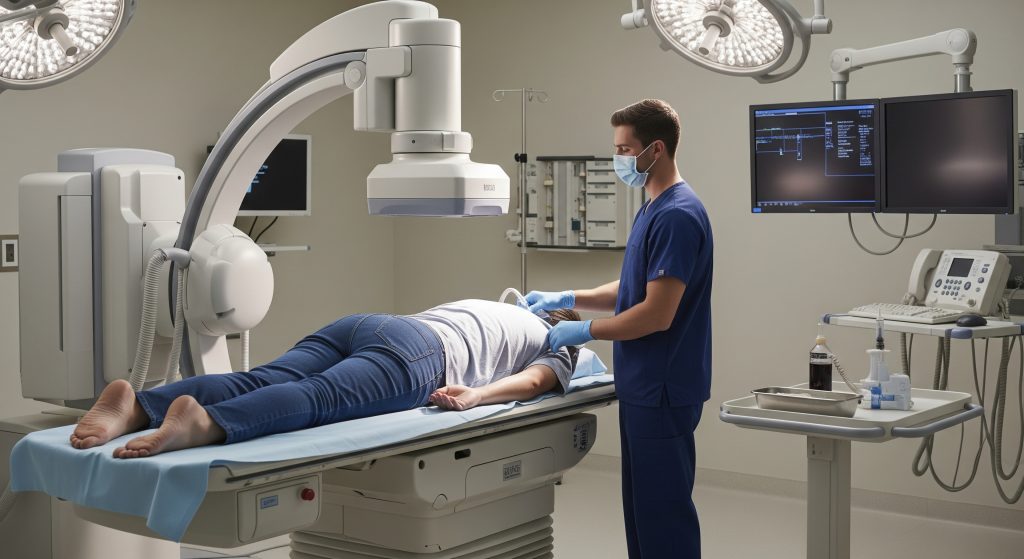
Conventional Myelography uses real-time X-ray guidance, called fluoroscopy, to place a fine needle into the spinal canal and introduce contrast. As RadiologyInfo explains, the contrast outlines the spinal cord, nerve roots, and lining, so structural narrowing or compression becomes clear. You then undergo targeted imaging while the contrast highlights the problem areas.
Accuracy at the needle stage matters. As PMC notes, fluoroscopy-guided lumbar punctures achieve high success by showing the landmarks as the needle advances. This is especially helpful if you have arthritis, prior surgery, or anatomical variation. In practice, that visual control is what keeps the myelogram procedure on track.
Conventional Myelography is often requested when MRI is not possible, or when metal hardware causes distortion. As Cleveland Clinic outlines, contrast imaging can reveal herniated discs, spinal stenosis, infections, or tumours that other scans may miss. This is the foundation on which more detailed CT Myelography builds, if required.
CT Myelography for Enhanced Detail
CT Myelography pairs contrast in the spinal fluid with high-resolution CT. The goal is to show fine anatomical detail and the exact relationship between bone, discs, and nerves. As RSNA summarises, this approach is particularly useful when MRI is contraindicated, and it can refine surgical planning with millimetre-level clarity.
It is also valuable in assessing cerebrospinal fluid dynamics and related abnormalities. As StatPearls notes, CT Myelography helps visualise CSF circulation within the canal and can improve detection of leaks. For lumbar spinal stenosis, sensitivity can exceed MRI in specific scenarios, as AJR reports, which explains why surgeons still request this test in complex cases.
There is an adjacent innovation story too. As UCHealth describes, simple devices that control position during imaging can sharpen detection of subtle CSF leaks. Technique matters. Small improvements in patient positioning and timing often make the diagnosis possible.
Dynamic CT Myelography for CSF Leak Detection
Dynamic CT Myelography targets a moving problem: where the spinal fluid escapes in real time. Protocols emphasise positioning, rapid imaging, and precise timing of contrast. As Mayo Clinic highlights, tailored approaches now localise CSF-venous fistulas more accurately, supporting treatment of spontaneous intracranial hypotension.
Operator technique is crucial. As AJNR points out, optimised positioning during dynamic Myelography can raise detection rates and cut repeat scans. Adjustments are made in real time as the contrast moves. The point is simple. Dynamic protocols use physics and gravity to expose the leak.
Parameter tuning also matters. As AJNR reports, altering contrast density and scan timing can improve conspicuity of CSF-venous fistulas. In practice, this turns a non-diagnostic test into a clear roadmap for targeted therapy.
MR Myelography as an Alternative
MR Myelography is a radiation-free technique that exploits fluid-sensitive MR sequences to depict CSF pathways. It is especially helpful when contrast injection is unsuitable or when soft-tissue detail is paramount. As PMC notes, MR-based techniques can evaluate spinal integrity when conventional methods fall short.
In nerve root and plexus disorders, the technique contributes directly to management. As PMC details, MR Myelography supports assessment of brachial plexus injuries and aids preoperative planning. It also assists in degenerative cervical myelopathy, as PMC outlines, by clarifying the structures compressing the cord.
Leak detection is another area of growth. As AJNR discusses, combining MR and CT Myelography can improve identification of complex CSF leaks. Three-dimensional MR Myelography can outperform 2D in visualising fine leaks, as ScienceDirect indicates. The practical takeaway is choice. The modality is adapted to the clinical question.
Common Conditions Diagnosed with Myelography
Myelography helps answer focused questions about nerve compression, canal narrowing, or CSF leakage. For cervical myelopathy, symptoms such as hand clumsiness and gait disturbance may prompt targeted imaging. As StatPearls summarises, cord compression in the neck is a frequent and serious diagnosis.
Tumour assessment is another example. As StatPearls notes, spinal metastases are common and can invade the canal, where Myelography assists surgical planning. For suspected CSF leaks with orthostatic headache, new protocols enhance localisation, as Mayo Clinic reports.
|
Condition |
How Myelography Helps |
|---|---|
|
Herniated disc |
Outlines nerve root compression where MRI is unclear. |
|
Spinal stenosis |
Shows canal narrowing and levels requiring decompression. |
|
CSF leak |
Localises the leak for targeted therapy or blood patch. |
|
Spinal tumours |
Clarifies canal involvement to guide surgery or radiotherapy. |
In short, Myelography answers specific anatomic questions when standard imaging leaves doubt. Precise questions. Precise answers.
The Complete Myelography Procedure
Pre-Procedure Preparation Requirements
Preparation is deliberately conservative and safety focused. Fasting for a period is common, and arrival is usually early for consent and checks. As UCSF Radiology advises, you should avoid food and drink for about six hours, take medicines with a small sip, and arrange a responsible adult to take you home.
Medication management varies by institution. As PMC observes, over 57% of practices do not stop seizure threshold-lowering drugs, and seizure rates are not higher. Modern non-ionic contrast has improved safety, though anticoagulants still require attention. As Johns Hopkins Medicine notes, blood thinners often need a pause to reduce bleeding risk, guided by your clinician.
Two practical rules stand. Confirm which medicines to hold and when to restart. Ensure transport home, because you may not be able to drive safely that day.
Step-by-Step Guide to the Injection Process
The myelogram procedure begins with identification, consent, and a brief review of your history. You will change into a gown and move to the fluoroscopy suite. Local anaesthetic is applied to the lower back to numb the skin.
-
Positioning: You are placed on the table, typically prone or on your side.
-
Skin preparation: The area is cleaned to maintain sterility.
-
Needle placement: Under fluoroscopy, a fine needle enters the spinal canal.
-
CSF confirmation: A small CSF return may confirm position.
-
Contrast injection: Contrast is gently injected into the subarachnoid space.
-
Distribution: You may be tilted to spread contrast.
-
Initial imaging: X-rays document contrast flow and any block.
The technical target is accurate placement at the lower lumbar levels. As PMC explains, injection at L3 or L4 is common, particularly when investigating CSF leaks. After injection, you may lie flat for about 30 minutes to optimise distribution before moving to CT. It is a careful sequence. Each step reduces ambiguity later.
If a CSF leak is suspected, your team may alter the sequence or position. As Mayo Clinic describes, this targeted approach increases the chance of spotting the leak during contrast movement.
Positioning and Imaging Techniques
Positioning is not an afterthought. It is the lever that improves visualisation of the canal, especially when narrowing is severe. As PubMed notes, a flexed sitting technique can show the lumbosacral canal below an apparent obstruction. In challenging stenosis, that view can be the difference between guesswork and clarity.
Novel patient positions and devices are expanding the toolkit. As PubMed suggests, upright CT Myelography can capture abnormalities missed in standard supine imaging. A new positioning device can also refine dynamic CT studies, as AJNR reports, improving efficiency and accuracy in suspected leaks.
Technique selection follows the clinical question. For suspected high-flow leaks, dynamic CT Myelography offers rapid series imaging, as PMC describes. For structural problems where MRI is not possible, CT Myelography merges contrast outlines with high-resolution detail, as RSNA explains. It is essentially contrast myelography paired with the sharpness of CT.
What Happens During the CT Scan Phase
CT follows once contrast has dispersed. The technologist positions you on the CT table and acquires rapid images of the spine. You will be asked to keep still while the scanner moves.
Position can be adjusted to exploit gravity when a fistula is suspected. As PMC notes, decubitus CT Myelography can reveal CSF-venous fistulas that do not appear in other positions. This is a targeted tactic. Small changes in posture can expose the leak path.
The scan itself is quick. Image review takes longer, as the radiologist inspects the canal, roots, and foramina frame by frame.
Expected Duration and Comfort Levels
Plan for a half day. The myelography procedure, review, observation, and travel time often add up. As RadiologyInfo notes, imaging and recovery generally take several hours, and aftercare is part of the process.
Discomfort is variable and usually manageable. As Cleveland Clinic explains, the procedural component is about an hour, with pressure or brief pain during the injection. Hydration and rest afterwards help. Patient accounts emphasise the value of clear expectations, as Spinal CSF Leak Foundation highlights, especially when headaches occur.
You may notice a positional headache later that eases when lying flat. As MSKCC advises, rest and fluids typically settle symptoms. Speak up if pain is severe or unusual. Comfort is a clinical signal, not an afterthought.
Recovery and Aftercare Following Your Myelogram
Immediate Post-Procedure Monitoring
After Myelography, you will be observed for a period in recovery. Staff monitor your neurological status and comfort, and ensure you can mobilise safely. Postural headache and nausea are the main early concerns.
Attention to CSF leak symptoms is deliberate. As PMC notes, early recognition of adverse neurological signs supports timely intervention. Hydration and positioning can reduce headache risk. Evidence on posture is mixed, but practical guidance is consistent. As PMC indicates, tailored positioning and fluids may help, while prolonged strict bed rest offers uncertain benefit.
Expect clear discharge instructions. They typically cover rest, fluids, transport, and warning signs that require escalation.
First 24-Hour Recovery Guidelines
The first day focuses on rest and steady hydration. As Eastern Radiologists advises, limit upright sitting, recline when comfortable, and sip fluids through the day. Caffeinated drinks can be helpful for some patients.
Short, light walks around the home are acceptable if you feel steady. As Teton Radiology suggests, supporting the head while resting for several hours may reduce headache incidence. Pace activities deliberately. Do not rush to normal intensity.
Managing Common Side Effects
The most frequent side effect is a post-dural puncture headache. It worsens when upright and improves when lying flat. As PMC reports, roughly 32% experience headaches after lumbar puncture, though severity varies considerably.
Care plans emphasise fluids, measured rest, and avoiding strenuous activity for a day. As RadiologyInfo notes, these steps often settle symptoms. Guidance from specialist clinics echoes the same approach, with added notes on nausea control and activity limits, as Ortho-Spine explains.
Some studies question prolonged bed rest as a preventive strategy, as PMC suggests. The practical approach remains balanced. Rest enough to be comfortable, hydrate well, and escalate care if headaches are severe or persistent.
When to Contact Your Healthcare Provider
Most symptoms settle with time and fluids. Certain red flags demand urgent review. Severe back or leg pain, new weakness, numbness, or bladder or bowel problems require prompt attention. Severe or unusual headache, fever, or wound issues also warrant escalation.
As Dr. Oracle summarises, neurological changes, incontinence, persistent vomiting, or atypical head pain should trigger immediate contact with your team. It is better to call early. Safety first.
Return to Normal Activities Timeline
Plan to scale back for the first 24 hours. Avoid driving on the day of Myelography. As Johns Hopkins Medicine notes, fluid intake should be increased, and strenuous activity avoided until you feel back to baseline.
Most people resume light desk work the next day if symptoms are mild. Arrange transport on the day itself in case sedatives are used, as Cleveland Clinic advises. Recovery is usually straightforward. But still, listen to your body and your discharge plan.
Making an Informed Decision About Myelography
There are two core questions to resolve. First, will Myelography change management. Second, is it the safest and most efficient way to answer the clinical question. If MRI already provides a complete and actionable picture, further testing may add little. If MRI is inconclusive, the targeted resolution of Myelography can be decisive.
Use this quick framework:
-
Clinical problem: Is the key uncertainty anatomical and potentially compressive.
-
Prior imaging: Is MRI impossible or non-diagnostic due to hardware or motion.
-
Decision link: Will results alter surgery, injection level, or conservative care.
-
Risk balance: Are you fit for the test, with medicines timed appropriately.
A brief example helps. Persistent leg pain after prior fusion with new weakness, but MRI is distorted by metal. Myelography clarifies the exiting nerve compression at one level and confirms patency elsewhere. The surgeon operates with confidence, not conjecture. That is the value proposition of Myelography, and of the myelogram procedure when the stakes are high.
Frequently Asked Questions
How does myelography compare to an MRI scan?
Myelography shows the canal outline with contrast. MRI shows soft tissues and cord signal without injection. When MRI is contraindicated or unclear, Myelography can provide sharper anatomic detail, especially around bone and hardware.
Can I drive myself home after a myelography procedure?
No. Arrange an adult to take you home. Sedation, positional headache, or delayed symptoms make driving unsafe on the day of testing.
What medications should I stop before my myelogram?
Anticoagulants often require a pause, guided by your clinician. Many centres do not stop seizure threshold-lowering drugs with modern contrast. Confirm an individual plan.
Is myelography safe during pregnancy?
It is generally avoided in pregnancy because it uses X-ray. Alternatives such as MRI or MR Myelography are considered first, subject to specialist advice.
How long does the contrast material stay in my body?
Modern water-soluble contrast disperses and is eliminated in urine within hours to days. Hydration supports clearance, though timing varies by kidney function.
Will I be sedated during the myelography procedure?
Light sedation may be offered, but many examinations use local anaesthetic only. The decision depends on anxiety, prior experience, and institutional practice.
What are the alternatives if I cannot have a myelogram?
MRI or MR Myelography are common alternatives. In specific scenarios, targeted CT, nerve root blocks, or watchful waiting may supply the needed information.
Key takeaways
-
Myelography is a precise test that answers targeted questions when MRI is inconclusive.
-
Technique and positioning strongly influence diagnostic yield, particularly for CSF leaks.
-
Plan transport, rest, and hydration. Escalate care if neurological symptoms arise.
If you have been advised to undergo contrast myelography, ask the team one question. How will the findings change the plan. Clarity on that point justifies the test and sets expectations for recovery.