


What Is Doppler Ultrasound? Understanding Its Use and Procedure
Dr. Beenish Khan


Conventional ultrasound shows anatomy. It does not show flow. That common advice ignores the core reason clinicians choose Doppler Ultrasound. You use it when the question is not just what a structure looks like, but how blood moves through it, how fast it travels, and in which direction. Flow answers physiology. Flow changes decisions.
Types of Doppler Ultrasound Examinations
Colour Doppler Imaging
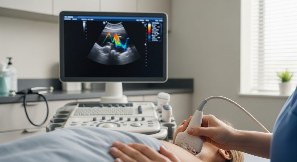
Colour Doppler overlays moving blood on the grey ultrasound image using a colour map. You see flow direction and relative speed at a glance. Red and blue indicate direction relative to the probe, not oxygenation. The colour scale bar on screen defines the convention used.
This modality is exceptional for quick orientation. It shows whether a vessel is patent, if a graft fills, or if a valve leaks. It is sensitive to angle and machine settings, so proper optimisation is essential for reliable impressions.
-
Rapid visual triage of flow presence and direction.
-
Useful for locating jets, stenoses, and shunts.
-
Angle dependent and subject to aliasing at higher velocities.
Power Doppler Ultrasound
Power Doppler displays the strength of moving signal rather than direction. It is more sensitive to low-velocity flow in tiny vessels. This makes it helpful in organs such as the kidney or thyroid, and in inflamed synovium.
Because it does not encode direction, the display is simpler. It is also more robust to angles. Motion artefact can appear, so deliberate probe handling matters.
-
Superior sensitivity to slow microvascular flow.
-
No direction information, only signal intensity.
-
Valuable when colour Doppler appears negative, but flow is suspected.
Duplex Ultrasound Technology
Duplex ultrasound combines B-mode imaging with spectral analysis in one view. You obtain structure and quantitative flow together. It is effectively anatomical plus functional data on one screen.
In vascular laboratories, duplex ultrasound underpins routine arterial and venous assessments. It allows precise measurements, including peak systolic velocity and end-diastolic velocity, with immediate correlation to the vessel wall appearance.
|
Feature |
Clinical value |
|---|---|
|
B-mode anatomy |
Maps plaque, thrombus, and lumen calibre |
|
Spectral waveform |
Quantifies velocity for grading stenoses |
|
Colour or power layer |
Guides sample placement and confirms patency |
Spectral Doppler Analysis
Spectral Doppler turns moving echoes into a velocity-time graph. The waveform encodes speed on the vertical axis and time on the horizontal axis. The result is a trace you can measure.
Indices such as resistive index and pulsatility index are derived from these traces. They help you appraise downstream resistance, distal disease, or transplant perfusion. Accurate angle correction and careful gate placement are essential for valid data.
-
Peak systolic velocity for grading arterial narrowing.
-
Resistive index to estimate parenchymal resistance.
-
Phasicity patterns to infer venous obstruction.
Continuous Wave Doppler
Continuous wave Doppler transmits and receives constantly. It can measure very high velocities without aliasing. It does not localise depth, so you sample all flow along the beam path.
Cardiology uses this method to quantify high-velocity jets, such as severe aortic stenosis. In general vascular practice, pulsed spectral methods are more common due to depth selectivity.
Clinical Applications and Medical Uses
Vascular Ultrasound for Blood Vessel Assessment
Vascular ultrasound is the workhorse for non-invasive vessel assessment. You can evaluate arteries and veins in the neck, limbs, abdomen, and grafts. Duplex and spectral tools provide flow quantification alongside clear anatomy.
-
Map plaque in carotid arteries and estimate stenosis grade.
-
Assess peripheral arterial disease and plan intervention.
-
Confirm venous patency before dialysis access creation.
-
Check bypass graft flow and detect early stenosis.
It is often the first test and, in many cases, the only test required. It is portable, safe, and repeatable.
Doppler Ultrasound for Renal Artery Stenosis
When hypertension is resistant or renal function declines, renal artery disease may be suspected. You can triage this scenario with Doppler Ultrasound before considering contrast imaging. Proper technique is central to meaningful results.
Key measures include peak systolic velocity at the stenosis and velocity ratios across segments. Accessory arteries and body habitus can complicate scanning, yet an experienced sonographer can still provide actionable data. If you require wording for prior authorisation, the phrase doppler ultrasound for renal artery stenosis is commonly used.
Pregnancy and Foetal Monitoring
In obstetrics, Doppler Ultrasound informs placental and foetal circulation. You will see it used for umbilical artery, middle cerebral artery, and ductus venosus profiles. These studies complement growth scans and clinical review.
-
Umbilical artery indices for placental resistance.
-
Middle cerebral artery to assess brain-sparing patterns.
-
Ductus venosus flow in selected high-risk cases.
These measures support timing of delivery decisions. They are interpreted within the broader clinical picture, not in isolation.
Cardiac Function Evaluation
Cardiac Doppler evaluates valve flow, intracardiac pressures, and shunts. Spectral and colour data quantify regurgitation and stenosis. Continuous wave methods capture very high jet velocities.
You can estimate gradients, measure stroke volume, and assess diastolic function from transmitral and pulmonary venous patterns. This applies in adult and paediatric echocardiography with appropriate protocols.
Deep Vein Thrombosis Detection
Doppler Ultrasound is the first-line test for suspected lower limb thrombosis. The cornerstone is compressibility of the vein on B-mode imaging. Colour and spectral data corroborate absent flow or collateralisation.
-
Non-compressible venous segment indicates thrombus.
-
Absent or damped spectral signals suggest obstruction.
-
Augmentation manoeuvres test venous responsiveness.
Upper limb studies follow similar principles. Pitfalls include calf thrombi below the knee and partial recanalisation that masks chronic clot.
Carotid Artery Disease Screening
Carotid duplex surveillance provides risk stratification in patients with cerebrovascular symptoms. You can measure plaque burden and derive a stenosis grade from velocities and ratios. The anatomical correlation helps you plan endarterectomy or stenting where indicated.
Screening strategies vary. Decisions are guided by symptoms, risk factors, and local pathways.
The Doppler Ultrasound Procedure
Pre-Examination Preparation Requirements
Most Doppler Ultrasound studies require no specific preparation. For abdominal vessels, fasting for 4 to 6 hours reduces bowel gas and improves visibility. Hydration may be requested before pelvic venous assessments.
Wear loose clothing and remove jewellery near the study area. Bring prior imaging reports if available, as comparison accelerates interpretation.
-
Fasting: helpful for abdominal aorta, mesenteric, and renal arteries.
-
Hydration: sometimes requested for pelvic venous reflux tests.
-
Medication: usually unchanged, unless your clinician advises otherwise.
Step-by-Step Examination Process
-
Identity confirmation and explanation of the test.
-
Positioning tailored to the vessel or organ of interest.
-
Application of gel to ensure good acoustic contact.
-
B-mode sweep to map anatomy and select windows.
-
Colour or power Doppler to locate and characterise flow.
-
Spectral sampling with angle correction for measurements.
-
Targeted manoeuvres, such as compression or augmentation, if needed.
-
Image capture and labelling for the report.
The sequence is orderly and reproducible. It allows you to correlate anatomy with function in real time.
Duration and Patient Positioning
Most examinations last between 20 and 45 minutes. Complex vascular mapping can take longer, especially in multi-vessel studies. The sonographer may adjust your position repeatedly to access optimal windows.
Common positions include supine, slight left lateral tilt, or upright for venous reflux assessment. Arm abduction facilitates subclavian views. Head rotation assists carotid access while avoiding excessive stretch.
Gel Application and Transducer Movement
The gel is water based and hypoallergenic. It eliminates air between the transducer and skin, improving transmission of sound. It does not stain clothing, though a wipe-down follows the scan.
The transducer glides along the skin and may apply brief pressure. Gentle pressure helps with venous compression and stabilises the image. For small vessels, very light touch prevents collapse and preserves flow.
What Patients Experience During Scanning
You will feel cool gel and light pressure from the probe. Some pressure over tender areas can be uncomfortable for a moment. There is no ionising radiation and no contrast injection in standard scans.
Breath holds or positional changes may be requested for short periods. The room is usually dim to improve screen visibility. Communication continues throughout, so you know what happens next.
Benefits, Limitations and Results Interpretation
Advantages Over Other Imaging Methods
-
No ionising radiation and no routine contrast material.
-
Real-time haemodynamic information alongside anatomy.
-
Portable, repeatable, and comparatively lower cost.
-
Immediate bedside decisions for urgent vascular questions.
In practice, Doppler Ultrasound often answers the clinical question quickly. A focused carotid study can triage a transient ischaemic event in minutes. That speed matters when time is brain.
Limitations and Contraindications
-
Operator dependence and learning curve for reliable measurements.
-
Acoustic shadowing from calcification can obscure the lumen.
-
Obesity or bowel gas reduces abdominal vessel visibility.
-
Very high velocities can alias with pulsed Doppler settings.
There are effectively no absolute contraindications for routine use. Caution applies over fresh wounds or severe pain sites. Sedation is rarely required and is not typical.
Pros vs Cons
-
Pros: Safe, rapid, functional, bedside capable.
-
Cons: User dependent, angle sensitive, limited by patient habitus.
Understanding Your Test Results
Your report blends qualitative impressions with quantitative metrics. It lists vessel segments, velocities, and waveform patterns. It correlates these with anatomical findings on B-mode imaging.
|
Term |
Meaning in context |
|---|---|
|
Peak systolic velocity |
Highest measured flow speed during systole at a given site |
|
Velocity ratio |
Stenosis grading by comparing peak speeds across segments |
|
Resistive index |
Estimate of downstream resistance within parenchyma |
|
Phasicity |
Respiratory variation in venous traces, reduced with obstruction |
|
Alias |
Wrap-around of colour or spectral display at higher speeds |
Reports usually state whether findings are normal, borderline, or abnormal. They may recommend correlation with clinical data or further imaging. Clear grading allows consistent follow-up and treatment planning.
Accuracy and Reliability Factors
Accuracy improves with standardised protocols, adequate training, and quality assurance. Angle correction must be set correctly, typically at 60 degrees or less. The sample gate should cover the central flow stream in laminar segments.
-
Consistent equipment calibration and preset use raise reliability.
-
Use of duplex ultrasound enables exact correlation of structure and flow.
-
Reproducible patient positioning and lab documentation reduce variation.
Roughly speaking, performance is highest in accessible vessels like carotids and femorals. It is lower where windows are limited, such as mesenteric arteries. That context frames interpretation and next steps.
Understanding Doppler Ultrasound’s Role in Modern Medicine
Doppler Ultrasound has become a core diagnostic tool across specialties. You rely on it to detect deep vein thrombosis, grade carotid disease, and evaluate graft patency. You also use it to assess foetal wellbeing and cardiac valve function.
It is non-invasive and delivers real-time physiology. It pairs anatomy with haemodynamics in the same sitting. That dual view reduces uncertainty and often avoids more invasive tests.
There are trade-offs. It is operator dependent and sensitive to technique. Yet with proper standards, it offers dependable information that drives timely care.
A practical principle: image the anatomy, map the flow, then measure what matters. In that order.
Use Doppler Ultrasound early when a flow question exists. It is basically fast, safe, and decision-focused. In many pathways, it is the first and best step.
Frequently Asked Questions
Is Doppler ultrasound safe during pregnancy?
Yes. Routine diagnostic Doppler Ultrasound uses sound waves, not ionising radiation. It is considered safe when used appropriately by trained clinicians. In pregnancy, it is applied with prudent settings and focused dwell times.
How long does a Doppler ultrasound examination typically take?
Most studies take 20 to 45 minutes. Complex vascular mapping or multi-territory assessments may extend to an hour. The duration depends on clinical questions and anatomy.
What’s the difference between regular ultrasound and Doppler ultrasound?
Regular ultrasound shows anatomy in greyscale. Doppler Ultrasound adds flow information such as speed and direction. Duplex ultrasound fuses both into one comprehensive examination.
Can Doppler ultrasound detect all blood clots?
No. It has high sensitivity for proximal limb veins. Small calf thrombi, pelvic clots, or partially recanalised chronic thrombus can be harder to prove. If suspicion remains, your clinician may arrange additional imaging.
Do I need special preparation before a vascular ultrasound?
Usually not. For abdominal vascular ultrasound, fasting for 4 to 6 hours helps reduce bowel gas. Wear loose clothing and follow any specific instructions from your imaging provider.
How accurate is Doppler ultrasound for detecting renal artery stenosis?
Accuracy is high in experienced hands, though not perfect. It depends on patient habitus, accessory arteries, and window quality. As part of a structured work-up, doppler ultrasound for renal artery stenosis provides valuable triage information.