


What Is an Echocardiogram Procedure and Why Is It Done?
Dr. Hriday Kumar Chopra
Disclaimer: The content shared here is for informational purposes only. Always consult a specialist doctor before attempting any treatment, procedure, or taking any medication independently.
Heart tests get pitched as complex or intimidating. The reality is more practical. An echocardiogram procedure is a structured, image-based assessment that shows how the heart looks and functions in real time. I focus here on what it is, how it works, when it is recommended, and how to prepare so the experience is predictable and useful.
Types of Echocardiogram Procedures
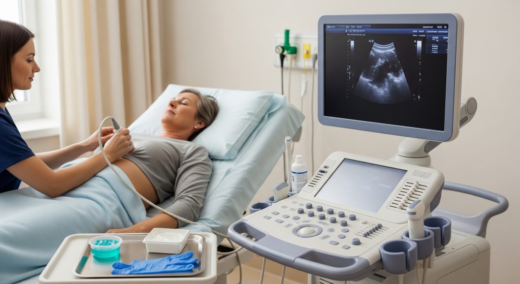
Transthoracic Echocardiogram (TTE)
I start with the standard test in most clinics. A transthoracic echocardiogram uses an ultrasound probe on the chest to capture moving images of chambers, valves, and the heart’s overall pump function. The sonographer applies gel, moves the probe in set windows, and records views that cardiologists recognise instantly. It is non-invasive, does not require sedation, and suits first-line evaluation in many cases.
In practice, a TTE often answers the immediate question. Examples include suspected valve disease, heart failure monitoring, or checking fluid around the heart. When a patient asks about an echocardiogram procedure, this is usually the one I describe first because it is quick and widely available.
-
Typical uses: valve assessment, chamber size, ejection fraction, pericardial effusion.
-
Comfort: generally comfortable, with brief pressure from the probe.
-
Limitations: image quality can be reduced by lung interference, body habitus, or chest wall anatomy.
Transesophageal Echocardiogram (TEE)
A transesophageal study brings the ultrasound probe closer to the heart by placing it in the oesophagus. As StatPearls notes, TEE provides high-resolution views that are valuable in unstable patients and intraoperative settings. I reserve this when TTE images are inconclusive or when finer detail is critical.
The process involves fasting and usually light sedation. A thin flexible tube carries the transducer down the throat. This reduces interference from lungs and ribs. It is particularly effective for complex valve problems, clots in the left atrium, and aortic pathology. It remains a key option within the wider echocardiogram procedure toolkit.
-
Strength: detailed visualisation of valves and posterior structures.
-
Preparation: fasting, sedation planning, and screening for oesophageal disease.
-
Use case: when TTE cannot show sufficient detail or surgery planning requires precision.
Stress Echocardiogram
A stress echocardiogram evaluates the heart under load. I use it to look for ischaemia, viability, or exertional symptoms that are hard to detect at rest. As StatPearls describes, it combines exercise or pharmacological stress with imaging to identify wall motion changes that suggest coronary artery disease.
There are two routes. Exercise on a treadmill or bike with quick imaging pre and post effort. Or medication such as dobutamine to simulate stress when exercise is not feasible. The principle is straightforward. If a region lacks adequate blood flow during stress, its movement will deteriorate transiently on the images. That signal guides next steps.
-
Applications: diagnosis of coronary disease, risk stratification, and preoperative assessment.
-
Format: exercise-based or medication-based, depending on fitness and safety.
-
Outcome: visual evidence of inducible ischaemia or reassurance if normal.
Doppler Echocardiogram
Doppler techniques measure blood flow speed and direction. I rely on this to estimate pressure gradients across valves, quantify regurgitation, and calculate pulmonary pressures. It is part of the echocardiogram procedure rather than a separate test. Spectral Doppler, colour Doppler, and tissue Doppler each add a layer of functional insight.
This matters for decision making. A valve that looks mildly abnormal may have severe haemodynamic impact on Doppler. The inverse can happen as well. Numbers guide treatment.
3D Echocardiogram
Three-dimensional imaging reconstructs valves and chambers in volumetric form. It can improve quantification and provide surgeons with meaningful anatomy for planning. I find 3D particularly useful for mitral valve interventions and complex congenital cases. It is not required in every echocardiogram procedure, but when morphology is central to the decision, 3D earns its place.
Why Doctors Recommend an Echocardiogram
Heart Conditions Diagnosed Through Echocardiogram
I order an echocardiogram procedure to clarify structure and function. The test identifies and grades valve stenosis or regurgitation, evaluates cardiomyopathies, and confirms pericardial effusion. It also assesses congenital defects like septal defects and complex outflow issues. In heart failure, it measures ejection fraction and fills in the diagnostic picture for HFpEF or HFrEF.
-
Valve disease: severity, mechanism, and impact on chambers.
-
Cardiomyopathy: dilated, hypertrophic, or restrictive patterns.
-
Pericardial disease: fluid, constriction, and tamponade features.
-
Pulmonary pressures: estimated through tricuspid regurgitation jet velocity.
Symptoms That Warrant an Echocardiogram
Certain symptoms justify imaging without delay. Chest discomfort with exertion, persistent breathlessness, unexplained fatigue, syncope, palpitations, or new murmur. I also consider swelling of legs with raised neck veins as a prompt. The aim is targeted. An echocardiogram procedure either confirms a structural cause or rules it out efficiently.
-
Exertional dyspnoea suggesting heart failure or valvular disease.
-
Palpitations with suspected cardiomyopathy or structural arrhythmia substrate.
-
Syncope where outflow obstruction or severe valve disease is possible.
Routine Screening and Follow-up Cases
Not every patient needs routine imaging. But follow-up timing matters when a condition is known. Mild valve disease may warrant yearly or two-yearly reviews. Dilated aortas require interval tracking. Stable cardiomyopathy usually needs periodic reassessment to adjust medication. An echocardiogram procedure is the backbone of that monitoring schedule.
For occupational fitness or preoperative assessment, I request imaging when clinical risk factors or symptoms suggest possible concern. The decision is pragmatic. Unnecessary tests add little value. Focus on cases where results will change management.
Echocardiogram for Different Age Groups
Age influences indications and interpretation. In children, congenital heart disease evaluation is common, including shunts and valve malformations. In adults, valve disease, hypertension effects, and coronary disease dominate. In older adults, diastolic dysfunction and degenerative valve pathology are frequent findings. The echocardiogram procedure adapts to the patient, not the other way round.
What to Expect During the Echocardiogram Procedure
Pre-procedure Preparation Steps
Preparation depends on the modality. For a standard transthoracic echocardiogram, I advise wearing a two-piece outfit and arriving a few minutes early. No fasting is required. Bring the medication list. For TEE, fasting and an escort for discharge are essential. For stress studies, comfortable footwear and clarity about any beta-blocker timing are important.
-
Medication review: confirm heart drugs and anticoagulants.
-
Consent: understand the purpose and any alternatives.
-
Logistics: allow time for check-in and recovery if sedation is planned.
Step-by-Step Procedure Process
A standard echocardiogram procedure follows a consistent sequence. This predictability helps reduce anxiety and improves image quality.
-
Check-in and brief history: the clinician confirms the clinical question.
-
Positioning: left lateral decubitus for most views, with ECG leads attached.
-
Imaging: gel applied, then parasternal, apical, subcostal, and suprasternal views.
-
Doppler assessment: colour, spectral, and tissue Doppler as indicated.
-
Measurements: chamber sizes, ejection fraction, valve gradients, and regurgitation.
-
Review: I confirm that the dataset answers the clinical question before ending.
In TEE, steps include throat anaesthesia, sedation, probe insertion, and controlled imaging runs. In stress studies, baseline images come first. Then exercise or pharmacological stress, followed by immediate post-stress imaging while the response is still evident.
Duration and Comfort Measures
Most transthoracic scans take 20 to 40 minutes. TEE and stress studies can take longer due to preparation and recovery. I explain positioning and breathing cues to reduce artefact. Short rests between views are provided if needed. The echocardiogram procedure prioritises image quality and patient comfort in equal measure.
-
Privacy: gowns and draping are standard.
-
Noise: the machine beeps for ECG and captures Doppler audio; it is normal.
-
Pacing: clear instructions reduce repeat images and shorten time on table.
Post-procedure Care and Results
After a transthoracic study, most patients leave immediately. For TEE, I advise no driving until sedation wears off and soft food once swallowing feels normal. For stress tests, cool-down and brief observation are standard. I deliver results in a structured report with images and measurements that link back to the clinical question. The echocardiogram procedure supports decisions, so clarity is essential.
Potential Risks and Safety Considerations
Risk is low for non-invasive imaging. Gel allergy is rare. Minor soreness from probe pressure can occur. TEE entails added considerations. Sedation effects, sore throat, or very rare oesophageal injury. Stress testing carries a small risk of arrhythmia or chest pain. I mitigate these risks through screening, monitoring, and immediate response protocols. The benefit usually outweighs the risk to a large extent.
Safety profile: low for transthoracic imaging, low to moderate for TEE and stress studies with proper monitoring.
Echocardiogram vs ECG and Other Heart Tests
Key Differences Between Echocardiogram and ECG
I am often asked about echocardiogram vs ecg. The difference is categorical. ECG records the heart’s electrical signals. Echocardiography shows structure and function with moving images and flow data. I use them together rather than in competition.
|
Test |
What it shows |
|---|---|
|
ECG |
Electrical activity, rhythm, conduction blocks, acute ischaemic changes. |
|
Echocardiogram |
Chamber size, valve anatomy, pump function, flows, pressures. |
|
When useful |
Arrhythmias, acute chest pain triage, baseline rhythm analysis vs structural disease evaluation. |
When to Choose Echocardiogram Over ECG
I choose an echocardiogram procedure when the question demands visual structure. For murmurs, heart failure, suspected valve disease, or pericardial fluid, imaging is decisive. ECG may be normal while echocardiography reveals significant pathology. The reverse holds in pure rhythm disorders, where ECG takes the lead.
-
Suspected valve disease: echocardiography first.
-
Unexplained breathlessness: echocardiography to check function and pressures.
-
Palpitations without structural suspicion: ECG and ambulatory rhythm monitoring.
Complementary Heart Testing Methods
Testing is not a one-test-fits-all sequence. I often combine modalities for completeness. An echocardiogram procedure may be followed by:
-
Cardiac MRI for tissue characterisation and precise volumes.
-
CT angiography for coronary assessment and aortic anatomy.
-
Ambulatory ECG monitoring for intermittent arrhythmias.
-
Blood tests such as BNP to support heart failure assessment.
Choice depends on the hypothesis. The goal is an accurate diagnosis with minimal redundancy.
Finding Echocardiogram Services Near You
Access matters. Patients often search for echocardiogram near me and compare facilities by speed, cost, and reporting quality. I recommend confirming accreditation, asking about consultant oversight, and checking whether Doppler and 3D are available when indicated. Availability of TEE and stress capability helps streamline care if escalation is needed.
Ask three practical questions. How soon can the study be scheduled. What is included in the fee. How quickly will a cardiologist review the images and report. A strong service answers clearly and delivers consistently.
Making Informed Decisions About Your Heart Health
Clarity reduces anxiety. The echocardiogram procedure is designed to answer specific clinical questions with visuals and measurements. It is not about images for their own sake. It is about matching a symptom or concern with reliable evidence and then acting on it. I encourage patients to bring their questions and share goals for activity, work, or sport. The result is a plan that respects both data and daily life.
Here is the practical approach I use. Define the clinical question in a single sentence. Select the modality that can answer it with the least burden. Prepare thoroughly to improve image quality and comfort. Review the results against the original question. If still uncertain, escalate to the next best modality. And stop when the decision is clear. Good medicine is often this direct.
Frequently Asked Questions
How long does an echocardiogram procedure take?
A standard transthoracic study takes about 20 to 40 minutes. TEE or stress imaging may take 45 to 90 minutes because of preparation and observation. Complex protocols can run longer, though I aim for efficiency without compromising image quality.
Is an echocardiogram painful or uncomfortable?
Pain is uncommon. Some pressure from the probe can be felt on the ribs. TEE may cause throat discomfort after sedation, which usually settles within a day. During a stress echocardiogram procedure, exertion is controlled and monitored continuously.
Can I eat before an echocardiogram?
Yes for standard transthoracic studies. No for TEE because fasting is required to reduce aspiration risk. For stress imaging, a light snack two to three hours before is reasonable, unless advised otherwise. Hydration helps with exercise performance and comfort.
How much does an echocardiogram cost in India?
Costs vary by city, facility, and modality. Prices are influenced by consultant reporting, stress or TEE capability, and add-ons such as 3D or contrast. I advise confirming inclusions in the quoted fee. Pricing can change depending on the source and regional factors.
How often should I get an echocardiogram?
Frequency depends on the condition. Mild valve disease may be reviewed annually or biennially. Cardiomyopathy and postoperative cases require scheduled follow-up. If symptoms change or new signs appear, bring the test forward. Routine repetition without clinical reason adds little value.
What’s the difference between 2D and 3D echocardiogram?
2D provides planar slices and is sufficient for most decisions. 3D reconstructs structures volumetrically and improves certain measurements and visualisation of complex valves. I add 3D when anatomy drives the decision, especially in mitral interventions or congenital assessments.
Can children safely undergo echocardiogram procedures?
Yes. Paediatric echocardiography is standard and safe. TTE is non-invasive and well tolerated. TEE may be used selectively with paediatric anaesthesia support. The focus remains on child comfort, minimal sedation, and clear diagnostic yield.
Final note. If a test is on the table, ask what decision it will change. The right echocardiogram procedure answers that question, then lets action follow.