


What Is a Stress Test Procedure? Full Heart Health Explainer
Dr. Hriday Kumar Chopra
Most advice about heart testing reduces everything to a single result. That is tidy, yet misleading. A modern Stress Test Procedure is best seen as a structured sequence that answers different clinical questions at once. I use it to probe exertional symptoms, quantify capacity, and reveal hidden ischaemia. It is basically a practical trial for the heart, under watchful eyes, with data captured at every stage.
Types of Stress Tests for Heart Health Assessment
The right Stress Test Procedure depends on symptoms, baseline ECG, and the diagnostic question. I match the modality to the patient and the decision at hand. Here is how the main options differ in purpose and data yield.
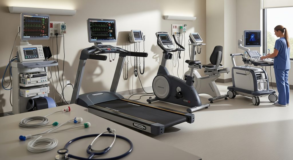
Treadmill Stress Test
A treadmill stress test assesses functional capacity and electrical changes under exertion. I use it when patients can exercise, and the resting ECG is interpretable. The Stress Test Procedure here focuses on ECG shifts, symptoms, and exercise time. That combination supports diagnosis and risk stratification.
-
Best for exertional chest discomfort with an interpretable ECG.
-
Quantifies fitness and symptom threshold alongside ECG changes.
-
Often the most economical first step when probability is intermediate.
I avoid a treadmill stress test for asymptomatic people seeking routine screening. The predictive value is low in that setting. A better route is risk factor management first, and targeted testing only when indicated. Clinical judgement still matters.
Nuclear Stress Test with Imaging
A nuclear stress test adds perfusion imaging to the Stress Test Procedure. It shows where blood flow is reduced during stress compared with rest. This helps when resting ECG is abnormal or when prior testing was equivocal.
-
Reveals regional perfusion deficits and global function.
-
Useful after an inconclusive treadmill study.
-
Guides decisions on invasive angiography or medical therapy.
In a cohort of 297 patients, gated SPECT after dipyridamole stress showed that post-stress ejection fraction fell in those with reversible defects, as Iran Journal of Nuclear Medicine reported. That pattern signals inducible ischaemia. It also refines risk estimates for events.
Pharmacological Stress Test Without Exercise
When exercise is not possible, a pharmacological stress test simulates exertion. Agents such as adenosine, dipyridamole, or regadenoson increase coronary blood flow. I pair this Stress Test Procedure with imaging to detect flow-limiting lesions.
-
Indicated for mobility limitations or contraindications to exertion.
-
Complements nuclear imaging or stress echocardiography.
-
Allows controlled haemodynamic stress in a monitored setting.
This approach ensures diagnostic completeness for those who cannot exercise. Safety protocols are tight, with immediate reversal options if needed.
Stress Echocardiogram
A stress echocardiogram uses ultrasound to assess wall motion under load. The Stress Test Procedure can involve a treadmill or dobutamine infusion. New or worsening wall motion abnormalities indicate ischaemia.
-
No radiation, and immediate bedside interpretation.
-
Useful when valve disease or structural issues are suspected.
-
Dobutamine protocols help when patients cannot run or cycle.
In practice, I choose stress echo when I need real-time mechanics and valve assessment. It is precise for territory level ischaemia and viable myocardium. A thoughtful choice, especially for younger patients.
Cardiopulmonary Exercise Test
The cardiopulmonary exercise test (CPET) integrates gas exchange with ECG and blood pressure. This Stress Test Procedure is about capacity and physiology, not only ischaemia. I use it for unexplained breathlessness, heart failure, and preoperative risk.
-
Measures peak oxygen uptake and ventilatory efficiency.
-
Separates cardiac from pulmonary limitations.
-
Supports rehabilitation targets and outcome prediction.
For a patient with heart failure and fatigue, CPET clarifies whether low fitness reflects cardiac output limits or deconditioning. That influences therapy and timing of referral. Precision, not guesswork.
How the Cardiac Stress Test Procedure Works
Every Stress Test Procedure follows a clear sequence. I explain the steps in advance so expectations are aligned. The method is rigorous, but the experience is usually straightforward.
Initial Preparation and Baseline Measurements
I start with a clinical review and clear test objectives. Baseline measurements include resting ECG, heart rate, and blood pressure. This baseline anchors the comparison during stress and recovery.
-
Confirm symptoms, medications, and prior test results.
-
Place electrodes and confirm signal quality.
-
Set termination criteria and safety signals.
Patient education is part of the Stress Test Procedure. I outline what patients will feel and when we stop. Clarity reduces anxiety.
Exercise Phase on Treadmill or Bike
Exercise begins at an easy workload. I increase speed and incline or resistance in small steps. The Stress Test Procedure aims to reach a sufficient cardiac workload without undue risk.
-
Continuous ECG and blood pressure monitoring throughout.
-
Symptom checks every stage, and on visible effort changes.
-
Immediate stop for concerning signs or patient request.
This phase reveals exertional thresholds. It also shows blood pressure trends and potential arrhythmias. Real-world performance under supervision.
Monitoring Heart Rate and Blood Pressure
Heart rate and blood pressure responses tell a coherent story. I evaluate the rate rise, peak values, and recovery. The Stress Test Procedure treats these as core safety and diagnostic metrics.
-
Inadequate rise may suggest chronotropic issues.
-
Excessive rise can indicate poorly controlled hypertension.
-
Drop in pressure with exercise is a red flag for ischaemia.
ECG changes only make sense in context. The haemodynamic profile provides that context. Together, they improve accuracy.
Recovery Phase and Cool Down
Recovery is deliberate. I lower intensity and keep monitoring for several minutes. The Stress Test Procedure continues until vitals return near baseline.
-
Gradual cool down prevents venous pooling and dizziness.
-
Heart rate recovery offers insight into autonomic tone.
-
Persistent symptoms trigger extended monitoring.
A structured cool down and observation period align with 2025 practice guidance, as American Heart Association outlined. I document time to recovery and any delayed ECG changes. Small details often matter.
Image Capture for Nuclear and Echo Tests
When imaging is planned, timing is everything. For a nuclear stress test, I or a colleague administers a tracer at peak stress or during pharmacological stress. The Stress Test Procedure then captures rest and stress images for comparison.
-
Stress echocardiography records wall motion at peak load.
-
Nuclear images display perfusion differences by territory.
-
Both approaches complement ECG findings.
Image quality depends on technique and patient cooperation. I ensure clear instructions and quick transitions. That protects diagnostic value.
Preparing for Your Stress Test
Preparation is an extension of the Stress Test Procedure. It prevents confounders and avoids avoidable delays. A short checklist helps.
Medications to Stop Before Testing
Certain drugs blunt or distort responses. I advise on temporary adjustments so results remain interpretable. Safety and accuracy guide these choices.
-
Some beta blockers and certain calcium channel blockers may be paused if safe.
-
Nitrates can mask ischaemia by relieving symptoms during exercise.
-
Discuss diabetic medications for the morning of the test.
Where nuclear imaging is planned, dipyridamole or adenosine may need to be avoided for at least 48 hours, and sildenafil type agents for about 24 hours, as The Heart Center notes. I individualise advice to comorbidity and urgency.
Fasting Requirements and Dietary Restrictions
I usually recommend light fasting for several hours before testing. Heavy meals impair performance and may trigger reflux. For nuclear protocols, caffeine can interfere with vasodilator agents.
-
No large meals within four hours of start time.
-
Avoid caffeine if pharmacological stress is planned.
-
Bring water for post-test rehydration.
These simple steps align the physiology with the Stress Test Procedure. It ensures a clean signal in the data.
What to Wear and Bring
Clothing should allow easy exercise and electrode placement. I suggest trainers and breathable layers. Bring a current medication list and any prior cardiology reports.
-
Loose top, shorts or track bottoms, supportive shoes.
-
No chest lotions on the day. They disrupt electrode contact.
-
Inhalers or nitro spray if prescribed, plus ID and insurance details.
Small preparation details speed the day along. The Stress Test Procedure then proceeds without avoidable interruptions.
Medical History and Consent Forms
I review cardiovascular history, procedures, and allergies. A signed consent confirms understanding and agreement. The Stress Test Procedure is explained in plain terms, with questions encouraged.
-
Record prior events, like infarction or stenting.
-
List all medications and doses.
-
Confirm emergency contacts and transport plans.
Good documentation supports safety and clinical clarity. It also protects future decision making.
Results, Risks, and Follow-Up Care
Interpreting a Stress Test Procedure requires synthesis. I integrate symptoms, ECG, haemodynamics, and any imaging outputs. Then I translate the meaning into a practical care plan.
Understanding Normal vs Abnormal Results
A normal result shows adequate exercise capacity, stable blood pressure, and no ischaemic ECG changes. Imaging, if used, shows uniform perfusion. That outcome supports ongoing prevention and routine follow-up.
-
Borderline patterns may need adjunct testing.
-
Abnormal changes cluster by coronary territory or workload.
-
Low capacity with symptoms can still be clinically significant.
An abnormal Stress Test Procedure can mean different things. Reduced exercise tolerance, clear ST changes, or regional perfusion defects point to obstructive disease. Context determines the next step.
Common Side Effects During Testing
Most patients feel breathless and tired for a short period. Transient dizziness or mild nausea can occur, especially with pharmacological stress. The Stress Test Procedure is monitored to detect and treat significant reactions promptly.
-
Chest tightness is assessed in real time.
-
Allergic reactions to tracers are rare and usually mild.
-
Serious complications are uncommon in supervised settings.
Risk is never zero. And yet, with screening and monitoring, it remains acceptably low for the diagnostic value gained.
When Additional Testing Is Needed
If the Stress Test Procedure indicates ischaemia, I often recommend another test to refine diagnosis. Choice depends on severity, symptoms, and overall risk. The aim is to define anatomy and guide therapy.
-
Stress echocardiography for wall motion detail.
-
Nuclear stress test for quantifying perfusion burden.
-
Coronary angiography if invasive clarification is warranted.
Invasive testing may be recommended when risk is high or symptoms are unstable. Non-invasive options still have a central role for most patients.
Treatment Options Based on Findings
Treatment flows from the pattern of findings and patient priorities. A normal Stress Test Procedure supports lifestyle optimisation and risk factor control. An abnormal study may trigger medication changes or procedural plans.
-
Medication: antiplatelets, statins, and antianginals where indicated.
-
Lifestyle: nutrition, exercise, sleep, and tobacco cessation.
-
Procedures: stenting or bypass for select anatomical disease.
I discuss benefits and trade offs with each route. The decision is shared, careful, and targeted to outcomes that matter.
Making Sense of Your Stress Test Journey
A Stress Test Procedure is not just a single appointment. It is a decision tool embedded in a broader care plan. Results refine risk, and they focus treatment. They also prompt overdue conversations about daily habits and long term goals.
-
Use the test to calibrate prevention, not only hunt for blockages.
-
Ask how capacity can improve with training and weight control.
-
Request a plain summary you can share with family and clinicians.
Think in phases. Preparation, testing, and follow up. Each step holds value. And together, they protect years of health.
Frequently Asked Questions
How long does a complete stress test procedure take?
Most visits take 60 to 120 minutes, depending on the protocol and imaging. The Stress Test Procedure itself may last 10 to 20 minutes of exercise. Recovery and image capture add time if nuclear or echo imaging is included.
Can I drive myself home after a nuclear stress test?
Usually yes. If a pharmacological agent is used, I advise waiting until symptoms fully settle. The Stress Test Procedure team will confirm readiness before discharge. Arrange a driver if unwell or if sedation was required for any reason.
What heart conditions can a stress test detect?
A Stress Test Procedure helps detect inducible ischaemia from coronary artery disease. It also reveals arrhythmias, exercise intolerance, and abnormal blood pressure responses. Imaging variants identify regional perfusion defects and wall motion changes.
Is a pharmacological stress test as accurate as a treadmill test?
For perfusion imaging, a pharmacological stress test is comparable in diagnostic yield when patients cannot exercise. Choice depends on clinical goals and contraindications. The Stress Test Procedure must achieve sufficient physiological stress either way.
How often should stress tests be repeated?
There is no fixed schedule. I repeat testing if symptoms evolve, risk changes, or treatment decisions require fresh data. A stable patient with a normal Stress Test Procedure often needs no routine repeat for some time.
What happens if I cannot complete the treadmill portion?
I stop the test and reassess. Options include switching to a pharmacological stress test with imaging. The Stress Test Procedure is flexible so diagnostic value is still achieved safely.
Are stress tests safe for elderly patients?
Yes, with careful screening and supervision. Protocols are adjusted for capacity and comorbidity. The Stress Test Procedure in older adults focuses on safety endpoints and clear stopping rules.