


What Is a Pelvic Ultrasound and Why It’s Done
Dr. Beenish Khan


Routine scans get sold as simple. They are not always simple when the question is urgent or the symptoms are vague. A Pelvic Ultrasound sits in that useful middle ground. It is noninvasive, detailed, repeatable, and fast. You can use it to investigate pain, evaluate bleeding, support fertility care, or monitor pregnancy. Here is how it actually works, why clinicians order it, and what you should expect from start to finish.
Types of Pelvic Ultrasound and How They Work
Transabdominal Pelvic Ultrasound
This is the surface scan across your lower abdomen. As STANDARD TREATMENT GUIDELINES OBSTETRICS & GYNAECOLOGY explain, a full bladder improves sound transmission and helps visualise fibroids and other reproductive structures. The approach is noninvasive and safe for repeat checks.
For context, the transabdominal route is often chosen first because it maps the pelvis broadly. As Pelvic Ultrasound – StatPearls – NCBI Bookshelf notes, both transabdominal and transvaginal methods avoid ionising radiation, which supports use in pregnancy and frequent follow up.
-
Best for an overview of the uterus, ovaries, bladder, and surrounding spaces.
-
Helpful when a uterine fibroid is suspected or bleeding needs quick evaluation.
-
Useful baseline before targeted internal imaging.
There are specialised uses too. As Transabdominal Ultrasound – StatPearls – NCBI Bookshelf outlines, clinicians use it widely across abdominal and pelvic organs without radiation exposure. That matters in pregnancy and in paediatrics. Researchers have also applied it to measure the lower uterine segment after a caesarean section, as The Use of Trans Abdominal Ultrasound reports. In rehabilitation, it can assess pelvic floor muscle function in real time, as shown by Transabdominal ultrasound measurement of pelvic floor.
Transvaginal Ultrasound
Transvaginal ultrasound is a closer, higher resolution view. A slim probe is placed in the vagina to capture detailed images of your uterus, endometrium, ovaries, and adnexa. As Sonography Transvaginal Assessment, Protocols, and Interpretation explains, this route provides superior detail for subtle pathology. It is especially useful when the question is precise.
In early pregnancy, clarity matters. As Pelvic Ultrasound – StatPearls highlights, transvaginal ultrasound is sensitive for ovarian cysts, fibroids, and ectopic pregnancy, without radiation. It also detects early abnormalities sooner than surface scanning. In complex venous symptoms, a protocolled approach improves diagnosis of pelvic congestion, as Transvaginal ultrasound approach for diagnosing pelvic venous disorders describes.
-
Often preferred when ectopic pregnancy is suspected.
-
Better for endometrial assessment and small ovarian lesions.
Roughly speaking, it outperforms the transabdominal approach for identifying ectopic pregnancies, as Comparison of the application value of transvaginal found. In practice, both methods complement each other. One maps the field. The other solves the riddle.
Doppler Ultrasound for Pelvic Assessment
Doppler evaluates blood flow patterns within pelvic vessels. That vascular detail can help distinguish benign from suspicious masses. As Obstetrics and Gynaecology | Official Website of VMMC & Safdarjung Hospital notes, it assists in diagnosing torsion and guides decisions in suspected ectopic pregnancy.
Flow assessment adds context to structure. As Pelvic Ultrasound – StatPearls – NCBI Bookshelf explains, Doppler characteristics can support differentiation between malignant and benign features and track cycle related vascular changes during fertility care. Early work demonstrated improved accuracy for ectopic complications when velocities are measured, as summarised by Doppler ultrasonography of the pelvis.
-
Confirms or challenges suspicion of ovarian torsion.
-
Supports evaluation of fibroids and other masses.
For a wide view of pelvic masses, Doppler offers a fuller picture, as Pelvic Ultrasound explains. Structure and flow together sharpen clinical decisions.
3D and 4D Pelvic Ultrasound Technology
3D builds volume datasets. 4D adds real time movement. In pelvic health, both have meaningful roles. As Three-dimensional/four-dimensional transperineal ultrasound shows, volumetric imaging has reshaped pelvic floor assessment and fetal evaluation.
Reliability matters for longitudinal care. Interobserver repeatability is high for pelvic floor muscle measurements at rest and during contraction, as Interobserver repeatability of three- and four-dimensional reports, though Valsalva assessments still need refinement. In obstetrics, 3D can offer prognostic insights for pelvic floor integrity during pregnancy, as 3D Ultrasound in Pelvic Floor details.
-
More precise pelvic floor evaluation in childbirth recovery.
-
Enhanced counselling with understandable 3D pictures.
It is not needed for every question. But when anatomy is complex, 3D helps you and your clinician see the same thing.
Why Pelvic Ultrasounds Are Performed
Diagnosing Gynaecological Conditions
This scan is a first line tool for common gynaecological questions. As Obstetrics and Gynaecology | Official Website of VMMC & sets out, Pelvic Ultrasound supports assessment of abnormal bleeding and pelvic pain using both abdominal and transvaginal routes.
For likely fibroids or endometrial concerns, ultrasound is often step one. As STANDARD TREATMENT GUIDELINES OBSTETRICS & confirms, it is a primary option for fibroid diagnosis and abnormal uterine bleeding. Protocols also encourage imaging in pelvic pain to narrow the differential, as STANDARD TREATMENT GUIDELINES MEDICINE advises.
-
Detect or characterise ovarian cysts and endometriosis.
-
Assess cervix and uterus when infection is suspected.
The modality is effective and avoids radiation, as Pelvic Ultrasound – StatPearls – NCBI Bookshelf notes. When precision is vital, transvaginal scans show high accuracy for endometriosis and outpatient triage, as The usefulness of transvaginal ultrasonography reports. That combination of access and accuracy is the appeal.
Monitoring Pregnancy and Foetal Development
In the first trimester, transvaginal ultrasound confirms an intrauterine pregnancy and checks viability. As Role of ultrasound in the evaluation of first-trimester pregnancies in the acute setting shows, measurements like mean sac diameter anchor gestational age and identify ectopic or failing pregnancies early.
Safety underpins its routine use. The technique is noninvasive and avoids ionising radiation. As Benefits and risks of ultrasound in pregnancy summarises, the benefits are substantial with proper technique. There are occasional misclassifications or discomfort, but the tool is widely considered safe.
-
Dating scans to set a reliable estimated due date.
-
Anatomy checks and growth tracking later in pregnancy.
Early dimensions can forecast risk. Gestational sac measurements correlate with outcomes, as Gestational sac diameter in very early pregnancy notes. Estimating weight later affects management plans, though estimates can be high depending on method and operator, as The accuracy of ultrasound estimation of fetal weight discusses. Innovations like 3D also support clearer communication with families, as CovenantHealth outlines.
Investigating Pelvic Pain and Abnormal Bleeding
Pelvic pain and irregular bleeding need structure and speed. Ultrasound delivers both. As Sonography Female Pelvic Pathology Assessment, Protocols details, a protocolled Pelvic Ultrasound evaluates fibroids, endometrium, and adnexa in routine and emergency settings.
In postpartum care, ultrasound can separate retained tissue from infection or normal changes. As Pelvic ultrasonography of the postpartum uterus explains, this supports timely management and reduces unnecessary procedures.
-
Combining transabdominal and transvaginal views improves accuracy.
-
Cycle timing can sharpen interpretation of the endometrium.
Exact timing may improve diagnostic yield for bleeding and ovulatory questions, as Panhandle OBGYN notes. For postmenopausal bleeding, evaluation is urgent since it is always abnormal, as Ultragyn advises. A fast, clear scan often changes the plan the same day.
Screening for Ovarian and Uterine Abnormalities
Ultrasound is central in detecting structural uterine and ovarian issues. As STANDARD TREATMENT GUIDELINES highlight, using both transvaginal and transabdominal routes improves diagnostic reach across gynaecological conditions.
It is affordable and noninvasive. As Pelvic Ultrasound – StatPearls – NCBI Bookshelf notes, it evaluates ovarian cysts and uterine fibroids effectively. In oncology pathways, ultrasound assists triage and surveillance of endometrial and ovarian lesions, as The Usefulness of Ultrasound Imaging in Gynecologic Oncology outlines.
-
Identify fibroids, polyps, and congenital uterine variants.
-
Assess ovarian morphology and cyst characteristics.
A caution. Routine screening with transvaginal ultrasound and CA-125 does not show a clear mortality benefit for ovarian cancer, as Ovarian, Fallopian Tube, & Primary Peritoneal Cancers indicates. Screening needs risk based judgement. But still, for symptoms or high risk history, targeted ultrasound remains invaluable.
Guiding Fertility Treatments and IVF
Ultrasound guides modern fertility care end to end. As Obstetrics and Gynaecology | Official Website of VMMC & Safdarjung Hospital explains, transvaginal ultrasound tracks follicles and endometrium to time interventions precisely.
In infertility workups, clinicians assess ovarian reserve and uterine factors. As Sonography Gynecology Infertility Assessment notes, adjuncts like saline infusion sonography expose cavity issues that could block implantation. Direct visualisation supports targeted treatment.
-
Measure endometrial thickness and pattern during stimulation.
-
Confirm follicle number and size before trigger.
Transvaginal ultrasound is the preferred technique for fertility anatomy and follicle assessment, as EviCore Pelvis Imaging Guidelines state. It also assists in identifying endometriosis or ovarian dysfunction that may impair outcomes, as Role of Transvaginal Sonography in the Diagnosis of Female Infertility describes. The net effect is better timing and fewer unknowns.
Emergency Pelvic Assessment
In acute pelvic pain or early pregnancy concerns, speed and accuracy matter. As STANDARD TREATMENT GUIDELINES MEDICINE emphasises, Pelvic Ultrasound is vital for suspected pelvic inflammatory disease, ectopic pregnancy, and related emergencies.
Point of care scanning can shorten emergency stays for early pregnancy complications. An early study showed high diagnostic performance for intrauterine pregnancy, with sensitivity 94 percent and specificity 100 percent, as Effect of Emergency Physician-Performed Pelvic Sonography reported.
-
Pregnancy status directs the imaging pathway in emergencies.
-
CT may follow if ultrasound is indeterminate or non-gynecological causes are suspected.
This aligns with the imaging triage described by Johns Hopkins Medicine. Ultrasound is first. CT or MRI comes next if the picture is still unclear, as Acute Pelvic Pain: Role of Imaging notes. Quick answers prevent harm. That is the goal.
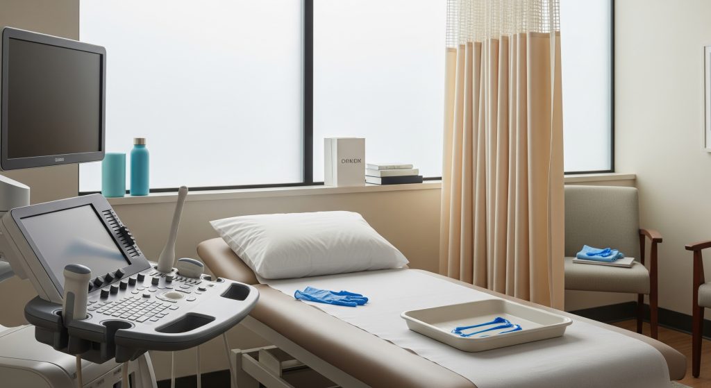
The Pelvic Ultrasound Procedure and What to Expect
Pre-Procedure Preparation Requirements
Preparation is simple. For a transabdominal Pelvic Ultrasound, you will usually arrive with a full bladder. As Midstate Radiology Associates advises, drinking about 1 quart of clear liquid 1 to 2 hours beforehand helps. Discomfort from fullness is common, so tell the technologist if it feels excessive.
Centres may vary slightly. As Cedars-Sinai notes, some ask for roughly 32 ounces one hour prior and request arrival 20 minutes early. Wear loose clothing, and expect to change into a gown, as Johns Hopkins Medicine suggests.
-
Bring previous reports if you have them.
-
Take medicines as normal unless told otherwise.
-
Ask if a full bladder is needed for your specific scan.
Step-by-Step Pelvic Ultrasound Procedure
The scan is straightforward. You will lie on a table. A clinician will apply gel and move a probe over your lower abdomen. For a transvaginal ultrasound, a covered probe is gently inserted into the vagina for closer views. As STANDARD TREATMENT GUIDELINES ENDOCRINOLOGY outlines, the internal approach often yields more detail for pelvic pathology.
For transabdominal views, a full bladder acts as an acoustic window. That improves visibility of the uterus and adnexa, as Sonography Female Pelvic Pathology Assessment, Protocols, and Interpretation describes. Real time images appear as sound waves reflect from tissues, allowing dynamic review of organs and sometimes blood flow, as Cleveland Clinic explains.
-
Check identity and confirm the clinical question.
-
Prepare bladder status as directed.
-
Perform transabdominal survey.
-
Proceed to transvaginal ultrasound if indicated.
-
Use Doppler selectively to assess vascularity.
-
Review images and confirm key measurements.
Duration and Comfort During the Scan
Most scans take 15 to 25 minutes, depending on whether both routes are needed. As Model Curriculum Handbook notes, transvaginal ultrasound is usually quicker and provides clearer images. Mild pressure or brief discomfort may occur during probe insertion.
The gel is often warm and the table is flat. The technologist keeps the room private and professional. As Patient Preparations | Pelvic Ultrasound suggests, alert the team if your bladder is uncomfortably full during the transabdominal portion. They can often pause and adjust.
Special Considerations for Different Patient Groups
Technique adapts to the person. In postmenopausal patients or those with complex anatomy, transvaginal imaging may be especially helpful. As Sonography Female Pelvic Pathology Assessment outlines, anatomical differences and risks across life stages influence the approach and the interpretation.
Communication underpins comfort and cooperation. As Gynecologic Pelvic Examination – StatPearls emphasises, a patient centred style improves the experience, particularly for sensitive exams. In adolescents and elderly women, tailored methods maintain accuracy, as Pelvic Ultrasound – StatPearls advises.
Transgender and nonbinary patients may face specific barriers. A gender affirming process and clear pre exam briefing reduce anxiety and improve care, as VolusonClub notes. Precision is clinical. Respect is clinical too.
Post-Procedure Instructions
You can generally resume normal activities immediately. There is no sedation, and no radiation exposure. If a transvaginal ultrasound was performed, minor spotting can occur. Drink water if you were very full and felt uncomfortable. If severe pain or bleeding occurs, contact your clinician promptly. Results are usually reported to your referrer, who will discuss next steps.
Understanding Pelvic Ultrasound Images and Results
Normal Pelvic Ultrasound Images
Normal findings vary with cycle phase. The endometrium changes in thickness and echogenicity across the month. As Pelvic Ultrasound – StatPearls – NCBI Bookshelf notes, that dynamism is expected and helps time fertility care.
Typical orientation shows a midline uterus with the cervix inferiorly and ovaries lateral to the uterus. As Sonography Female Pelvic Pathology Assessment summarises, this pattern provides a reliable map for comparisons.
|
Structure |
Usual appearance |
|---|---|
|
Uterus |
Anteverted or retroverted; homogeneous myometrium without focal masses |
|
Endometrium |
Thin in early follicular; trilaminar near ovulation; thicker in luteal phase |
|
Ovaries |
Multiple small follicles; dominant follicle pre-ovulation; corpus luteum post-ovulation |
|
Adnexa |
No masses; clear spaces without free fluid, except small physiological amounts |
Examples of normal transvaginal views include an anteverted uterus and average ovarian sizes, as illustrated by Radiopaedia. Standard dimensions and indications are also compiled by Female pelvic ultrasound | Radiopaedia.
Common Findings and Their Significance
Many Pelvic Ultrasound reports list familiar items. Cysts, fibroids, polyps, adenomyosis, suspected endometriosis, or an early intrauterine pregnancy. As Pelvic Ultrasound – StatPearls outlines, ultrasound is core to diagnosing ovarian cysts, fibroids, and ectopic pregnancy.
-
Simple ovarian cysts are common and often self resolving.
-
Fibroids can distort the cavity and affect bleeding or fertility.
-
Endometriosis may be suggested by endometriomas or specific signs.
Combining transabdominal and transvaginal views gives a comprehensive survey, as Sonography Female Pelvic Pathology Assessment, Protocols, and Interpretation describes. For pelvic pain, recognised sonographic features help plan surgery and preserve fertility, as Pelvic pain and endometriosis discusses.
Ultrasound rarely answers everything. It usually answers the first and most important question fast.
When Further Testing Is Needed
Occasionally findings are unclear or carry red flags. Indeterminate masses, suspicious vascularity, or atypical endometrial features can trigger MRI, CT, repeat scans, or biopsy. As Pelvic Ultrasound – StatPearls notes, ambiguous masses or possible malignancy warrant escalation. The next test depends on the question. Not all complexity is cancer. But caution is prudent.
Discussing Results with Your Healthcare Provider
Ask three things. What was found. What it likely means. What happens next. Request to see key pelvic ultrasound images if that helps you understand the plan. Clarify whether follow up is time based, symptom driven, or both. If you are in fertility care, confirm how findings change dosing, timing, or add ons. Keep the report for your records, especially if you travel between clinics.
Making Informed Decisions About Pelvic Ultrasounds
A Pelvic Ultrasound is often the fastest safe way to clarify a pelvic concern. It is basically the triage tool that many pathways depend on. The method is noninvasive, repeatable, and widely available. Yet the details matter. Transvaginal ultrasound often provides decisive information. Doppler adds context. 3D helps in selected pelvic floor or complex anatomy cases.
-
Confirm the clinical question before booking the scan.
-
Ask whether transvaginal imaging will be used and why.
-
Bring prior reports for comparison and continuity.
If you are weighing choices, consider accuracy, operator expertise, and your comfort. The right test at the right moment saves time and protects health. That is the point.
Frequently Asked Questions
Is a pelvic ultrasound painful?
Most people describe mild pressure rather than pain. A full bladder can feel uncomfortable during the surface scan. Transvaginal ultrasound may cause brief pressure. Severe pain is not expected, so alert the sonographer if it occurs.
How long does a transvaginal ultrasound take?
It typically takes 10 to 15 minutes, though total visit time can be 20 to 30 minutes when both methods are used. Complex cases may take longer for complete documentation and measurements.
Can I have a pelvic ultrasound during my period?
Yes. Bleeding does not automatically prevent imaging. For some questions, timing outside menstruation is preferred to assess the endometrium. If bleeding is heavy, bring appropriate supplies and inform the team.
What’s the difference between a pelvic ultrasound and a pelvic MRI?
Ultrasound uses sound waves and provides real time imaging at low cost. MRI uses magnetic fields and offers high soft tissue contrast without radiation. Ultrasound is first line. MRI follows when detail or problem solving is required.
How accurate are pelvic ultrasound results?
Accuracy is high for many conditions, especially with transvaginal ultrasound and experienced operators. Some findings remain indeterminate and need follow up imaging or biopsy. Accuracy also varies with body habitus and bowel gas.
Do I need a full bladder for a transvaginal ultrasound?
No. A full bladder is usually needed for transabdominal scanning only. For transvaginal ultrasound, you will often be asked to empty your bladder before the internal portion begins.