


Understanding Knee MRI Scans: Uses, Side Effects, and Cost in India
Dr. Beenish Khan


The common advice is to get an X-ray first and see how it goes. That often delays a clear diagnosis when soft tissues are involved. For knee pain that does not add up on X-ray, an early knee MRI scan can save time, reduce guesswork, and guide targeted treatment. I will explain the scan types available in India, when and why clinicians request them, the knee MRI procedure, key preparation points, pricing factors, and practical ways to decide confidently.
Types of Knee MRI Scans Available in India
Standard Knee MRI Without Contrast
For most knee complaints, I recommend a non contrast knee MRI scan first. It produces high quality detail of cartilage, menisci, ligaments, tendons, marrow, and joint lining. The sequences are optimised to reveal common patterns of injury and inflammation. In practice, this is the workhorse study for suspected meniscal or ligament tears, unexplained swelling, and persistent pain after a twist or fall.
Non contrast studies suit initial assessment because they avoid extra steps, reduce potential risk, and still answer the big questions. Here is why. Most internal derangements show characteristic signals without dye. The images are diagnostic in a large majority of day to day cases. Contrast can be reserved for specific clinical questions.
-
Best for routine sports injuries and degenerative findings.
-
Efficient to schedule and complete in a single sitting.
-
Fewer contraindications and fewer workflow dependencies.
For clarity, I reserve contrast only when the clinical picture points to synovial disease, suspected infection, or certain tumour like processes. Otherwise, standard protocols deliver decisive information.
Knee MRI with Contrast Enhancement
Contrast enhanced knee MRI is not the default. It becomes valuable when I need to evaluate synovitis, characterise infection, or assess a mass with more confidence. Intravenous gadolinium shortens or enhances signal in vascular tissues, which helps separate inflamed synovium from joint fluid and highlights areas of abnormal perfusion. That extra layer can refine a surgical plan or confirm the extent of disease.
Pre assessment is simple but necessary. I confirm any prior reactions to contrast agents, review renal function if relevant, and discuss breastfeeding guidance when applicable. The core protocol mirrors the non contrast set, with additional sequences after injection to exploit the enhancement pattern. It is basically the same examination with a sharper look at vascularised structures.
-
Consider if infection, synovial proliferation, or a tumour is suspected.
-
Useful for postoperative evaluation when scar must be separated from recurrence.
-
Requires cannulation and brief monitoring after the scan.
Most patients tolerate gadolinium well. Even so, I keep contrast use purposeful rather than routine.
Bilateral Knee MRI Examination
When symptoms are bilateral or when comparison adds value, scanning both knees in one appointment is efficient. A bilateral knee MRI scan aligns sequences side by side in the reporting workflow, which helps me judge subtle cartilage thinning, early marrow changes, or meniscal morphology differences without relying on memory.
Patients often ask whether two single knee studies are better. In many centres, bilateral protocols are streamlined and may be more economical than booking two separate visits. The choice is practical. If both knees hurt or if systemic disease is on the table, I consider bilateral imaging.
-
Reduces repeat visits and compresses total reporting time.
-
Useful for systemic arthropathy or symmetrical overuse patterns.
-
Can support surgical planning by providing an internal control side.
There is one caution. If metalwork is present on one side only, I adjust sequences to minimise artefact while keeping the comparison valid.
1.5 Tesla vs 3 Tesla Knee MRI
Scanner field strength matters for resolution and certain artefacts. A 1.5T system is highly capable for routine knee imaging. A 3T system can provide finer detail and faster imaging options, though it can exaggerate some artefacts. The right choice depends on the clinical question, the coil quality, and technologist expertise.
|
Feature |
1.5T Knee MRI |
3T Knee MRI |
|---|---|---|
|
Typical use |
Standard diagnostics |
High detail questions and advanced cartilage mapping |
|
Image detail |
Adequate for most findings |
Sharper depiction of small structures |
|
Artefact sensitivity |
More forgiving |
Higher susceptibility in metal or near implants |
|
Availability |
Wide across India |
Expanding in tertiary centres |
If cartilage mapping, subtle osteochondral lesions, or complex revision surgery planning is on the agenda, I prefer 3T. If implants or motion are likely issues, 1.5T can be wiser.
Wide-Bore MRI for Claustrophobic Patients
Wide bore systems increase the gantry diameter and reduce the closed in feeling. This small design change significantly improves comfort for anxious or larger patients. It is not only about comfort. Less anxiety usually means less motion and fewer repeats. Better images follow.
-
Consider early for known claustrophobia or prior aborted scans.
-
Supports padding and careful positioning to keep the knee still.
-
Pairs well with music, eye masks, and simple breathing cues.
And yet, even a standard scanner can succeed with clear coaching and a calm, predictable process. Comfort is a technique, not just a machine.
Common Uses and Medical Conditions Diagnosed
Ligament Injuries (ACL, PCL, MCL, LCL)
Ligament injuries are the classic reason to order a knee MRI scan. A pivot or deceleration injury suggests ACL damage. A dashboard strike suggests PCL involvement. Valgus or varus stress patterns point to MCL or LCL tears. Clinical tests guide suspicion, but MRI clarifies tear grade, location, associated bone bruises, and any meniscal involvement. That detail changes management.
-
ACL: pivot shift mechanisms, concurrent lateral meniscal tears are common.
-
PCL: posterior tibial translation with posterior capsule strain.
-
MCL and LCL: fibre disruption along femoral or tibial attachments, with oedema tracks.
-
Multi ligament injury: consider posterolateral corner and neurovascular risk.
I share reports that map each injured fibre group. Surgeons plan grafts and tunnel positions around that map. Rehabilitation teams set safer milestones with fewer surprises.
Meniscal Tears and Cartilage Damage
When locking or catching is the main complaint, the meniscus is suspect. MRI reveals tear type, location, and whether fragments displace into the gutters. For cartilage, the scan shows focal defects, delamination, and bone marrow response. Those features anchor decisions between conservative therapy, arthroscopic repair, or restorative techniques.
Here is the practical value. A small, stable tear with low grade chondral change can do well with physiotherapy and load management. A bucket handle tear with a flipped fragment demands urgent attention. Same joint. Very different plan.
Bone Fractures Not Visible on X-rays
Occult fractures hide on X-ray, especially when lines are hairline thin or overlap anatomy. MRI shows bone marrow oedema and subtle cortical breaches, so stress injuries are found earlier. That means immobilisation or activity change can start before a full fracture develops. Better still, it avoids a false sense of security after a normal radiograph.
I also look for subchondral insufficiency fractures that mimic meniscal pain. Missing these delays offloading and risks collapse.
Arthritis and Joint Degeneration
In osteoarthritis, MRI captures cartilage loss, osteophytes, synovitis, effusions, and bone marrow lesions in one view. In inflammatory arthritis, it highlights pannus, erosions, and capsular changes. The advantage is not a prettier picture. It is the ability to separate structural pain drivers from incidental findings and track them through treatment.
-
Targets specific pain generators for injections or braces.
-
Monitors early response to disease modifying therapy.
-
Supports shared decisions about timing of surgery.
Arthritis is not only wear and tear. It is pattern and pace. MRI shows both.
Tumours and Infections in Knee Joint
Red flags like fever, night pain, or a palpable mass change the brief. Contrast enhanced MRI helps delineate marrow involvement, soft tissue spread, and joint communication. While biopsy is often needed for a final label, the scan guides the safest approach and helps avoid contamination of compartments during sampling.
In infection, I review marrow signal, synovial enhancement, and adjacent soft tissue changes. In neoplasia, I document compartmental boundaries. The report is a roadmap for the next clinical step.
Pre-Surgical Planning and Post-Surgery Monitoring
Surgeons want precise answers: what is torn, where is the stump, how large is the defect, and what else is unstable. I structure the report around those questions. Postoperatively, MRI can differentiate expected healing changes from complications like graft failure, arthrofibrosis, or recurrent meniscal tears. Timing matters, so I align follow up imaging intervals with the operative plan.
One more point. I call out incidental but relevant findings like popliteal cysts, small loose bodies, or vascular variants. Small notes. Large value during arthroscopy.
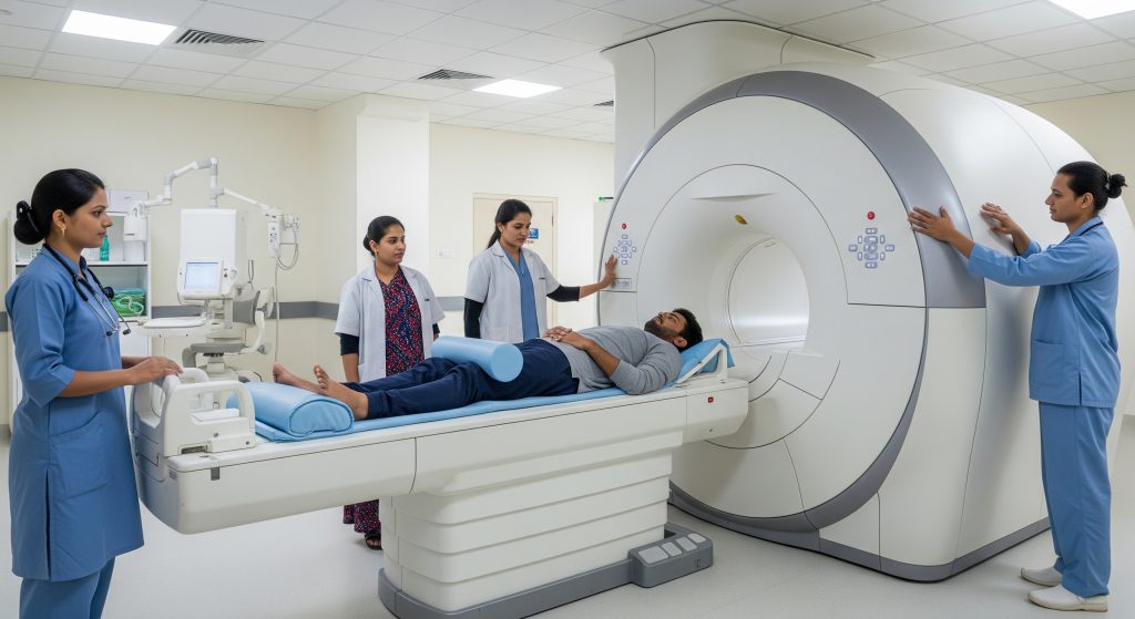
Knee MRI Procedure and Preparation
Pre-Scan Requirements and Clothing Guidelines
I ask patients to arrive in comfortable, metal free clothing. Changing into a gown is often required to avoid zips, snaps, or stray metallic fibres. A brief questionnaire screens for implants, prior surgery, allergies, and pregnancy. It also captures the exact pain site so positioning suits the problem.
-
Bring prior imaging and reports for comparison.
-
Remove jewellery, hairpins, and fitness trackers.
-
Carry any implant cards or operative notes.
Simple preparation removes avoidable artefacts and keeps the workflow smooth. That is the essence of good knee MRI scan preparation.
Fasting Instructions for Different MRI Types
For a non contrast knee MRI scan, fasting is not usually required. For contrast studies, I prefer a light gap to reduce nausea risk and to simplify any unexpected decisions. Regular medications continue unless advised otherwise by the treating team. Diabetic patients using certain agents should confirm timing with their clinician.
Hydration helps. Comfortable patients move less, and stillness is image quality.
Metal Objects and Implant Safety Checklist
Safety in MRI is non negotiable. I run a quick but thorough checklist to identify contraindications or adjust protocols. If in doubt, documentation is requested and checked.
|
Pacemaker or defibrillator |
Confirm MRI conditional status and required settings |
|
Cochlear implant |
Many are incompatible or require special handling |
|
Cerebral aneurysm clips |
Model and material must be verified before entry |
|
Orthopaedic hardware |
Usually safe, but can cause artefact near the knee |
|
Shrapnel or metal fragments |
Orbital X-ray may be needed before scanning |
|
Insulin pumps and patches |
Remove prior to scanning and store safely |
Most implants these days are MRI conditional to an extent. The key is to know the exact model and follow its label.
Step-by-Step Scanning Process
-
Registration and safety screening are completed at reception.
-
I review the clinical question and any prior images.
-
The technologist positions the patient supine, with the knee in a dedicated coil.
-
Pads and straps are used to reduce movement and improve comfort.
-
Localiser images are acquired, then diagnostic sequences follow.
-
If contrast is planned, a cannula is placed and post contrast sets are acquired.
-
Images are checked for quality before the patient leaves the scanner room.
-
I interpret the study and prepare a structured report for the referring clinician.
The sequence mix depends on the scanner and the clinical indication. The aim is consistent planes, thin slices, and robust fat suppression. That is how detail is preserved.
Duration and Patient Positioning
The knee MRI scan time depends on scanner strength, protocols, and whether contrast is used. Most dedicated knee studies fit comfortably into a single session without rushing. Good positioning is simple. The knee sits centred in the coil, with minimal rotation and stable padding under the calf and ankle. Some cases benefit from slight flexion to open the joint spaces.
Motion is the enemy. I advise patients to relax, focus on slow breathing, and treat each sequence as a short stillness interval. Short, deliberate rest. Better images.
Managing Claustrophobia During the Scan
Claustrophobia is common and manageable. I discuss the process beforehand and agree a simple hand signal for stopping. Music through headphones helps. So does an eye mask and a cool room. Wide bore scanners reduce the enclosed feeling for most patients.
-
Book a trial run if prior scans were difficult.
-
Ask about a mild anxiolytic through the referring doctor if needed.
-
Schedule at a calm time of day to reduce stress.
It sounds like a small thing. It is not. Comfort directly improves diagnostic quality.
Cost Analysis and Factors Affecting Price in India
City-Wise Price Comparison Across Metro Cities
Pricing for a knee MRI scan varies across metros due to rental costs, staffing, scanner availability, and local demand. Centres in Mumbai and Delhi often price at the higher end. Chennai, Kolkata, Hyderabad, Bengaluru, and Pune sit at mixed levels depending on neighbourhood and hospital brand.
|
Metro city |
Typical relative price band |
|---|---|
|
Delhi NCR |
Medium to higher |
|
Mumbai |
Higher |
|
Bengaluru |
Medium to higher |
|
Chennai |
Medium |
|
Kolkata |
Medium |
|
Hyderabad |
Medium |
|
Pune |
Medium |
Local competition and scanner mix influence the final figure. A premium tertiary centre will usually cost more than a standalone imaging clinic.
Government vs Private Hospital Pricing
Government hospitals often offer subsidised rates. Access can be constrained by eligibility, appointment queues, and narrower time windows. Private hospitals and accredited diagnostic chains provide faster scheduling, broader time slots, and more comfort features. The premium reflects speed and service level rather than a difference in image physics.
In practice, I advise patients to balance clinical urgency, convenience, and the need for specific scanner capabilities before deciding where to book.
3T MRI vs 1.5T MRI Cost Differences
3T scanners require higher capital and maintenance expenditure, which often translates to a higher price point per study. If the clinical question benefits clearly from 3T detail, the premium can be justified. If not, a well run 1.5T study provides excellent value for money.
-
Select 3T for small cartilage defects, complex revisions, or research protocols.
-
Select 1.5T for standard ligament and meniscal evaluation near implants.
The right match reduces repeat scans and avoids hidden costs from inconclusive results.
Insurance Coverage and Cashless Facilities
Outpatient knee MRI scan coverage varies by policy. Many plans cover imaging that is prescribed by a registered specialist and tied to a documented diagnosis pathway. Cashless facilities depend on network tie ups and pre authorisation rules. I encourage patients to request a formal referral note with a clear clinical question. Insurers process specific indications faster.
Self pay remains common for straightforward cases. For hospital admissions, scans are often billed within the admission package when clinically indicated.
Online Booking Platforms and Discount Options
Aggregators and hospital sites sometimes offer promotional pricing for off peak slots. Flexibility on timing can reduce cost without compromising quality. The crucial point is to verify accreditation, radiologist expertise, and the scanner model. A small discount does not compensate for poor protocols.
-
Check whether reporting is by an on site musculoskeletal radiologist.
-
Confirm turn around time for the report and images.
-
Ask if contrast or repeat sequences incur an extra fee.
Transparent quotes avoid surprise charges. It is a straightforward request that protects the budget.
Making Informed Decisions About Knee MRI Scans
Choosing the right knee MRI scan is less about brand and more about fit. Fit between the clinical question, the scanner, and the protocol. If the issue is a routine twist injury, a non contrast study on a competent 1.5T system is appropriate. If the issue is a complex cartilage defect or revision surgery, 3T may add value. If infection or synovial disease is suspected, contrast helps. The logic is simple. Match question to tool.
Two final levers matter just as much. Positioning and stillness. The best scanner will not overcome a poorly positioned knee or restless movement. Good setup, calm coaching, and a clear plan elevate image quality and reduce repeats.
As a rule, I advise patients to ask three questions before booking. Which scanner and coil will be used. Who will report the study and with what musculoskeletal experience. What protocol adjustments are planned for the specific complaint. Those answers reveal quality.
And one more consideration. Time. Early, accurate imaging can prevent months of trial and error treatment. That is not indulgence. It is clinical efficiency.
Frequently Asked Questions
How much does a knee MRI scan cost in Delhi NCR in 2026?
Prices vary by centre type, scanner strength, and whether contrast is used. Large private hospitals and premium centres tend to charge more than standalone clinics. Government hospitals may offer subsidised options with longer wait times. For a realistic estimate, request a written quote specifying scanner type, contrast use, and reporting time. That avoids last minute add ons.
Can I eat before my knee MRI appointment?
For a non contrast knee MRI scan, eating normally is acceptable. For contrast studies, I prefer a light gap to minimise nausea risk and to streamline consent. Keep regular medications unless advised otherwise by your clinician.
Is knee MRI safe for patients with pacemakers or metal implants?
Many implants are MRI conditional under specific settings. Safety depends on the exact device model, lead type, and institutional protocols. Provide implant cards for verification. If an implant is not compatible, alternative imaging or special workflows are considered. Safety screening is mandatory and protects everyone.
How long does a typical knee MRI scan take to complete?
Most knee studies are completed in a single session. The knee MRI scan time depends on scanner strength, motion control, and whether contrast is used. Allow adequate buffer for screening and positioning so the images are acquired without rush.
What is the difference between 1.5T and 3T MRI for knee imaging?
1.5T provides excellent routine imaging and is widely available. 3T offers finer detail and can shorten some sequences, but is more sensitive to artefact. I choose based on the clinical question, implant presence, and the need for advanced cartilage or quantitative sequences.
Will I need contrast dye for my knee MRI scan?
Not usually. Contrast is reserved for suspected infection, synovial disease, tumour assessment, or specific postoperative questions. If requested, the team will discuss consent, prior reactions, and brief aftercare. The decision is clinical, not automatic.
Can I get my knee MRI report on the same day?
Many centres provide preliminary reports the same day and final reports within a short time frame. Turnaround depends on case volume, the need for prior comparisons, and whether complex findings require correlation with the referrer. Ask for expected timelines when booking to plan follow up accordingly.