


Understanding Fracture Surgery and Healing Stages
Dr. Neetan Sachdeva
Set the cast and wait. That old advice still circulates. For unstable injuries and high-demand patients, it is poor guidance. I approach fracture surgery as a precise intervention with clear aims: restore alignment, provide stable fixation, and enable safe, timely rehabilitation. The science is robust, but the art is in choosing the right method for the right fracture at the right time. In the sections below, I outline practical choices, the fracture healing stages, and the recovery milestones that actually drive outcomes.
Types of Fracture Surgery and Treatment Options
When I plan fracture surgery, I prioritise stability, biology, and function. Technique is not a badge of honour. It is a tool to solve a mechanical and biological problem with the least collateral damage.
Open Reduction Internal Fixation (ORIF)
Open reduction internal fixation allows me to visualise the fracture, restore anatomy, and secure it with plates, screws, or specialised implants. It is particularly helpful for displaced articular injuries and complex periarticular patterns where precise joint congruity matters. In practice, I select approaches that protect soft tissue and blood supply, then fix fragments in a sequence that restores length, rotation, and alignment.
ORIF is not a single playbook. For example, ankle fractures that are out of alignment often demand anatomical reduction to prevent post-traumatic arthritis. Hip and proximal humeral fractures may benefit from rigid fixation that supports early motion and reduces stiffness. I also counsel patients that implants may remain indefinitely, unless they cause symptoms or interfere with later care. This is part of modern fracture surgery: plan for function now and for options later.
-
Strengths: anatomical reduction, rigid stability, early joint motion.
-
Limitations: soft tissue exposure, infection risk, potential implant irritation.
-
Typical candidates: displaced intra-articular fractures, unstable diaphyseal fractures with comminution, non-unions.
External Fixation Methods
External fixation uses percutaneous pins or wires connected to an external frame. I use it to stabilise fractures where soft tissue is compromised or when rapid provisional control is vital. It buys time. With severe open fractures, it maintains alignment while plastic surgery addresses coverage and contamination. In wrists with swollen, blistered skin, a spanning frame can protect the joint while swelling resolves.
Modern options include uniplanar frames, circular constructs, and hybrid external fixation that combines rings and rods. Each offers adjustable stability. The mechanical concept is simple but effective: stabilise the bone while leaving wounds accessible for care. Blood loss is minimal. Adjustments are straightforward. For the right injury, it is elegant in its simplicity.
-
Strengths: rapid application, soft tissue access, minimal additional trauma.
-
Limitations: pin tract care, patient comfort, limited rotational control in some frames.
-
Typical candidates: contaminated open fractures, limb damage with swelling, temporary stabilisation for polytrauma.
Closed Reduction Procedures
Closed reduction aims to realign the fracture without surgical exposure. I pair it with immobilisation or percutaneous fixation when the fracture is stable after reduction. The technique relies on traction, gentle manipulation, and careful fluoroscopic checks. Sedation or local anaesthesia is usually sufficient, which reduces risk and recovery time.
When a stable pattern is reducible and congruent, closed methods can shorten hospital stay and avoid implant-related issues. In paediatric fractures, the biology is forgiving, and closed reduction often excels. As Egyptian Journal of Hospital Medicine reported, a series of 20 children with radial head injuries achieved excellent outcomes in 16 cases after closed reduction with elastic nailing, supporting this conservative-first mindset where stability permits.
-
Strengths: minimal invasiveness, lower infection risk, faster basic recovery.
-
Limitations: risk of loss of position, need for vigilant follow up.
-
Typical candidates: stable, reducible fractures that hold alignment in a cast or brace.
Intramedullary Nailing
Intramedullary nailing (IM nailing) places a load-sharing rod inside the canal of long bones. It offers strong fixation through central alignment and interlocking screws. For femur and tibia fractures, this method combines biology and mechanics well: minimal periosteal stripping with robust stability. It is my default for many diaphyseal patterns.
Outcomes are consistently favourable. In segmental long bone injuries, open intramedullary nailing achieved a union rate of 91.7% within 4 months, with no major complications, as documented by the Open Intramedullary Nailing report. In a separate femoral cohort, there were no cases of non-union among 30 patients, underscoring reliability in appropriately selected fractures, as noted by the Clinical outcomes of intramedullary femoral nailing system.
-
Strengths: biologically friendly, strong mechanical control, early mobilisation.
-
Limitations: canal width constraints, entry-point morbidity, hardware prominence.
-
Typical candidates: diaphyseal femur and tibia fractures, select humeral and subtrochanteric patterns.
Bone Grafting Techniques
Bone grafting supports biology when the defect or non-union biology is insufficient. I choose the graft based on three qualities: osteogenesis, osteoinduction, and osteoconduction. Autograft provides cells and growth factors. Allograft adds scaffold without donor site pain. Synthetic options (for example, calcium phosphates) can fill defects and support new bone formation when combined with stable fixation.
I deploy grafts to bridge voids, augment comminuted articular surfaces, or restart biology in atrophic non-unions. The rule remains constant. Mechanics first, then biology. Without stable fixation, grafts underperform.
Fracture Healing Stages and Recovery Timeline
Understanding fracture healing stages helps set expectations and reduce anxiety. I frame recovery in phases that link biology to function and to what patients can safely do each week.
1. Inflammatory Phase (0-2 Weeks)
Immediately after injury, bleeding and clot formation trigger an inflammatory cascade. Pain and swelling peak early. My aims are simple: protect the reduction, manage pain, and maintain circulation and nerve function. Elevation, ice, and safe immobilisation matter much more than heroics. Early complications begin here, so I check wounds, compartments, and neurovascular status often.
-
Priorities: soft tissue care, oedema control, DVT prevention, baseline ROM in unaffected joints.
-
Typical activity: protected limb rest, gentle isometrics, basic breathing exercises.
2. Soft Callus Formation (2-6 Weeks)
Fibrocartilaginous callus bridges the fracture. Pain reduces, and controlled motion becomes safer. I introduce guided physiotherapy to restore range without stressing the fixation. Depending on stability, partial weight bearing may start. The principle is cautious progress, not bravado.
-
Focus: gradual ROM, swelling reduction, re-education of gait with aids.
-
Red flags: escalating pain, new deformity, wound drainage.
3. Hard Callus Formation (6-12 Weeks)
Mineralisation converts soft callus into hard callus. On radiographs, the gap begins to blur. Functionally, strength returns unevenly. I often increase load at this stage, provided alignment holds and pain remains controlled. It is basically biology giving permission for more work.
-
Focus: progressive resistance, proprioception drills, careful step-up in tasks.
-
Checks: serial X-rays, hardware position, gait symmetry.
4. Bone Remodelling Phase (3-9 Months)
Remodelling reshapes bone along stress lines. Capacity increases gradually. Patients feel strong but are not fully recovered. This is where many overreach and irritate tissues. I emphasise patient-specific targets: return to work, sport mechanics, endurance. The goal is robust function, not just a healed X-ray.
-
Focus: sport or job-specific drills, higher loads, endurance work.
-
Outcome marker: pain-free function in daily tasks and targeted activities.
Factors Affecting Healing Speed
Several variables shift the timeline. Age, smoking, diabetes, poor nutrition, high-energy trauma, infection risk, and suboptimal stability all slow progress. Fixation choice interacts with biology too. For example, an IM nail in a diaphyseal tibia may allow earlier weight bearing than a tenuous bridge plate in severe comminution. Here is why: load sharing encourages adaptive callus when alignment and rotation are secure.
-
Favourable factors: stable fixation, early but safe mobilisation, good protein and vitamin D intake.
-
Unfavourable factors: nicotine exposure, poor glucose control, extensive soft tissue damage.
Types of Bone Fractures Requiring Surgery
I group the types of bone fractures that commonly necessitate an operative plan by stability, displacement, and joint involvement. Not every unstable fracture demands immediate operation, but most need timely, structured intervention.
Compound and Open Fractures
When bone communicates with the external environment, the priority is decontamination, soft tissue management, and stabilisation. I frequently use external fixation at first, then definitive fixation once the soft tissue envelope is ready. Early antibiotics and irrigation are critical.
Displaced Fractures
Displacement that alters length, axis, or rotation can produce long-term dysfunction. Operative reduction restores biomechanics and reduces the risk of malunion. ORIF or IM nailing is selected to match the pattern and patient demands.
Comminuted Fractures
Multiple fragments require bridge constructs that protect biology while restoring alignment. I plan fixation to share load and allow controlled micromotion. Bone grafting may fill voids or buttress joint surfaces when needed.
Spiral and Oblique Fractures
Long spiral or oblique patterns accept lag screws well, either alone or under a neutralisation plate. In the femur or tibia, intramedullary nailing is often preferred for load sharing and alignment control.
Intra-articular Fractures
Articular congruity is non-negotiable. Small step-offs can trigger arthritis. I prioritise anatomic reduction of the joint surface and stable fixation that permits early controlled motion. Navigation or arthroscopy may help in select cases.
Pathological Fractures
When bone quality is impaired by tumour or metabolic disease, standard rules shift. I stabilise more robustly and consider cement augmentation. Oncological planning and medical therapy proceed in parallel. The fixation must outlast the biology.
Post-Surgery Care and Rehabilitation
Fracture surgery sets the stage. Rehabilitation delivers the function. I clarify expectations in writing, because small daily behaviours add up.
Immediate Post-Operative Management
In the first 48 hours, I focus on pain control, limb protection, and soft tissue status. Elevation, ice, and gentle toe or finger motion keep swelling in check. I review neurovascular signs and compartments, particularly after high-energy trauma.
|
Item |
What I Look For |
|---|---|
|
Wound |
Dry dressing, no active bleed, no tense swelling |
|
Sensation |
Intact light touch, no new numbness |
|
Perfusion |
Warm skin, brisk capillary refill |
|
Alignment |
Maintained reduction on check X-ray |
Pain Management Strategies
I employ multimodal analgesia to reduce opioid load: paracetamol, NSAIDs if bone and soft tissue risk is acceptable, nerve blocks where indicated, and ice. Sleep and nutrition are part of analgesia too. Poor sleep worsens pain perception. In select cases, I use short opioid courses with a taper plan from day one.
-
Checklist: bowel regimen, clear taper, realistic pain targets, night-time routine.
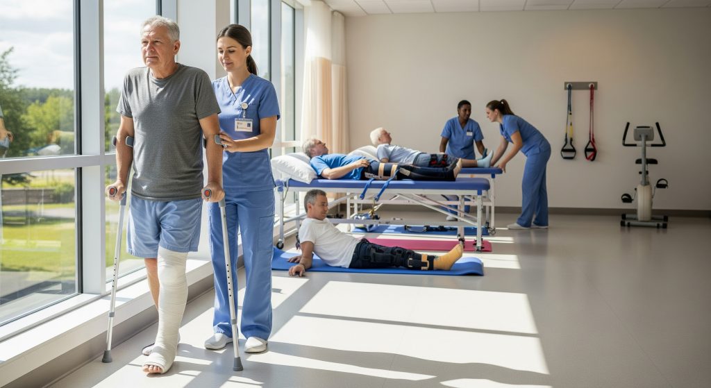
Physical Therapy Protocols
Physiotherapy starts early, often day one. Protocols are fixation and fracture specific. For ORIF around a joint, I emphasise early controlled ROM to prevent stiffness. After IM nailing of the femur, gait training progresses as comfort and alignment allow. I also track simple metrics: straight leg raise, knee flexion, single-leg balance. Small wins demonstrate progress and build confidence.
Weight-Bearing Progression
Weight bearing is a prescription, not a guess. I specify non-weight bearing, toe-touch, partial, or as tolerated based on the construct and biology. WBAT (weight bearing as tolerated) is safe after many nails, while tenuous bridge plating may need longer protection. I adjust at each review using pain, function, and X-ray evidence as anchors.
-
Rule: load follows stability and symptoms.
-
Tool: crutches now, cane next, unassisted when gait is symmetric.
Monitoring for Complications
Complications are rare with sound technique, but vigilance matters. I screen for infection, deep vein thrombosis, complex regional pain, hardware failure, loss of reduction, and non-union. Laboratory markers (CRP, ESR) and imaging support the clinical picture. Early detection simplifies treatment. Late discovery complicates everything.
Small changes in pain or function are often the first signal. Listen to the pattern. Then investigate.
Making Informed Decisions About Fracture Surgery
Patients make better decisions when options are framed by goals, not by tools. I start with functional aims: walk without aids, return to work, protect the joint. Then I map options: open reduction internal fixation for articular precision, intramedullary nailing for load sharing, external fixation to protect soft tissue, or closed reduction where biology and stability allow. Each path has trade-offs. Acknowledge them openly.
Two final principles guide me. First, technique must serve biology. Stable fixation that preserves blood supply accelerates the fracture healing stages and makes rehabilitation practical. Second, function drives timelines. A builder’s tibia is not a desk worker’s wrist. The plan must reflect activity, risk tolerance, and support at home. This is careful medicine. And still, it is personal.
Frequently Asked Questions
How long does fracture surgery typically take?
Most procedures take 45 to 120 minutes, depending on fracture complexity and the method chosen. Polytrauma and periarticular reconstructions can extend longer due to positioning, reduction, and implant steps. Time in the theatre does not predict outcome alone. The quality of reduction and stability does.
What are the risks associated with fracture surgery?
General risks include infection, bleeding, nerve or vessel injury, thromboembolism, stiffness, non-union, and hardware irritation. The actual risk profile depends on the injury, comorbidities, and timing. Strong perioperative protocols and meticulous soft tissue handling reduce these risks considerably.
When can normal activities resume after fracture surgery?
Light daily tasks usually resume within 2 to 3 weeks, with modifications. Driving follows safe control of the limb and cessation of sedating analgesia. Higher demand tasks return between 8 and 16 weeks, aligned to radiographic and functional milestones. Sport-specific drills wait until strength and proprioception are symmetrical.
How do surgeons decide between different fixation methods?
I match the mechanical problem and the biology to the construct. Articular precision points to open reduction internal fixation. Diaphyseal load sharing favours intramedullary nailing. Severe soft tissue injury often needs external fixation. Stable, reducible patterns may succeed with closed reduction. Patient goals refine the final choice.
What signs indicate improper fracture healing?
Persistent pain after expected milestones, progressive deformity, hardware loosening, or a visible gap on serial X-rays suggest delayed union or non-union. Worsening redness, heat, or drainage implies infection. Any new neurological change requires prompt assessment. Early review prevents escalation.
Can all fractures heal without surgery?
No. Many undisplaced and stable patterns heal reliably without an operation. However, displaced, unstable, open, and intra-articular fractures often need fracture surgery to restore alignment and function. The decision is not ideological. It is evidence, mechanics, and patient goals.
Quick reference
|
Primary aim of fracture surgery |
Restore alignment and stability to enable safe, timely rehabilitation |
|
Key phases |
Inflammation, soft callus, hard callus, remodelling |
|
Useful jargon |
IM nail (intramedullary nail), WBAT (weight bearing as tolerated), ROM (range of motion) |
|
Secondary phrases to know |
open reduction internal fixation, fracture healing stages, types of bone fractures |
Final note: Fracture surgery is a means to an end. The end is durable, confident function, achieved through sound mechanics and patient-centred rehabilitation.