


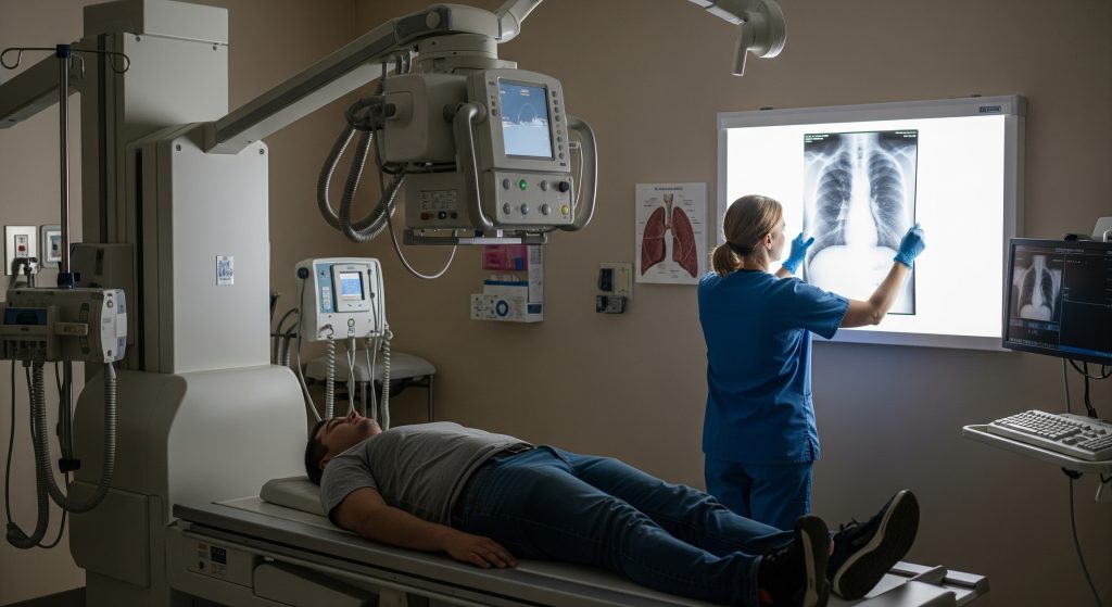


Pneumothorax X-ray Explained: What It Shows About a Collapsed Lung
Dr. Beenish Khan


Common advice says every chest film tells the story on first glance. That is rarely true. A careful, structured read of a Pneumothorax X-ray prevents misses and helps you act quickly when a collapsed lung is evolving. This guide shows you what to look for, how to size what you see, and how those findings guide management. It is basically a practical map, not a scatter of textbook trivia.
Key X-ray Signs of Pneumothorax
1. Visible Lung Edge
On a Pneumothorax X-ray, the visible lung edge appears as a sharp, visceral pleural line separated from the chest wall by air. As NCBI – NIH notes, that separation confirms air in the pleural space and the measured distance helps estimate size, with more than 2 cm typically classed as large. Ultrasound often complements your film when doubt remains. As Lung Point Sign in Ultrasound Diagnostics of Pneumothorax explains, identifying a lung point on ultrasound is highly specific and pinpoints the pleural interface.
-
Look for a crisp, white pleural line with no lung markings peripheral to it.
-
Measure the shortest distance from the lung edge to the chest wall at the hilum level.
-
Correlate with clinical status before calling it small or large.
2. Absent Lung Markings
Absent peripheral vascular markings beyond a pleural line is a classic Pneumothorax X-ray cue. As StatPearls – NCBI Bookshelf describes, the key is the combination of a sharp line plus lucency, not lucency alone. Emphysema and even large bullae can mimic the appearance. The pattern tends to be regional in those conditions, with subtle linear textures lingering in the field. The clinical picture matters here. A careful differential spares you from overcalling a collapsed lung in severe COPD.
3. Hyperlucency in Pleural Space
Hyperlucency suggests air where lung should be. On a Pneumothorax X-ray, the lucent area between chest wall and lung edge lacks vascular markings and often looks strikingly uniform. As Demystifying the persistent pneumothorax notes, other processes can enhance lucency or mimic it, including exposure factors or large effusions that shift the lung. Where doubt persists or underlying disease is suspected, CT clarifies the plane and the cause. In some cases, as Spontaneous Pneumothorax: A Review reports, marked hyperlucency may point to a secondary event on top of chronic lung pathology.
4. Mediastinal Shift
Mediastinal shift signals pressure trouble, not just trapped air. On a Pneumothorax X-ray, deviation of the heart and trachea away from the affected side suggests rising intrathoracic pressure. As StatPearls – NCBI Bookshelf highlights, tension physiology compresses venous return and destabilises circulation. In the same vein, Tension Pneumothorax – StatPearls emphasises that progressive shift is an emergency that warrants immediate decompression.
Mediastinal shift is a late sign. Treat clinical tension before waiting for the film that proves it.
5. Deep Sulcus Sign
In the supine position, air collects anteriorly and basally, exaggerating the costophrenic angle. The deep sulcus sign shows an abnormally deep, radiolucent costophrenic recess on the affected side. You may also see a lucent diaphragm outline that looks unusually sharp. On a Pneumothorax X-ray taken supine, this sign can be your earliest clue in trauma or ventilated patients.
-
Compare both sulci for asymmetry.
-
Scan for associated features like a sharp cardiac border or low anterior lung markings.
6. Tension Pneumothorax Features
Tension pneumothorax is a clinical diagnosis supported by imaging. On a Pneumothorax X-ray, you may see a completely collapsed lung, depressed hemidiaphragm, and mediastinal shift. As Tension Pneumothorax – StatPearls sets out, causes include trauma and ventilation barotrauma, and hallmark signs include hypotension, unilateral breath sounds, distended neck veins, and tracheal deviation. Immediate needle decompression followed by definitive tube thoracostomy is the sequence.
Reading Pneumothorax X-rays: Different Views and Techniques
Upright Chest X-ray Interpretation
An upright film remains the most informative Pneumothorax X-ray for routine assessment. Air rises to the apex, often revealing a visible pleural line and peripheral lucency. Take a systematic approach. Check tracheal position, then scan apices, lung edges, and costophrenic angles. Confirm side markers, especially in mirrored mobile images. The most subtle apical stripes hide in busy films, so magnify the corners if needed.
-
Apical lucency with a fine white pleural line is the key pairing.
-
Peripheral lucency without vessels should match that line and the clinical story.
Supine X-ray Findings
In a supine Pneumothorax X-ray, air redistributes anteriorly and basally. Expect indirect signs rather than apical lines. The deep sulcus sign, a sharp cardiac silhouette, and a lucent hemidiaphragm are more telling patterns. Look for hemithorax asymmetry in overall density. Small volumes can look deceptively benign. Radiographic subtlety does not negate clinical urgency.
Lateral View Assessment
A lateral film may reveal an anterior pleural line that is invisible on the frontal view. This is useful when a Pneumothorax X-ray in the PA or AP view is equivocal. Inspect the retrosternal clear space and the anterior costophrenic recess. A thin linear edge can confirm suspicion and prevent an unnecessary CT in stable cases.
Expiratory Film Advantages
Expiratory imaging reduces lung volume and increases the relative contrast between aerated pleural space and compressed lung. A Pneumothorax X-ray obtained on expiration can accentuate small air pockets that disappear on inspiration. It also helps distinguish bullae from true pleural air when the edge behaviour differs between phases.
Small Pneumothorax Detection
Small volumes are easy to miss among overlapping ribs and clavicles. Train your eye to search the apices, the parasternal region, and the lateral recess. If a standard Pneumothorax X-ray remains inconclusive and suspicion is high, bedside ultrasound can help identify a missing lung sliding sign or a lung point. CT remains a reserve option in complex trauma or when procedural planning requires precision.
Size Estimation Methods
Size estimation guides the urgency of pneumothorax treatment. A consistent method matters more than a perfect method. On a Pneumothorax X-ray, estimate the shortest distance from the lung edge to the chest wall at the hilum level. Alternatively, use approximate percentage rules to stratify small, moderate, and large volumes.
|
Method |
How to apply |
|---|---|
|
Linear distance |
Measure lung edge to chest wall. More than 2 cm often indicates a large pneumothorax. |
|
Rule of twos |
If the pleural gap is roughly 2 cm at the apex and laterally, consider escalation. |
|
Percent estimate |
Use visual quartiles. Up to roughly 20 percent is small, above that is moderate to large. |
These are estimates. Clinical status and gas exchange data refine the decision in practice.
Types of Pneumothorax and Their Causes
1. Primary Spontaneous Pneumothorax
This affects individuals without known lung disease, often tall adults with subpleural blebs. A Pneumothorax X-ray typically shows an apical air pocket with a visible edge. Smoking increases risk to a degree that is clinically relevant. Recurrence is common, and recurrence risk influences your definitive plan.
2. Secondary Spontaneous Pneumothorax
Secondary events occur in the setting of COPD, interstitial disease, cystic lung disorders, or infections. The same Pneumothorax X-ray signs apply, yet patients decompensate faster due to reduced reserve. Oxygenation may fall with a small volume. Early intervention is therefore more likely, even for modest separations.
3. Traumatic Pneumothorax
Penetrating or blunt trauma can introduce air into the pleural space directly or via lung laceration. Rib fractures are often present. On a Pneumothorax X-ray, search for subcutaneous emphysema, rib discontinuity, and a deep sulcus in supine trauma films. Positive pressure ventilation increases the risk of tension physiology. Act promptly when haemodynamics shift.
4. Iatrogenic Pneumothorax
Procedures carry risk. Central venous line placement, lung biopsy, thoracentesis, and even acupuncture have been implicated. A post procedure Pneumothorax X-ray is routine in higher risk interventions. Most iatrogenic pneumothoraces are small and respond to observation, though not invariably.
Risk Factors and Prevention
-
Smoking and tall body habitus raise primary risk.
-
COPD, cystic lung disease, and infections raise secondary risk.
-
Barotrauma during ventilation raises tension risk.
-
Procedural risk falls with ultrasound guidance and experienced operators.
When you discuss pneumothorax causes with a patient, keep advice concrete. Smoking cessation reduces recurrence risk and overall pulmonary decline. Scuba diving and unpressurised flights should be avoided until cleared, especially after a collapsed lung episode.
Treatment Decisions Based on X-ray Findings
Conservative Management Criteria
Observation is appropriate for a stable patient with a small separation and manageable pneumothorax symptoms. A clear, small pleural gap on a Pneumothorax X-ray, good oxygenation, and minimal dyspnoea favour this option. High flow oxygen accelerates pleural air absorption. You should arrange timely reassessment and provide a fallback plan.
-
Stable observations and minimal pain.
-
Small radiographic size and no mediastinal shift.
-
Reliable follow up within 24 to 48 hours.
Needle Aspiration Indications
Needle aspiration suits moderate pneumothoraces in stable patients, especially in primary spontaneous cases. On the Pneumothorax X-ray, a separation around the mid zone with symptoms that hinder activity is a reasonable threshold. The immediate goal is symptom relief and re-expansion. Failure proceeds to tube drainage.
Chest Tube Placement Guidelines
Insert a chest drain when the pneumothorax is large, symptomatic, or complicated by underlying disease. A Pneumothorax X-ray showing a wide pleural gap, or any signs of mediastinal shift, justifies intervention. In ventilated patients, act early. Small bore drains often suffice, though situational judgement applies.
Drain choice is not an identity. It is a tool. Match the tube to the air leak and the patient’s physiology.
Follow-up X-ray Monitoring
After any intervention, confirm position and effect on a Pneumothorax X-ray taken promptly post procedure. Subsequent films document re-expansion and detect complications like re expansion oedema. In ambulatory observation, repeat imaging ensures the volume is stable or shrinking and aligns with symptom improvement.
Resolution Timeline
Spontaneous resolution can be slow, roughly speaking at a few percentage points of volume each day. A Pneumothorax X-ray taken at intervals shows the lung edge creeping back to the wall. The timeline varies by volume, oxygen supplementation, and ongoing air leak. Prepare patients for a staged recovery, not an instant fix.
Emergency Intervention Signs
Certain features demand immediate action. A Pneumothorax X-ray with mediastinal shift, a collapsed hemithorax, or rapidly rising ventilator pressures signals danger. Clinical markers dominate here. Hypotension, worsening hypoxia, and unilateral breath sounds should trigger decompression without waiting for imaging in the unstable patient.
-
Haemodynamic instability or rising lactate.
-
Expanding subcutaneous emphysema in ventilated patients.
-
Tracheal deviation and distended neck veins.
The radiograph informs care. The physiology dictates speed.
Understanding Your Pneumothorax X-ray Results
When your report arrives, read the language with intent. A Pneumothorax X-ray report usually notes side, size estimate, and any shift. It may comment on tubes, lines, or associated fractures. It should link the appearance to likely pneumothorax treatment options and next steps. If terminology feels opaque, ask for a plain language summary pinned to action. That is not a courtesy. It is good medicine.
-
If you see small separation and stable observations, observation plus oxygen may be appropriate.
-
If you see moderate size and symptoms, needle aspiration might be offered.
-
If you see large separation or instability, chest drain or emergency decompression follows.
A short example helps. A young person with pleuritic pain and a small right apical gap might leave with observation and safety netting. An older person with COPD, dyspnoea, and a moderate left pleural gap will likely require drainage despite similar measured size. Same Pneumothorax X-ray framework. Different risk, different decision.
Frequently Asked Questions
How quickly does pneumothorax show on X-ray?
Air appears immediately, but detectability depends on volume, position, and exposure. An upright Pneumothorax X-ray typically reveals apical air promptly. In supine imaging, early signs are indirect, such as a deep sulcus or sharp cardiac border. If the clinical picture suggests tension, treat first and image after stabilisation.
Can a small pneumothorax be missed on X-ray?
Yes, especially on AP portable films with overlapping structures. A small apical pneumothorax can hide beside clavicles and ribs. An expiratory Pneumothorax X-ray or a lateral view can help, and bedside ultrasound increases sensitivity. When suspicion remains high, CT may be warranted in selected cases.
What percentage of lung collapse requires treatment?
Thresholds vary by guideline and by patient reserve. A small, primary pneumothorax may be observed, sometimes up to roughly 20 percent. A similar percentage in COPD may require drainage. Use the Pneumothorax X-ray size estimate as a guide, then prioritise symptoms, oxygenation, and stability.
How often should follow-up X-rays be taken?
In ambulatory observation, repeat imaging is often performed within 24 to 48 hours to confirm stability. After intervention, a post procedure Pneumothorax X-ray confirms tube position and lung re-expansion. Frequency then tapers with clinical improvement. More frequent imaging is justified if symptoms worsen.
Can pneumothorax heal without treatment?
Many small primary events resolve with observation and oxygen. The body reabsorbs pleural air over days to weeks. Your Pneumothorax X-ray trend should show progressive re-expansion. Secondary cases and symptomatic patients are less tolerant of watchful waiting, so treatment thresholds are lower.
What activities should be avoided after pneumothorax diagnosis?
Avoid air travel, scuba diving, and strenuous exertion until cleared. Pressure changes and heavy strain risk recurrence or deterioration. Your team will clear gradual activity as the Pneumothorax X-ray shows sustained resolution. Smoking cessation reduces recurrence risk to a meaningful degree.