


Pelvic Sonography Explained: What to Expect and Why It’s Used
Dr. Beenish Khan
Routine advice often says all pelvic scans are the same. That assumption wastes time and sometimes money. Pelvic Sonography is a suite of precise methods that answer different clinical questions. Used well, it accelerates diagnosis and spares you unnecessary procedures. Used poorly, it leads to repeat visits and unresolved symptoms. This guide explains what will happen, why each approach is chosen, and how the results inform care.
Types of Pelvic Sonography and How They Work
Transabdominal Ultrasound Procedure
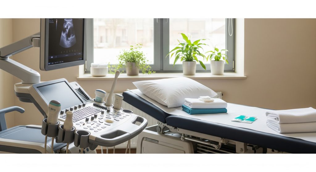
Transabdominal ultrasound evaluates your pelvic organs through the lower abdomen with a handheld probe and gel. As Johns Hopkins Medicine explains, gel removes air between the probe and skin to sharpen real-time images. You will usually be asked to arrive with a full bladder because urine creates an acoustic window that improves clarity. As StatPearls notes, the absence of radiation makes this approach suitable for reproductive-age patients. As STANDARD TREATMENT GUIDELINES OBSTETRICS & GYNAECOLOGY outline, it visualises the uterus and ovaries well and is often paired with transvaginal imaging for a fuller assessment.
-
Best for an overview of uterus, ovaries, bladder, and surrounding spaces.
-
Non-invasive and generally comfortable.
-
Often the first step before a more targeted view.
Transvaginal Ultrasound Method
Transvaginal ultrasound places a slim, covered probe in the vagina to bring the transducer closer to the uterus and ovaries. This proximity increases resolution, especially for early pregnancy, endometrium, ovaries, and adnexa. You can expect privacy protections, careful consent, and a clear explanation before the scan begins. The bladder is typically emptied to improve comfort and visualisation. In practice, this modality complements transabdominal ultrasound when detail is required near the uterus or ovaries. It is especially useful for suspected cysts, fibroids, or early pregnancy concerns.
-
High-detail images of endometrium and ovaries.
-
Useful for early pregnancy assessment and follicle tracking.
-
Short procedure with minimal discomfort for most patients.
Transrectal Sonography Option
Transrectal ultrasound (TRUS) is primarily used for the male pelvis and the prostate. As StatPearls describes, TRUS offers real-time imaging for diagnosing prostate disease and for guiding biopsies. As RadiologyInfo clarifies, a small probe is inserted into the rectum to view the prostate and nearby structures with high detail. Infection control measures are standard. As National Guidelines emphasise, strict protocols mitigate procedural risks. TRUS can also support targeted procedures beyond prostate imaging. As PubMed reports, endoscopic ultrasound-guided transrectal drainage is a minimally invasive option for selected pelvic abscesses.
How Sound Waves Create Images
Ultrasound uses high-frequency sound waves to generate images based on echoes. As Model Curriculum Handbook outlines, differences in tissue density change how waves reflect and propagate. The system interprets these echoes to build the image on screen. As Ultrasound Physics and Instrumentation notes, frequency influences resolution and penetration. Higher frequencies show fine detail but penetrate less deeply. In pelvic sonography, that trade-off guides probe choice.
Ultrasound converts micro-echoes into anatomy in motion. That is the power of real-time imaging.
Doppler Ultrasound for Blood Flow
Doppler techniques measure blood flow within vessels and tissues. As Model Curriculum Handbook notes, this improves diagnosis in suspected ectopic pregnancy or ovarian torsion by showing flow patterns. Colour Doppler visualises direction and velocity of flow, aiding decisions about masses and the placenta. As Doppler ultrasonography of the pelvis explains, vascular patterns help distinguish benign from malignant masses. In practice, Doppler is added when vascular information will change management.
Common Reasons for Pelvic Ultrasound Examination
Pregnancy Monitoring and Foetal Assessment
Pelvic Sonography underpins antenatal care from viability checks to growth reviews. As NCBI Fetal Assessment details, each trimester has protocols to maximise detection of anomalies and track development. First trimester scans evaluate location, dating, and heart activity. As NCBI 1st Trimester notes, it is essential for ruling out ectopic pregnancy and early complications. High-risk pregnancies require closer surveillance. As PMSMA outlines, factors like severe anaemia or multiples prompt targeted scanning. Advances continue to refine anomaly detection. As PMC reports, modern systems improve assessment when pain or atypical symptoms occur.
Investigating Pelvic Pain Causes
Acute and chronic pelvic pain have overlapping symptoms. Imaging clarifies the cause when examination is inconclusive. As PMC describes, endometriosis, torsion, and pelvic inflammatory disease often require urgent or thorough assessment. Transvaginal ultrasound adds sensitivity for adnexal pathology in acute pain. As PMC Sonographic Findings notes, the right technique can prevent delayed care and protect fertility. Roughly speaking, chronic pain also merits systematic evaluation, including ultrasound, when the history suggests endometriosis or PID.
Detecting Ovarian Cysts and Masses
Pelvic ultrasound is the first-line test for suspected cysts or masses. As StatPearls explains, it characterises size, structure, and suspicious features that shape follow-up. Many cysts are benign and self-limiting. As StatPearls Ovarian Cyst notes, some can rupture or twist, requiring prompt care. Distinguishing benign cysts from malignancy guides urgency. As OCRA clarifies, symptom overlap means imaging plus clinical review is vital. Early referral is prudent when features are concerning.
Evaluating Uterine Abnormalities
Uterine structure affects bleeding patterns, fertility, and pregnancy outcomes. As NCBI Female Pelvic Pathology states, pelvic ultrasound is the first-line test for fibroids, polyps, and congenital anomalies. Transvaginal imaging offers superior endometrial detail. As StatPearls adds, Doppler can assist when vascular information will change management. Anomalies may contribute to recurrent loss. As STANDARD TREATMENT GUIDELINES document, septate uteri carry higher miscarriage risk and may need 3D imaging or hysteroscopy.
Checking for Fibroids
Fibroids are common and often incidental. As STANDARD TREATMENT GUIDELINES estimate, they affect a large proportion of reproductive-age women and can cause heavy bleeding or pressure. Ultrasound defines number, size, and location, which drives treatment choice. As PMC notes, clear reporting reduces surgical complications. Contrast-enhanced techniques may align closely with MRI for mapping in selected cases. As PubMed shows, correlation for size and location can be high.
Assessing Infertility Issues
Pelvic Sonography informs infertility assessment without radiation and with strong diagnostic value. As NCBI Infertility Assessment explains, ultrasound evaluates ovarian reserve markers, uterine contour, and sometimes tubal patency using saline infusion sonography or HyCoSy. These techniques identify structural factors that hinder implantation or sperm transport. As EMRO suggests, it is a cost-effective first imaging step that shapes IUI or IVF planning.
Emergency Conditions Requiring Immediate Scan
When severe pelvic pain strikes, time matters. Ultrasound is the first-line test in many emergency pathways. As Johns Hopkins Imaging advises, pregnancy testing and urgent scanning help exclude ectopic pregnancy, torsion, or haemorrhagic cysts. PID and ruptured cysts are common culprits. As Mayo Clinic notes, ultrasound supports early diagnosis to protect future fertility. Ectopic pregnancy requires swift confirmation. As Mayo Clinic details, prompt imaging prevents rupture and major bleeding.
What to Expect During Your Pelvic Ultrasonography
Pre-Examination Preparation Steps
Preparation is simple. For a transabdominal ultrasound, you are usually asked to drink water beforehand. As Cedars-Sinai advises, about **32** ounces one hour before is common to fill the bladder. This improves acoustic windows and image quality. As NCBI Female Pelvic Pathology adds, comfortable clothing helps. The transvaginal approach generally requires an empty bladder to improve comfort.
-
Bring prior reports if available.
-
Take regular medicines unless told otherwise.
-
Arrive a little early for consent and questions.
Clothing and Positioning Requirements
Loose clothing simplifies access to the lower abdomen. You may change into a gown depending on the area scanned. As RadiologyInfo outlines, positioning differs by method. Transabdominal scans are done lying on your back. For transvaginal ultrasound, you lie with knees bent and the probe is gently inserted. As Johns Hopkins Medicine notes, you may be asked to empty your bladder before the transvaginal part.
Duration and Comfort Levels
Most examinations take **15 to 30** minutes. You may feel pressure on a full bladder or mild internal discomfort during the transvaginal part. As Kaiser Permanente summarises, pain is uncommon and preparation is minimal. Some centres offer self-insertion for comfort. As Pink Door Imaging notes, this option can increase your sense of control. Warm gel helps on the abdomen to improve contact.
Communication with Your Sonographer
Clear communication improves both comfort and image quality. Sonographers will explain each step and invite questions. As NCBI Female Pelvic Pathology notes, simple language and rapport reduce anxiety and improve cooperation. Attentive demeanour matters. As BB Imaging highlights, managing tone and acknowledging concerns shape your experience. You can request pauses, clarification, or a chaperone.
Receiving Your Results
Images are reviewed by a radiologist or qualified clinician, then reported to your referrer. As NCBI TVUS Interpretation notes, collaboration between the sonographer and reporting clinician ensures a coherent interpretation. Timelines vary by clinic. As Mayo Clinic explains, results generally arrive within a few days. For ovarian or adnexal findings, you might see O-RADS terms in the report. As RadiologyInfo outlines, O-RADS categorises risk and suggests follow-up intervals.
Benefits, Limitations and Special Considerations
Advantages Over Other Imaging
Pelvic Sonography offers real-time imaging with no radiation. As Johns Hopkins Medicine confirms, that safety profile suits pregnancy and repeat follow-up. Ultrasound is cost-effective and mobile. As PMC notes, it excels in soft tissue evaluation and can guide procedures. It also integrates Doppler for vascular insights. As Model Curriculum Handbook adds, this combination raises diagnostic confidence for pelvic pathology.
|
Imaging option |
Key consideration |
|---|---|
|
Pelvic ultrasound |
Real-time, no radiation, bedside availability |
|
MRI |
Superior soft tissue contrast, higher cost, longer exam |
|
CT |
Fast overview in emergencies, uses ionising radiation |
What Pelvic Sonography Cannot Detect
Not every condition is visible. Early superficial endometriosis often evades detection. As STANDARD TREATMENT GUIDELINES state, small implants may not be seen, while endometriomas are usually visible. Deep infiltrating disease may require MRI or specialist review. As PMC explains, subtle abnormalities or purely hormonal problems also fall outside ultrasound’s scope. Imaging is powerful. It is not omniscient.
Safety During Pregnancy
Diagnostic ultrasound is considered safe when used prudently. As ACOG advises, choose imaging judiciously to answer a clinical question. Keep exposure as low as reasonably achievable while capturing required information. As AIUM emphasises, examinations should be performed by trained professionals who understand safety indices and settings.
Considerations for Different Age Groups
Technique adapts to age and clinical context. Postmenopausal imaging focuses on endometrial thickness, adnexal masses, and symptoms like bleeding. As StatPearls outlines, both transabdominal and transvaginal methods are relevant, selected for comfort and diagnostic value. In older adults, avoiding radiation remains beneficial. As Johns Hopkins Medicine notes, understanding age-related anatomy changes supports accurate interpretation.
Making Informed Decisions About Pelvic Sonography
Your clinician recommends Pelvic Sonography to answer a specific question. The question determines the method, sequence, and whether Doppler is added. A practical pathway might be: start transabdominal for overview and then switch to transvaginal ultrasound for detail. Add Doppler if flow data could change decisions. Request a clear, actionable report. Ask which findings need surveillance and which require referral.
-
Clarify the clinical question before your appointment.
-
Confirm preparation, including bladder instructions.
-
Discuss comfort options and chaperoning.
-
Understand next steps if results are indeterminate.
Simple rule. The right scan, at the right time, with the right protocol.
Frequently Asked Questions
Is pelvic ultrasound painful or uncomfortable?
Most patients report pressure or mild discomfort rather than pain. A full bladder can feel uncomfortable during the transabdominal part. For the transvaginal ultrasound, the probe is slim and lubricated. Short breathing pauses and clear communication usually suffice. If anything hurts, say so immediately. Adjustments can be made.
How long does a typical pelvic sonography take?
Expect **15 to 30** minutes for most studies. Complex cases, Doppler assessments, or combined approaches take longer. Reporting time is separate. Results are typically issued within a few days, though urgent findings are escalated faster.
Can I eat before my transabdominal ultrasound?
Yes, unless your provider advises fasting for a combined abdominal study. Hydration is the key preparation. Drink the requested volume of water to achieve a comfortably full bladder.
What’s the difference between pelvic ultrasound and CT scan?
Pelvic ultrasound uses sound waves and offers real-time views without radiation. CT uses X rays and is faster for broad surveys in acute settings. Ultrasound excels for reproductive organs. CT is reserved for specific indications where cross-sectional anatomy or urgent triage is required.
How accurate is pelvic ultrasonography for detecting problems?
Accuracy is high for many structural issues like fibroids, cysts, and pregnancy assessment. It is arguably limited for tiny or superficial endometriosis and hormonal problems. When the picture is unclear, MRI or follow-up scanning may be recommended. Method and operator skill both matter.
Will I need both transabdominal and transvaginal ultrasound?
Often, yes. The approaches answer different parts of the same question. Transabdominal gives overview. Transvaginal provides detail near the uterus and ovaries. Your clinician will choose based on symptoms, age, pregnancy status, and the specific question at hand.
This page explains Pelvic Sonography, including transabdominal ultrasound, transvaginal ultrasound, pelvic ultrasound, and pelvic ultrasonography, in formal UK English.