


Leg MRI Explained: Procedure, Timing, and Benefits
Dr. Beenish Khan


The common advice says X-rays are enough for most leg issues. That view ignores how injuries actually present and how often symptoms mask deeper damage. A leg MRI gives a clear view of soft tissue, bone marrow, cartilage, and small structures that standard imaging can miss. I will explain what to expect from the leg MRI procedure, how long it takes, and how to think about costs and results. Precision is the goal. Comfort and speed matter too.
Top Benefits of Leg MRI for Accurate Diagnosis
Detection of Ligament and Tendon Injuries
When the knee twists or the ankle rolls, clinical tests can be suggestive but not conclusive. A leg MRI visualises ligaments and tendons in detail, which helps confirm partial tears, complete ruptures, and chronic degeneration. This matters for treatment planning. A stable partial tear may need rehabilitation, while a complete rupture may need surgery. I often use the scan to check for concurrent injuries around the main site, because multi-structure involvement is common in sport. One example is a runner with an apparent Achilles tendinopathy who also has paratenon inflammation and subtle retrocalcaneal bursitis. The images show the full picture.
Identification of Bone Fractures and Stress Injuries
Not all fractures are visible on X-ray in the first days. Early stress injuries can hide in plain sight. A leg MRI highlights bone marrow oedema and subtle cortical changes, which helps distinguish a stress reaction from a true stress fracture. This distinction is important for load management and return-to-sport timelines. It is also protective. Missing a femoral neck stress fracture, for example, risks progression to a complete fracture with serious consequences.
Assessment of Cartilage Damage and Joint Problems
Cartilage injuries are a frequent cause of persistent pain after a pivot or an awkward landing. A leg MRI shows focal cartilage defects, subchondral bone response, and loose bodies. These details guide whether conservative care will work or whether arthroscopy is sensible. In complex knees, I review sequences that show both structure and cartilage composition, because composition changes can precede obvious thinning. For joint decisions, early clarity reduces trial-and-error.
Diagnosis of Soft Tissue Masses and Infections
Soft tissue lumps often turn out to be benign, but rapid growth, deep location, or pain warrants a closer look. A leg MRI characterises a mass by its borders, internal signal, and relationship to vessels and nerves. That informs referrals and surgical planning. For suspected infection, I look for oedema, collections, and any involvement of bone or joint spaces. The scan can map the extent of inflammation, which shortens the path to the right treatment.
Imaging does not treat an injury. It sharpens the diagnosis so the treatment is precise.
Early Detection of Bone and Soft Tissue Abnormalities
Timely detection changes outcomes. A leg MRI can reveal occult bone injuries and early inflammatory changes that are not yet obvious on other tests. It also helps distinguish aggressive lesions from benign ones when bone tumours or tumour-like lesions are suspected. That distinction is critical for staging and urgency. When symptoms are vague yet persistent, early imaging can prevent months of ineffective therapy. Less guesswork. More targeted action.
Complete Leg MRI Procedure Step-by-Step
Pre-Procedure Preparation Requirements
Preparation is simple. I advise clothing without metal, removal of jewellery, and an honest pre-scan disclosure about implants, pacemakers, or metal fragments. Share any history of kidney problems or previous reactions if contrast might be used. If claustrophobia is a concern, mention it early. The team can offer strategies such as a wider-bore scanner or, where appropriate, light sedation. Arrive a little early, carry previous reports, and keep phones and cards out of the scan room. It is basically a checklist that prevents delays.
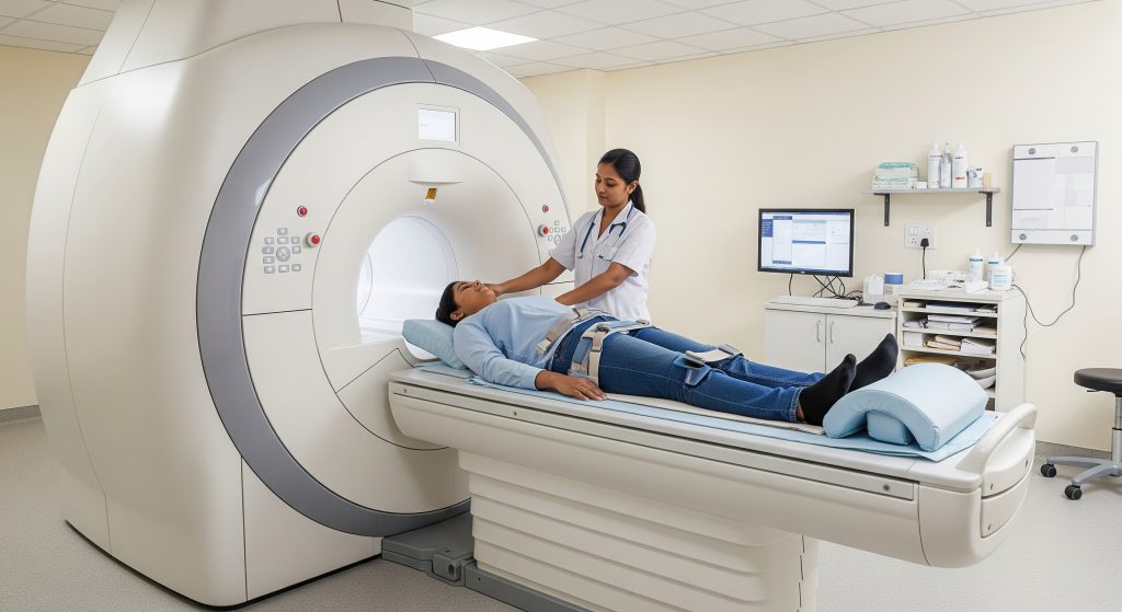
Patient Positioning and Equipment Setup
Positioning determines image quality. For a lower leg or ankle, I position the limb with supportive pads to limit motion. Dedicated coils are placed to optimise signal capture around the area of concern. Subtle comfort adjustments help patients stay still. This avoids repeats. For those with anxiety or pain, I build in extra time for positioning and breathing cues. Good setup protects accuracy.
Scanning Process and Duration
The scanner acquires multiple sequences, each tuned to highlight different tissues. Expect rhythmic tapping noises and short periods where staying still is essential. Most leg MRI acquisitions are completed within a standard session. As Healthline notes, scans often run 30 to 60 minutes, sometimes up to 90 minutes if the area is complex. Movement extends time and reduces clarity. I typically build in rest moments between sequences for comfort.
Contrast Administration When Necessary
Contrast is not always needed. When I am assessing a mass, infection, or postoperative changes, a gadolinium-based contrast agent may be recommended to highlight active tissue. Prior to contrast, renal function and allergy history are reviewed. During administration, the agent is given through an intravenous line and the scan continues. Mild sensations such as a metallic taste can occur. Serious reactions are uncommon, but screening remains essential.
Post-Procedure Guidelines
After a non-contrast leg MRI, normal activities can resume immediately. If contrast was used, hydration helps clearance. If sedation was used, an escort and rest are prudent. The images are reviewed by a radiologist, who prepares a report for the referring clinician. If I see an urgent finding during reporting, I expedite communication. The aim is timely decisions with no gaps in follow-up.
Timing Factors and Scan Duration
Average Scan Time for Different Leg Areas
Scan time depends on which part of the leg is assessed and how many sequences are required. A focused ankle study tends to be shorter than a complex multi-joint review. As Healthline outlines, typical MRI durations cluster around 30 to 60 minutes and can extend to 90 minutes for more detailed work. In practice, a leg MRI that involves both joints and long bones usually sits toward the higher end. Focused follow-ups are shorter.
Factors Affecting Total Procedure Time
Several operational and patient factors shift the clock. Additional sequences, repeat images due to motion, and the need for contrast all add minutes. The scanner strength and protocol design matter as well. As PMC reported, the technologist factor can account for notable variability, with roughly a 29 percent swing observed across exams. Inpatient studies also tend to run longer than outpatient sessions due to logistics. Small details compound and either save time or stretch it.
Report Generation Timeline
Turnaround times vary by facility and urgency. Many centres provide reports the same day for urgent cases, while routine studies are completed within a short window. Complex findings may benefit from sub-specialist review before finalisation. I recommend clarifying expectations during booking. It avoids unnecessary follow-up calls and aligns clinical appointments with likely report availability.
Emergency vs Routine Scheduling
True emergencies take priority. Acute neurovascular compromise, suspected septic arthritis, or pain with red flags usually escalates scheduling. Routine follow-up for chronic pain is batched in standard slots. As Large-Scale Assessment notes, variability in scan time and scheduling efficiency influences how quickly emergency and routine cases move through. The practical point is simple. When symptoms meet urgent criteria, state that clearly during referral.
Leg MRI Cost in India 2024-25
City-wise Price Comparison
Pricing varies by city, facility tier, and protocol complexity. Major metros often command higher fees due to higher operating costs, denser demand, and broader sub-specialist availability. Tier 2 and Tier 3 cities may offer lower fees, though access to 3T systems and rapid reporting can be limited in smaller centres. Government institutions can be more affordable but may have longer queues.
|
City Tier |
Typical Relative Price Level |
|---|---|
|
Metro hubs (Delhi, Mumbai, Bengaluru) |
High |
|
Tier 2 cities (Pune, Jaipur, Lucknow) |
Moderate |
|
Tier 3 and semi-urban |
Lower to moderate |
|
Government hospitals |
Lower, often with longer wait |
For clarity, the fee also depends on whether contrast is used, the need for bilateral imaging, and whether sedation is required. I advise patients to ask for the full package quote up front. It avoids surprises.
Private Hospital vs Diagnostic Centre Rates
Private hospitals usually charge more than independent diagnostic centres because of higher overheads and broader on-site services. When contrast is required or when sub-specialist reporting is requested, the fee increases further. Turnaround time can be faster in premium settings, which is sometimes worth the differential in complex cases. If the clinical question is straightforward, a reputable diagnostic centre can be a cost-effective choice without compromising diagnostic quality.
Insurance Coverage and Reimbursement
Health insurance in India often covers MRIs performed during inpatient admissions. Outpatient scans are covered under some plans, but not all. Policies differ on pre-authorisation requirements, especially when contrast is used. The practical move is to get a written estimate, the referral note, and pre-approval where possible. I also recommend using network centres to simplify claims. The net effect is faster reimbursement and fewer administrative loops.
Ways to Reduce MRI Costs
-
Ask whether a 1.5T scanner is sufficient for your question. It often is, and fees can be lower than 3T.
-
Request a protocol focused on the clinical question to avoid unnecessary sequences.
-
Use network facilities under your insurer for cashless or simplified claims.
-
Enquire about weekday or off-peak slots, which some centres price more competitively.
-
Carry previous imaging so the radiologist can avoid repeating what is already known.
-
Confirm whether contrast is truly needed for your case before booking.
When comparing quotes, match scope to scope. A low headline price that excludes contrast or reporting fees can become expensive later. For search purposes and clarity with administrators, I explicitly reference leg MRI cost in India in documentation and estimate requests. It ensures like-for-like comparisons.
Making an Informed Decision About Leg MRI
The decision to book a leg MRI rests on one question. Will the result change management? If yes, the scan is worth it. If the plan is unchanged regardless of findings, reconsider. A sensible pathway is to align symptoms, exam findings, and prior imaging with a tightly worded MRI question. For example, persistent lateral knee pain after a pivot with equivocal tests might justify a question such as: rule out lateral meniscal tear, assess chondral surface, evaluate ITB friction. That invites a focused protocol.
Two final considerations shape quality. First, choose a centre with musculoskeletal reporting expertise. Sub-specialist eyes reduce the risk of missed secondary injuries. Second, read the report with context. Imaging is a tool, not a verdict. The best outcomes come when the clinician, the radiologist, and the patient align on next steps.
If you need a crisp summary for the referral letter, this is the core: a leg MRI helps confirm ligament or tendon injury, detects occult fractures, maps cartilage damage, and characterises masses and infection, with scan times typically under an hour and report delivery aligned to urgency. That clarity speeds appropriate care.
Frequently Asked Questions
Is leg MRI painful or uncomfortable?
A leg MRI is non-invasive and should not be painful. Some patients feel discomfort from holding still or from the confined space. Ear protection reduces noise. If lying flat is difficult, I request cushions and position changes between sequences. The team can pause if you need a short break.
Can I get a leg MRI with metal implants?
Many implants are MRI-conditional, meaning scanning is possible with specific precautions. Provide the implant card or details in advance. The facility will confirm safety with the device documentation. If the implant is not compatible, alternative imaging or adjusted protocols are considered. Safety is prioritised over speed.
How long does it take to receive leg MRI results?
Urgent findings are flagged quickly. Routine reports are usually delivered within a short time frame set by the imaging centre. Complex cases may require sub-specialist input, which can add review time. When scheduling, I request expected timelines and coordinate the follow-up appointment accordingly.
Do I need to fast before a leg MRI?
Fasting is typically not required for a standard leg MRI. If contrast or sedation is planned, specific instructions may apply. The facility will advise on food, fluids, and medication timing. Follow their checklist to avoid delays on the day.
What conditions can a leg MRI diagnose?
A leg MRI helps diagnose ligament tears, tendon injuries, stress fractures, cartilage defects, meniscal tears, soft tissue masses, and infections. It also detects early bone marrow changes and post-operative complications. The modality is versatile and precise for lower limb assessment.