


How to Identify and Manage Injuries at the Medial Epicondyle of Humerus
Dr. Neetan Sachdeva
Resting everything on imaging or gadgets is popular advice. It misses the point. I start with structure, load, and patterning around the medial epicondyle of humerus, because that is where most diagnostic clarity appears first. Get the pattern right and treatment plans become simpler and safer.
Common Injuries and Identification Signs at the Medial Epicondyle
1. Medial Epicondylitis (Golfer’s Elbow)
I treat medial epicondylitis as a tendon problem, not a pure inflammatory flare. At the medial epicondyle of humerus, degenerative change often develops within the common flexor tendon origin after repetitive wrist flexion and pronation. Patients describe focal tenderness and aching that worsens with gripping or resisted wrist flexion. Grip feels weaker. Sometimes there is ulnar nerve irritability if swelling or mechanics crowd the cubital tunnel.
The practical differential matters. Medial elbow pain can also reflect ulnar neuritis, UCL irritation, or a cervical referral. I rely on load tests and palpation mapping around the medial epicondyle of humerus, then add nerve bias tests if symptoms tingle or radiate. When symptoms sharpen with resisted pronation and stretch into wrist extension, tendinopathy is likely. When symptoms shock with elbow flexion and prolonged typing, nerve irritation enters the picture.
-
Pain site: precise tenderness at the medial epicondyle of humerus.
-
Pain behaviour: worse with gripping, wrist flexion, and pronation under load.
-
Possible accompaniment: subtle ulnar neuritis, especially with prolonged elbow flexion.
In clinic, I sometimes use ultrasound to confirm tendon texture or to visualise the UCL in dynamic valgus. Still, the story and examination drive the decision. A good history usually beats a rushed scan.
2. Medial Epicondyle Fractures
Fractures at the medial epicondyle of humerus appear after valgus stress or a fall. In younger athletes, a powerful pull from the forearm flexor muscles can avulse the apophysis. Swelling, bruising, and marked tenderness occur at the medial epicondyle of humerus with painful movement. Range is reduced and the joint may feel unstable if the UCL or capsule is involved.
In paediatric practice, medial epicondyle injuries are not rare. As StatPearls notes, these fractures comprise about 12% of elbow fractures in children, and they can accompany elbow dislocation. That single figure justifies a low threshold for imaging after a fall with valgus pain. I screen the ulnar nerve as well, because transient neurapraxia is possible after traction or swelling.
-
Nondisplaced injuries may be managed without surgery.
-
Displacement, incarceration, or frank instability pushes the case toward fixation.
-
High-demand throwers often require stricter alignment to protect valgus stability.
3. Ulnar Collateral Ligament Tears
UCL injury presents differently. Patients report medial pain during late cocking or acceleration phases. Valgus stress testing at 30 degrees of flexion reproduces deep medial pain at the medial epicondyle of humerus. The endpoint feels soft or asymmetric. Throwers may describe loss of velocity and command. I check for ulnar nerve symptoms because chronic valgus overload can irritate both structures.
A partial tear can masquerade as stubborn tendinopathy. The clue is valgus testing that hurts more than tendon loading, and pain localised just distal to the medial epicondyle of humerus along the UCL path. In such cases, MRI or ultrasound with dynamic valgus is appropriate to quantify fibre integrity.
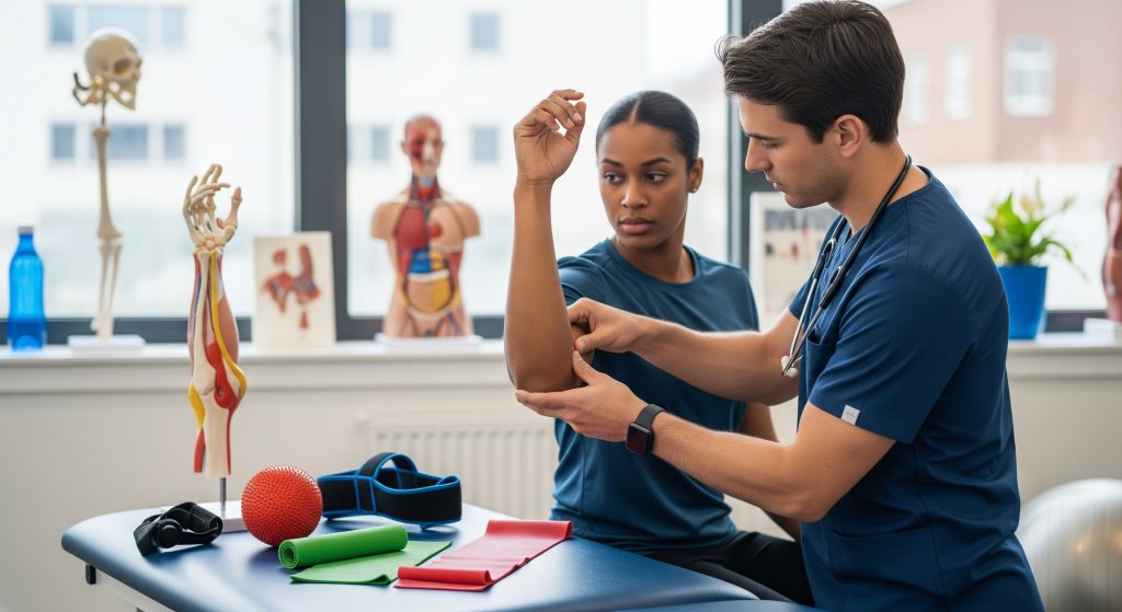
Key Physical Examination Tests
-
Palpation grid: map the medial epicondyle of humerus, the flexor-pronator mass, and the UCL line.
-
Resisted wrist flexion and pronation: reproduces pain in tendinopathy.
-
Passive wrist extension stretch: elicits focal pain at the medial epicondyle of humerus.
-
Valgus stress at 30 to 70 degrees: screens UCL pain or laxity.
-
Moving valgus stress test: sensitive in throwers with UCL pathology.
-
Tinel at cubital tunnel and elbow flexion test: screens ulnar neuritis.
When findings conflict, I prioritise the dominant pain generator first. Treat the clearest lesion, then re-test. Layered injuries are common around the medial epicondyle of humerus.
Diagnostic Imaging Requirements
Radiographs are my starting point after trauma or suspicion of avulsion at the medial epicondyle of humerus. Oblique views help. For soft tissue injuries, ultrasound can identify hypoechogenicity, neovascular changes, and dynamic UCL gapping. MRI provides cross-sectional detail when surgical planning is likely.
|
Study |
Primary utility |
|---|---|
|
X-ray |
Rules out fracture or apophyseal avulsion at the medial epicondyle of humerus. |
|
Ultrasound |
Assesses tendon texture and dynamic UCL integrity in real time. |
|
MRI |
Defines partial vs complete tears and associated cartilage or bone oedema. |
I reserve advanced imaging until clinical questions remain or when management hinges on structural clarity.
Treatment and Management Strategies
Conservative Management Options
Most tendinopathies at the medial epicondyle of humerus respond to structured conservative care. I start with load reduction, isometrics for pain relief, and progressive tendon loading. Bracing can unload the tendon origin during work tasks. NSAIDs help pain, although they do not remodel tendon. A night elbow splint can calm coexisting nerve irritability.
I review activity modification in precise terms. Reduce gripping sets, avoid maximal wrist flexion under load, and control the rate of return. As Mayo Clinic outlines, conservative strategies should lead, with surgery considered only after roughly 6 months of failed nonoperative care. That timeline aligns with tendon remodelling windows in practice.
-
Relative rest and ice in the acute pain window.
-
Counterforce brace for heavy, repetitive tasks.
-
Isometrics early, then eccentric-concentric progression for the forearm flexor muscles.
-
Task and tool adjustments to reduce peak wrist flexion torque.
For UCL sprains without gross instability, I use a hinged brace, staged throwing cessation, and gradual plyometrics before throwing programs. Fractures of the medial epicondyle of humerus that are nondisplaced can be immobilised briefly, then rehabilitated carefully to avoid stiffness.
Physical Therapy Protocols
Rehabilitation succeeds when it is both simple and specific. I structure tendon care around graded exposure. The early aim is pain-calibrated isometrics, then slow eccentrics, then heavy slow resistance. I cue neutral wrist positioning initially to calm pain at the medial epicondyle of humerus, then progress into functional arcs. Joint mobilisation, proprioceptive drills, and grip training round out the plan.
-
Pain control and isometrics: 5 sets of 45 seconds at a tolerable level.
-
Eccentric emphasis: wrist flexion eccentrics 3 times weekly, 3 sets of 8 to 12 reps.
-
Heavy slow resistance: barbell or cable wrist flexion with strict form.
-
Energy storage and release: medicine ball tosses, rebounder drills, and controlled deceleration.
I also address upstream drivers. Shoulder external rotation strength and scapular control protect the elbow during throwing. Poor trunk timing increases valgus at the medial epicondyle of humerus. A whole chain approach prevents recurrence.
Surgical Interventions
Surgery is reserved for specific indications. For tendinopathy, this is rare. Debridement and repair are options after comprehensive rehabilitation fails. For UCL tears, reconstruction or repair with augmentation suits high-demand throwers with clear instability or avulsion patterns. For fractures, open reduction and internal fixation protect stability when displacement or fragment entrapment exists at the medial epicondyle of humerus.
Pros vs Cons of Surgery
-
Pros: Restores stability or tendon integrity, allows higher load tolerance, resolves mechanical catching.
-
Cons: Surgical risks, rehabilitation time, potential stiffness, and variable return-to-sport timelines.
The threshold is functional. If patients cannot meet task demands safely despite optimal conservative care, I discuss operative paths with clear expectations.
Pain Management Techniques
Pain relief should support, not replace, the loading plan. I use activity pacing, topical NSAIDs, and short courses of oral analgesia when needed. For focal tendon pain at the medial epicondyle of humerus, isometric holds are often effective. Injections can modulate pain, though I weigh short-term relief against tendon biology. For nerve irritability, night splinting and neural glides help. The goal is calm tissue and consistent loading.
Recovery Timeline and Rehabilitation
Acute Phase Management
The first 2 to 3 weeks set the tone. I reduce provocative loads, use ice as needed, and begin isometrics within pain limits. Gentle mobility of the elbow joint, wrist, and shoulder keeps stiffness away. If a fracture at the medial epicondyle of humerus is present and immobilised, I protect until clearance, then progress range cautiously.
|
Primary goal |
Reduce pain while maintaining safe movement at and around the medial epicondyle of humerus. |
|
Key actions |
Load control, brace if indicated, isometric strength, and neural comfort. |
|
Stop signals |
Night pain escalation, sharp valgus pain, or spreading paraesthesia into the hand. |
Progressive Strengthening Exercises
Strength returns in stages. Tendon responds to intensity more than variety. I build from isometrics to eccentrics to heavy slow resistance, and finally to elastic energy tasks. The anchor remains controlled discomfort that resolves within 24 hours. I cue forearm flexor muscles to work through midrange first, then terminal range.
-
Wrist flexion bodyweight isometrics against a table edge, 5 by 45 seconds.
-
Slow eccentrics with a dumbbell, 3 by 10, twice weekly to start.
-
Grip dynamometer sets to track meaningful progress.
-
Medicine ball chest pass and overhead throw when pain allows, protecting the medial epicondyle of humerus with planned volume.
This is where most programmes fail. Loads creep up too quickly or rest is too sparse. I hold the weekly increase to modest steps. Consistency beats hero sessions.
Return to Activity Guidelines
Return criteria must be objective. I look for pain free palpation at the medial epicondyle of humerus, symmetric grip strength, and sport specific drills without pain during or after. Throwers complete a staged interval throwing programme with scheduled rest days. Manual workers demonstrate sustained task tolerance at typical shift durations. For fractures, radiographic healing guides the pace, not enthusiasm.
-
Baseline pain control and full elbow extension and flexion.
-
Strength within 10 percent of the opposite side.
-
Completion of work or sport drills at target volume without symptom spikes.
-
Clearance after shared decision making on risk tolerance.
If symptoms relapse, I step back one phase and compress the volume. Not everything is a failure. Sometimes the plan just needs one quieter week.
Long-term Monitoring
Relapse prevention is straightforward. Monitor weekly training loads, maintain grip and wrist strength, and keep shoulder and trunk capacity high. I reassess technique twice per season in throwers. Any return of focal tenderness at the medial epicondyle of humerus is addressed early with isometrics and reduced volume for a brief window.
For workers in repetitive hand tasks, I recommend tool and handle changes, micro breaks, and training on wrist neutral strategies. Small changes repeated daily protect the structure more than any single hack.
Prevention and Risk Factors
Biomechanical Risk Factors
The elbow is a passenger when the shoulder and trunk underperform. Limited shoulder external rotation or thoracic mobility drives valgus load to the medial epicondyle of humerus. Weak scapular stabilisers raise forearm demand. Excessive wrist flexion torque during gripping tasks overloads the tendon origin. Poor deceleration mechanics in throwing increase stress on the UCL and the medial epicondyle of humerus.
-
Inadequate proximal strength and timing creates distal overload.
-
High acute workload spikes predict flare ups more than weekly volume.
-
Tool geometry and handle friction alter wrist angles and tendon load.
Proper Technique Training
Technique is prevention in disguise. I coach neutral wrist alignment during lifts and gripping. For throwers, I cue earlier trunk rotation and better front leg bracing. This reduces late valgus stress at the medial epicondyle of humerus. I also teach a simple warm up sequence: shoulder activation, forearm isometrics, and light plyometrics before maximal efforts.
In office tasks, keyboard height and mouse contour reduce sustained wrist flexion. Small ergonomic gains add up. The aim is fewer minutes at end range across the week.
Protective Equipment Guidelines
Counterforce straps can offload the tendon origin when tasks cannot change. A hinged brace protects the UCL after sprain. Gloves with better grip reduce the wrist flexion moment. For youth sport, I support pitch count policies and two sport seasons, not four. The medial epicondyle of humerus benefits from strategic rest.
|
Counterforce strap |
Reduces tensile load at the medial epicondyle of humerus during repetitive tasks. |
|
Hinged elbow brace |
Protects against valgus stress while healing a UCL sprain. |
|
Ergonomic tools |
Encourage neutral wrist positions and lower grip force demands. |
Conclusion
The shortest path to recovery is precise identification, not a blanket protocol. Define the primary pain generator at the medial epicondyle of humerus, confirm or exclude instability, and load the tissue with intent. Manage the chain, manage the volume, and watch for early signals. The elbow rewards disciplined, incremental work. That is the through line across sport and manual tasks alike.
Frequently Asked Questions
What activities most commonly cause medial epicondyle injuries?
Repetitive wrist flexion and pronation, heavy gripping, and valgus loading during throwing are typical drivers. Manual tasks with poor tool geometry or long unbroken sets add risk. When these stack, the medial epicondyle of humerus takes the strain.
How long does recovery from medial epicondylitis typically take?
Most cases improve within 8 to 12 weeks with structured loading and activity modification. Some require several months for full strength and tolerance. If the medial epicondyle of humerus remains tender beyond that, I reassess technique, volume, and the programme’s progression.
When should surgery be considered for medial epicondyle fractures?
Surgery enters the discussion with displacement, fragment entrapment, or instability. High demand athletes may favour fixation to protect valgus stability. Nondisplaced fractures of the medial epicondyle of humerus often heal well with conservative care.
Can children develop medial epicondyle injuries differently than adults?
Yes. The apophysis is weaker than tendon in younger athletes, so avulsion is more likely than pure tendinopathy. Pain localises at the medial epicondyle of humerus and may follow a fall or a forceful throw. Early imaging is sensible after acute swelling and deformity.
What are the warning signs that require immediate medical attention?
Acute deformity, inability to move the elbow, significant numbness in the ring and little fingers, or severe night pain. Rapid swelling after a fall also warrants urgent review. These signs point to possible fracture, dislocation, or nerve compromise at the medial epicondyle of humerus.
Supporting terms for indexing: elbow anatomy, elbow joint ligaments, forearm flexor muscles.