


How Does Lung Cancer Spread? An Explainer You Shouldn’t Miss
Dr. Kunal Luthra
Conventional advice says metastasis is chaotic and unpredictable. I disagree. The routes are systematic, the timing follows patterns, and with the right framework, lung cancer spread can be explained in clear terms that inform real decisions. This explainer sets out those routes, the typical patterns by stage, what symptoms to expect as disease extends, and what actually influences the pace and path of spread. It is designed for fast comprehension and careful reading, both.
Primary Pathways of Lung Cancer Spread
1. Local Invasion into Nearby Tissues
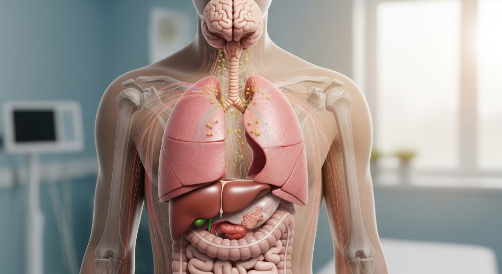
I describe local invasion as the ground game. Cancer cells breach their immediate boundaries and infiltrate neighbouring structures such as the pleura, chest wall, pericardium, or mediastinum. This aspect of lung cancer spread starts with cells losing adhesion, then moving through tissue planes, and finally creating small pockets of tumour in adjacent sites. In practice, this can cause pleuritic pain, hoarseness from recurrent laryngeal nerve involvement, or a pleural effusion that complicates treatment planning.
-
Typical direction: outwards from the primary tumour into the same lobe, then adjacent lobes or chest structures.
-
Common consequences: persistent chest pain, new hoarseness, breathlessness from effusion, or superior vena cava obstruction.
-
Imaging clues: spiculated margins crossing planes, pleural thickening, or chest wall involvement.
Local invasion is often underestimated. It is the reason a seemingly confined tumour can still cause major symptoms and compromise resection margins.
2. Lymphatic System Spread
This is the relay network. Tumour cells enter lymphatic vessels, seed the hilar nodes, then the mediastinal stations, and occasionally the supraclavicular nodes. Lymphatic transit underpins many staging decisions and determines suitability for surgery or the need for combined chemoradiation. In this pattern of lung cancer spread, nodal disease can precede obvious size growth of the primary lesion, which is why nodal mapping is critical.
-
Typical sequence: peribronchial and hilar nodes, then ipsilateral mediastinal nodes, and sometimes contralateral or supraclavicular nodes.
-
Clinical hints: a persistent cough that worsens, chest discomfort not explained by infection, or unexplained systemic fatigue.
-
Decision impact: nodal burden influences both prognosis and the extent of radiation fields.
Here is why the detail matters. Micrometastatic nodal disease changes the treatment intent and often the entire pathway of care.
3. Bloodstream Metastasis
Hematogenous spread is the express route for lung cancer spread. Cells enter pulmonary veins, then systemic circulation, and establish new sites in organs like the brain, liver, and bones. This mechanism is particularly relevant in more aggressive tumours and at advanced stages. Once haematogenous spread is established, local control alone is no longer sufficient.
-
Common targets: brain, liver, adrenal glands, and skeletal system.
-
Typical clues: neurological changes, bone pain, pathologic fractures, or abnormal liver enzymes.
-
Implication: systemic therapy becomes the cornerstone, with local measures reserved for symptom control or oligometastatic scenarios.
Metastatic seeding by blood is not random. It reflects cell biology, cell-surface adhesion profiles, and microenvironments that allow growth.
4. Direct Seeding Through Airways
Less discussed, but real. Tumour fragments can detach into the bronchial tree and implant in downstream mucosa. This airways-based lung cancer spread can present as endobronchial lesions distinct from the primary tumour. It is uncommon, yet it explains multifocal endobronchial disease in the same lung without widespread nodal or distant involvement.
-
Clues on bronchoscopy: multiple mucosal nodules along the airflow path.
-
Clinical effect: worsening wheeze, localised airway obstruction, or recurrent post-obstructive infections.
-
Therapeutic options: endobronchial debulking, stenting, or localised radiation for palliation.
When the airway is the conduit, symptoms often arrive before imaging shows dramatic change. Timing matters.
Common Metastatic Sites for Lung Cancer
Patterns vary by histology and mutation profile, but the usual list is consistent. Brain, bone, liver, and adrenal glands account for most distant sites of lung cancer spread. Skin, pericardium, and gastrointestinal tract metastases occur, but less frequently. The brain tends to be a preferred site in adenocarcinoma. The chest wall and pleura are often involved with locally invasive squamous tumours. Adrenal metastasis is common and sometimes solitary, which raises targeted treatment possibilities.
-
Brain – headaches, seizures, cognitive change.
-
Bone – focal pain, fractures, spinal cord compression risk.
-
Liver – right upper quadrant discomfort, fatigue, abnormal liver tests.
-
Adrenal – often asymptomatic, occasionally adrenal insufficiency if bilateral.
A short example to anchor this: a patient with a 2.5 cm upper lobe adenocarcinoma presents with new headaches and visual aura. Brain MRI confirms solitary parietal metastasis. The distant site came first in the symptom timeline, yet the biology was set months earlier.
Stages of Lung Cancer and Spread Patterns
Stage I: Localised Tumour
Stage I disease is confined to the lung and has not involved lymph nodes. Lung cancer spread at this point is primarily local microscopic infiltration, if present at all. Surgical resection, with or without adjuvant therapy, is standard for suitable candidates. Recurrence risk depends on margin status, grade, and molecular profile.
-
Spread profile: minimal, often within the same lobe.
-
Goal: curative intent with complete resection or stereotactic radiotherapy if surgery is unsuitable.
-
Monitoring focus: margins, vascular invasion on histology, and early imaging follow-up.
At Stage I, halting lung cancer spread relies on local control and addressing occult risks flagged by pathology.
Stage II: Regional Lymph Node Involvement
Stage II includes involvement of nearby lymph nodes or deeper chest wall invasion. Here, lung cancer spread tracks along lymphatic pathways or into adjacent structures. Treatment typically combines surgery with adjuvant chemotherapy or radiotherapy. Nodal disease at this stage increases recurrence risk and requires disciplined follow-up.
-
Spread profile: hilar or intrapulmonary nodes, limited local extension.
-
Therapy goal: eradicate micro-metastatic nodal disease and secure local control.
-
Key decision: adjuvant therapy based on nodal burden and pathological risk factors.
The pivot from Stage I to II is small on imaging but large in prognostic weight. And yet, cure is still achievable.
Stage III: Advanced Local Spread
Stage III spans a spectrum. From extensive nodal disease within the mediastinum to invasion of major structures. Lung cancer spread here involves complex nodal stations and often unresectable local extension. Combined chemoradiation becomes the backbone in many cases, sometimes followed by consolidation immunotherapy depending on response and eligibility.
-
Spread profile: multi-station mediastinal nodes, chest wall or mediastinal organ invasion.
-
Therapy goal: disease control with curative intent in selected patients or strong palliation.
-
Complexity: careful staging with PET-CT and invasive nodal sampling is often required.
Stage III is not a single scenario. It is many. The nuance lies in nodal mapping and resectability assessments.
Stage IV: Distant Metastasis
Stage IV marks the presence of distant disease. Lung cancer spread to the brain, liver, bones, or adrenal glands defines this stage. Systemic therapy is primary, guided by histology and biomarkers. Local treatments are added for control of symptoms or oligometastatic disease.
-
Spread profile: haematogenous dissemination to one or multiple organs.
-
Therapy goal: disease control, symptom relief, and quality of life with survival extension.
-
Decision drivers: molecular targets, PD-L1 status, burden of disease, and performance status.
Once distant sites are involved, the therapeutic question shifts from can it be removed to how best to control and prioritise.
TNM Classification System Explained
I use TNM for consistency and precision. T describes tumour size and local features. N describes the pattern and extent of nodal involvement. M confirms or excludes distant lung cancer spread. Together, they delineate the formal stage and indicate treatment routes.
|
Component |
Definition |
|---|---|
|
T – Tumour |
Size, number of nodules, and invasion of nearby structures. |
|
N – Nodes |
Which lymph node stations are involved and on which side. |
|
M – Metastasis |
Presence of distant disease, single or multiple sites. |
Think of TNM as the map. It shows where the cancer is and hints at where it may go next.
Recognising Lung Cancer Symptoms at Different Spread Stages
Early-Stage Lung Cancer Symptoms
Early disease is often quiet. Many patients report a persistent cough, a subtle change in breathing, or mild chest discomfort. Haemoptysis may appear intermittently. Lung cancer symptoms at this point overlap with more common respiratory issues, which explains delayed presentations. A key sign is a cough that changes character and does not settle with routine therapy.
-
Persistent or changing cough over weeks.
-
Localised chest ache, worse on deep breath.
-
Unexplained fatigue or slight weight loss.
At Stage I or II, symptoms mostly reflect local irritation or early airway compromise rather than distant lung cancer spread.
Symptoms of Regional Spread
When nodes and nearby structures are involved, the symptom profile broadens. Patients may notice hoarseness, dysphagia, or swelling of the face and neck with venous congestion. These lung cancer symptoms suggest pressure effects and nodal bulk rather than just tumour size. Recurrent infections downstream of an obstructed bronchus are also common.
-
Hoarseness from nerve involvement.
-
Recurrent chest infections due to airway blockage.
-
Facial swelling or cyanosis in superior vena cava obstruction.
The pattern is mechanical. The symptoms reflect where the lung cancer spread exerts pressure or blocks airflow.
Signs of Brain Metastasis
Neurological symptoms demand urgent attention. Headaches, seizures, dizziness, and cognitive change raise concern for brain involvement. Differentiating these from stroke-like syndromes can be complex and often requires CT or MRI for accurate diagnosis. Unilateral weakness or new-onset seizures should trigger immediate evaluation, especially when lung cancer spread is already suspected.
-
Red flags: new morning headaches, focal weakness, seizures.
-
Assessment: urgent imaging and neurological review.
-
Treatment context: local radiotherapy or surgery alongside systemic therapy.
Once the brain is involved, symptom control is as vital as tumour control. Both matter.
Bone Metastasis Indicators
Bone metastases typically present with focal, persistent pain that worsens at night or with movement. A pathologic fracture may be the first sign. Localised tenderness over ribs or spine is common, and spinal involvement brings risk of cord compression. Here, the lung cancer spread communicates through pain before imaging confirms the site.
-
Focal bone pain unresponsive to simple analgesics.
-
Fractures with minimal trauma, especially ribs or long bones.
-
Neurological changes with spinal lesions.
Early radiation to painful sites can offer rapid relief. It also prevents further skeletal compromise in many cases.
Liver Spread Symptoms
Liver involvement can be quiet at first. Fatigue, reduced appetite, and a vague fullness under the right ribs are common. Jaundice is uncommon unless biliary obstruction occurs. Blood tests may show rising liver enzymes before symptoms escalate. In this scenario, lung cancer spread declares itself biochemically first and clinically later.
-
Right upper quadrant discomfort or fullness.
-
Unexplained fatigue with abnormal liver tests.
-
Occasional nausea or early satiety.
Monitoring trends, not single values, gives better insight into hepatic involvement over time.
Factors Influencing How Lung Cancer Spreads
Type of Lung Cancer
Histology shapes behaviour. Non-small cell lung cancer (NSCLC) is heterogeneous, and adenocarcinoma often shows early hematogenous lung cancer spread to the brain or bones. Squamous cell carcinoma is more locally aggressive with chest wall involvement. Small cell lung cancer (SCLC) tends to disseminate rapidly, with frequent early nodal and distant spread.
-
Adenocarcinoma: distant seeding to brain and bone is common.
-
Squamous: stronger pattern of local invasion and airway involvement.
-
SCLC: early systemic dissemination and high initial chemo-sensitivity.
Different types, different playbooks. I match strategy to biology and not the other way around.
Genetic Mutations and Biomarkers
Genomics adds precision. EGFR, ALK, ROS1, KRAS, MET, RET, and BRAF are not just labels. They signal preferred routes of lung cancer spread and responsiveness to targeted therapy. PD-L1 expression informs the likelihood of immunotherapy benefit. To an extent, these markers also correlate with metastatic patterns and organ tropism.
-
Driver mutations: often associated with specific metastatic profiles.
-
Actionable targets: define therapy choice and sometimes the tempo of spread.
-
PD-L1: guides immunotherapy and shapes combination strategies.
Precision medicine is not optional anymore. It clarifies prognosis and often slows progression materially.
Lung Cancer Causes That Affect Spread
Causes and exposures influence tumour biology. Tobacco smoking remains the dominant factor among lung cancer causes, and it is linked with squamous histology and locally invasive disease. Radon exposure and occupational carcinogens add risk in specific settings. Air pollution is implicated in adenocarcinoma in urban cohorts. These upstream causes shape the downstream pattern of lung cancer spread by altering mutation signatures and immune microenvironments.
-
Smoking: strong association with local invasion and central tumours.
-
Radon and asbestos: long-latency risks with additive effects.
-
Air quality: linked with adenocarcinoma patterns and diffuse parenchymal change.
Risk factors are not just about causation. They hint at behaviour and potential resistance mechanisms.
Speed of Progression Timeline
The pace varies. SCLC often progresses in weeks to months. Some NSCLC subtypes with indolent profiles may evolve over years. Roughly speaking, growth rates depend on histology, genetics, host immunity, and the microenvironment. Earlier in the disease course, lung cancer spread may be microscopic and clinically silent. Later, it becomes evident through symptoms or imaging.
-
Rapid tempo: SCLC and high-grade tumours.
-
Intermediate: most NSCLC without actionable drivers.
-
Slower: selected low-volume, driver-positive tumours on targeted therapy.
There is a rule of thumb I use. Fast symptoms plus rising biomarkers usually equal faster spread, though not without exceptions.
Understanding Your Lung Cancer Journey
I approach this journey in four steps. First, stage the disease with accuracy. Second, define the biology with histology and biomarkers. Third, map symptoms to the likely routes of lung cancer spread so nothing important is missed. Fourth, match therapy to risk and goals, balancing disease control with quality of life.
-
Stage precisely. That means appropriate imaging, nodal sampling when ambiguous, and a clear TNM classification.
-
Profile the tumour. Actionable mutations and PD-L1 are more than academic details.
-
Correlate symptoms. For example, new headaches in an adenocarcinoma demand brain imaging.
-
Decide and review. Start the plan, monitor objectively, and adjust early rather than late.
Two short examples make this concrete. A Stage II squamous tumour with hoarseness and chest wall pain benefits from aggressive local control and adjuvant therapy. A Stage IV adenocarcinoma with an EGFR mutation and limited brain lesions may do best with targeted therapy plus focused brain treatment. Different routes of lung cancer spread, different strategies, same disciplined logic.
I keep one principle close. Control what is controllable and anticipate what is likely next. That is how the journey stays organised, humane, and effective.
Frequently Asked Questions
How quickly does lung cancer spread to other organs?
Timing varies by histology, genetics, and host factors. SCLC can disseminate within a few months. Many NSCLC tumours take longer, and the pace slows further when an effective targeted therapy is available. The earliest phase of lung cancer spread is microscopic and not visible on scans, which is why careful staging is essential.
Can lung cancer spread without symptoms?
Yes. Asymptomatic spread is common, particularly to the adrenal glands or liver in early phases. Lung cancer symptoms often appear only when an organ is large enough to be disrupted or an airway is obstructed. Surveillance imaging and biomarker trends help reveal silent progression.
What percentage of lung cancers spread before diagnosis?
Estimates vary by cohort and methodology. Many registries report a significant minority presenting with distant disease at first diagnosis, depending on access to screening and primary care. As far as current data suggests, screening programmes reduce late-stage presentation in eligible populations.
Does small cell lung cancer spread faster than non-small cell?
Generally, yes. SCLC is characterised by rapid growth and early dissemination. NSCLC displays broader variability, with some subtypes showing slower lung cancer spread, especially when driver mutations are targetable.
Can lung cancer spread be prevented or slowed?
It can be slowed, and sometimes significantly. Early detection, complete local control when feasible, and timely systemic therapy are key. Targeted therapy and immunotherapy have transformed outcomes in selected cases. Supportive measures, smoking cessation, and comorbidity management contribute to resilience.
Which organs does lung cancer spread to first?
There is no universal order, but common early sites include brain, bone, liver, and adrenal glands. Adenocarcinoma often shows a predilection for brain and bone. Squamous tumours frequently demonstrate strong local invasion before extensive distant spread.
How is lung cancer spread detected and monitored?
Imaging is central, using CT, PET-CT, and MRI for the brain when indicated. Biopsy confirms uncertain lesions. Serial assessments track response. Objective measures and symptom review together provide the clearest signal of change in lung cancer spread over time.