


Explainer: Understanding Comminuted Fractures and Healing Process
Dr. Rajeev K Sharma
Simple fracture advice often promises quick fixes and tidy timelines. That guidance unravels when you face a Comminuted Fracture. You are dealing with multiple fragments, competing priorities, and a higher risk profile. It is basically orthopaedic problem solving under pressure. With the right framework, you can understand the options, set realistic expectations, and make informed decisions that stand up to scrutiny.
Types of Comminuted Fractures and Their Classification
Displaced Comminuted Fractures
In a displaced injury, fragments no longer align with the original bone axis. You see a gap, angulation, or rotation. This pattern usually requires reduction and stabilisation to restore length and alignment. The logic is straightforward. Mechanics drive biology. If fragments move, blood vessels shear and callus struggles.
-
Typical causes include high-energy trauma such as road collisions or falls from height.
-
Neurovascular assessment is essential, especially near joints or in the forearm and ankle.
-
Imaging often includes CT to define fragment geometry and plan fixation.
This is a Comminuted Fracture that seldom tolerates simple casting. Internal or external control is usually necessary to protect soft tissue and early healing.
Nondisplaced Comminuted Fractures
Here, the bone is broken into pieces, yet alignment remains acceptable. You still have multiple fragments, but the cortex holds shape. Management may be conservative if stability is sufficient and skin integrity is intact. However, vigilance is mandatory. Secondary displacement can appear after swelling subsides.
For a nondisplaced Comminuted Fracture, immobilisation and close follow up can work. You must confirm stability with early repeat imaging. Any drift in alignment should trigger escalation to operative care.
Open Versus Closed Comminuted Fractures
Open injuries breach the skin and expose bone. Closed injuries do not. The distinction is critical for infection risk and timing of surgery. With open Comminuted Fracture patterns, urgent antibiotics and irrigation come first. Soft tissue status dictates the fixation sequence and the need for staged procedures.
|
Term |
Definition |
|---|---|
|
Open fracture |
Skin and soft tissue breach with direct communication to bone. |
|
Closed fracture |
Skin intact with no external communication. |
|
Gustilo grade |
Classification of open injuries by wound size, contamination, and soft tissue damage. |
An open Comminuted Fracture often needs staged fixation and possible soft tissue coverage. A closed injury may proceed to definitive fixation sooner, provided swelling allows safe incision placement.
Highly Comminuted Fractures
Highly comminuted describes a bone shattered into many small pieces. Reduction becomes less about perfect anatomic reconstruction and more about functional alignment. In practice, surgeons respect biology. They bridge segments, protect the periosteum, and accept small gaps that will fill with callus.
This is the archetypal complex fracture. Stabilise the main fragments and let controlled micromotion stimulate healing. Overhandling fragments risks devascularisation and delayed union.
Common Locations for Comminuted Fractures
High-energy mechanisms cluster in predictable sites:
-
Distal radius after a fall onto an outstretched hand, especially in osteoporotic bone.
-
Tibial shaft and plateau after road traffic collisions.
-
Calcaneus and talus after axial loading falls.
-
Proximal humerus in older adults with low-energy twists or falls.
-
Acetabulum and distal femur in polytrauma scenarios.
Each site influences the Comminuted Fracture strategy. Joint involvement pushes towards stable fixation to allow early motion. Diaphyseal injuries may tolerate relative stability and callus formation.
Treatment Options for Comminuted Fractures
Surgical Interventions for Complex Fractures
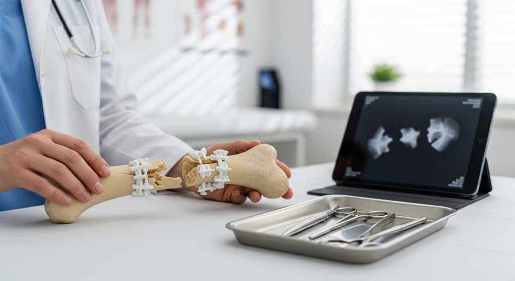
Operative treatment aims to restore alignment, length, and rotation, while preserving blood supply. For a complex fracture, sequencing matters. You stabilise, decontaminate if open, then reconstruct. ORIF is common. The acronym means open reduction and internal fixation. It implies exposure, alignment, and placement of implants to hold fragments.
Staged approaches are common with swelling, contamination, or soft tissue compromise. A temporary external fixator may precede definitive internal fixation once tissues recover. This stepwise plan reduces complications and respects the biology of a Comminuted Fracture.
Internal Fixation Methods
Internal fixation delivers stability from within the limb. Options include plates, screws, intramedullary nails, and specialised devices:
-
Locking plates create fixed-angle constructs that support weak metaphyseal bone.
-
Intramedullary nails provide load sharing in long bones and permit early weight bearing in selected cases.
-
Lag screws compress articular fragments where precise joint reduction is essential.
-
Cerclage and K-wires can supplement plate or nail constructs for small fragments.
Choice depends on fracture morphology, bone quality, and soft tissue condition. A Comminuted Fracture across a joint surface often receives a combination: articular reconstruction plus metaphyseal bridging.
External Fixation Techniques
External fixation stabilises bone using pins connected to an external frame. It protects soft tissue and allows access for wound care. Techniques include uniplanar bar frames and circular frames. Circular systems can gradually realign fragments and correct deformity.
Use external fixation when swelling is severe, wounds are contaminated, or bone loss precludes immediate internal fixation. In some cases, it serves as definitive management for a Comminuted Fracture, especially with segmental defects or infection risk.
Bone Grafting Procedures
Bone grafting addresses defects and encourages healing. Autograft from the iliac crest provides osteogenic cells and growth factors. Allograft fills volume but offers less biological activity. Synthetic substitutes and bone morphogenetic proteins may supplement limited autograft supply.
Indications include metaphyseal voids, nonunion, and highly comminuted metaphyseal fractures after reduction. Graft choice balances biology, volume, and donor site morbidity. In a Comminuted Fracture with articular depression, structural grafts can restore joint support.
Non-Surgical Management Approaches
Non-operative care suits stable, closed, and nondisplaced patterns. It relies on immobilisation, pain control, and scheduled imaging. Functional bracing can permit early joint movement while maintaining alignment. The approach must be disciplined.
-
Confirm stability with serial radiographs during the first two to three weeks.
-
Escalate to surgery if displacement appears, pain remains uncontrolled, or function deteriorates.
-
Educate the patient about warning signs: numbness, increasing deformity, or skin compromise.
Non-operative paths can succeed for a nondisplaced Comminuted Fracture. They require close oversight and a willingness to change course promptly.
Healing Process and Recovery Timeline
Three Stages of Bone Healing
Bone heals through three overlapping stages:
-
Inflammation: Haematoma forms, cytokines recruit cells, and pain peaks over several days.
-
Repair: Soft callus develops, then mineralises into hard callus over weeks to months.
-
Remodelling: Bone reshapes along stress lines, often for many months after union.
Mechanical environment influences each stage. Relative stability encourages callus. Absolute stability promotes direct bone healing with minimal callus. For a Comminuted Fracture, relative stability is often deliberate to allow bridging callus.
Expected Recovery Timeframes
Timeframes vary by site, age, and stability. Roughly speaking, diaphyseal long bones unite in 12 to 20 weeks. Metaphyseal areas may heal faster. Complex periarticular injuries often take longer to recover function than to achieve union.
|
Region |
Typical union window |
Functional recovery |
|---|---|---|
|
Tibial shaft |
16 to 24 weeks |
6 to 12 months for near-full function |
|
Distal radius |
8 to 12 weeks |
3 to 6 months for grip and rotation |
|
Proximal humerus |
10 to 16 weeks |
6 to 12 months for overhead reach |
|
Calcaneus |
12 to 20 weeks |
9 to 18 months for impact activities |
For planning discussions, you can reference Comminuted Fracture recovery time as a range rather than a promise. Healing is biological and variable. Set staged milestones instead of fixed dates.
Factors Affecting Healing Speed
Outcomes hinge on modifiable and fixed variables. The following factors consistently shift timelines:
-
Age and bone quality, including osteoporosis or osteopenia.
-
Smoking status and nicotine exposure, which impair blood flow and osteoblast function.
-
Diabetes control and vascular disease, affecting perfusion and immunity.
-
Soft tissue injury severity and contamination in open wounds.
-
Quality of reduction and stability in the Comminuted Fracture construct.
Some factors you cannot change. Many you can. Optimise what you control and monitor what you cannot.
Physical Therapy and Rehabilitation Phases
Rehabilitation moves in phases aligned to biology and fixation strength. The aim is to regain motion, strength, and proprioception without jeopardising union. Protocols should individualise progression while following broad principles.
-
Protection and motion: Reduce swelling, maintain adjacent joint mobility, and start isometrics as tolerated.
-
Controlled loading: Introduce assisted range, begin partial weight bearing if permitted, and progress to closed chain drills.
-
Strength and endurance: Advance resistance, add balance tasks, and restore gait or functional patterns.
-
Return to activity: Sport or work-specific drills, impact testing, and readiness assessment.
Your therapist will integrate surgeon restrictions with functional goals. A stable Comminuted Fracture with robust fixation can accept earlier motion. A tenuous construct needs more caution.
Monitoring Progress Through Follow-up Care
Follow up tests your assumptions against biology. The essentials are simple and systematic:
-
Scheduled radiographs to assess callus and alignment.
-
Wound surveillance for infection and soft tissue healing.
-
Neurovascular checks and compartment assessments when indicated.
-
Load progression decisions anchored to pain and imaging, not the calendar.
Document each step. Adjust plans when the Comminuted Fracture shows lagging callus or delayed motion. Precision matters, but so does patience.
Potential Complications and Risk Factors
Immediate Post-Surgery Complications
Early complications concentrate around the wound and the implant. The main concerns are bleeding, infection, and compartment syndrome. Anaesthetic risks apply as usual. Implant risks include hardware malposition or loss of fixation in weak bone.
For an open Comminuted Fracture, the infection risk is higher. A staged irrigation-debridement pathway and timely antibiotics reduce that risk to an extent. Early escalation for rising pain or tense swelling is essential.
Long-Term Complications
Delayed union and nonunion sit at the top of the list. Malunion can limit function if alignment is poor. Post-traumatic arthritis follows joint surface incongruity. Hardware irritation is common, especially over the tibia or radius, and may require removal.
There is a contrarian point often raised. Some argue that aggressive early motion always prevents stiffness. It helps, but not always. Excessive stress on a fragile Comminuted Fracture can push a stable construct into failure. Balance is the discipline.
Risk Factors for Poor Outcomes
-
High-energy trauma with severe soft tissue damage.
-
Uncontrolled diabetes, peripheral vascular disease, or immunosuppression.
-
Smoking or nicotine replacement use during healing.
-
Osteoporosis and poor baseline nutrition, including low vitamin D.
-
Suboptimal reduction or inadequate fixation strategy.
Mitigate what you can before and after surgery. The cumulative effect is real and measurable.
Special Considerations for Children
Paediatric bone heals faster and remodels more aggressively. Growth plates introduce unique patterns and risks. Avoid crossing physes with hardware whenever possible. In many cases, elastic stable intramedullary nails provide adequate control with low irritation.
A paediatric Comminuted Fracture still demands precise alignment in joints. Malreduction across a joint line does not remodel meaningfully. Protect the physis and the articular surface. Function tomorrow depends on those decisions today.
Prevention Strategies for Complications
Complication prevention starts with planning and continues through rehabilitation. A concise checklist helps teams stay aligned:
-
Align fixation choice with fracture biology and soft tissue status.
-
Load manage by phase. Advance only when healing markers support the step.
-
Address modifiable risks early: nicotine, glucose control, vitamin D, and protein intake.
-
Use clear return-to-work criteria and staged activity ladders.
-
Educate on red flags: fever, wound changes, increasing deformity, or persistent night pain.
Maybe that is the point. Prevention is a workflow, not a slogan.
Conclusion
A Comminuted Fracture is not just another entry on the list of bone fracture types. It is a structural problem with biological constraints and lived consequences. Classify the pattern accurately. Match fixation to biology. Respect soft tissue. Then manage loading with discipline. Do this, and outcomes improve, even when the injury is messy and the timetable is long.
If you remember one thing, remember this. Mechanics drive biology, and biology drives recovery. Plan for both and progress with intent.
Frequently Asked Questions
How long does it typically take to recover from a comminuted fracture?
Union often occurs between 12 and 24 weeks, depending on site and stability. Functional recovery usually extends beyond union. Expect staged gains over 6 to 12 months for major weight-bearing bones. Frame Comminuted Fracture recovery time as a range, and revisit it as healing data arrives.
What is the difference between a comminuted and segmental fracture?
Both involve multiple fragments. A comminuted pattern creates several pieces at one site. A segmental fracture features at least two distinct breaks that create an intermediate segment. Treatment principles overlap, but segmental injuries may favour bridging constructs and careful load sharing.
Are comminuted fractures more common in certain age groups?
Yes, patterns cluster. High-energy comminution is common in younger adults after trauma. Low-energy comminution appears in older adults with osteoporosis, especially at the distal radius and proximal humerus. Bone quality and mechanism shape the pattern more than age alone.
Can a comminuted fracture heal without surgery?
It can, if alignment is acceptable and stability is sufficient. Nondisplaced patterns with intact soft tissues may be managed in a cast or functional brace. Close monitoring is essential. If alignment drifts, operative stabilisation becomes the safer path for the Comminuted Fracture.
What lifestyle changes support healing after a comminuted fracture?
-
Cease nicotine entirely during healing. This includes patches and vaping.
-
Prioritise protein, vitamin D, and calcium to support bone turnover.
-
Manage glucose if diabetic and optimise sleep for recovery.
-
Follow weight-bearing and exercise guidance precisely. Do not jump stages.
Small, consistent habits compound. They shorten the path from fixation to function.
Comminuted Fracture content includes complex fracture insights, bone fracture types context, comminuted fracture treatment options, and comminuted fracture recovery time guidance.