


Echocardiogram Procedure Explained: What to Expect and Why It Matters
Dr. Hriday Kumar Chopra
Disclaimer: The content shared here is for informational purposes only. Always consult a specialist doctor before attempting any treatment, procedure, or taking any medication independently. Treatment costs and pricing may vary depending on the patient’s condition, medical requirements, hospital, and other factors.
Cardiac imaging advice often starts with a sweeping claim that one test fits every situation. It does not. Choosing the right echocardiogram procedure depends on the clinical question, your risk factors, and the detail required. In this guide, I set out the types available, how the appointment unfolds, practical preparation, what the findings mean, and how to manage echocardiogram cost without compromising care.
Types of Echocardiograms Available
Different questions demand different tools. I match the echocardiogram procedure to the decision at hand, whether that is screening valve disease, clarifying a murmur, or stress testing symptoms that appear only on exertion.
Transthoracic Echocardiogram (TTE)
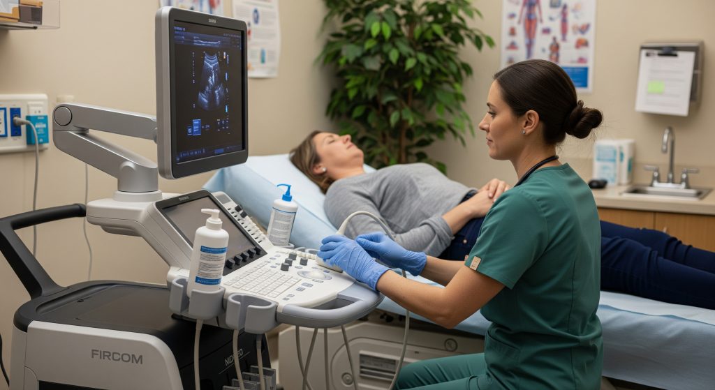
A standard heart ultrasound performed over the chest wall. It is non-invasive, widely available, and usually the first choice. I use it to assess chamber sizes, pump function, valve motion, and pericardial fluid. In shorthand, you will see terms like LV (left ventricle), RV (right ventricle), and EF (ejection fraction). EF is the percentage of blood the LV pumps out with each beat.
-
Strengths: fast, comfortable, and rich baseline data.
-
Limitations: image quality can drop with obesity, lung disease, or rib spacing.
When clarity is adequate, a TTE answers most routine questions on structure and function. This echocardiogram procedure is my default starting point.
Transesophageal Echocardiogram (TEE)
TEE places a small ultrasound probe in the oesophagus to obtain high-resolution images. It is particularly useful when transthoracic views are inadequate, for suspected valve infection, or when searching for clots in atrial fibrillation. Sedation is given, and fasting is required. The result is a sharper look at valves, the atria, and the upper aorta. In urgent settings, this echocardiogram procedure can be decisive.
Stress Echocardiogram
This option evaluates the heart under strain, either with exercise or medication. I use it to investigate chest pain, exercise breathlessness, and equivocal ECG changes. Images are captured at rest and at peak stress to reveal new wall motion abnormalities, which are a hallmark of reduced blood flow. When patients cannot exercise, dobutamine is used to mimic exertion. A stress echocardiogram often clarifies whether symptoms reflect coronary disease or something else.
Doppler Echocardiogram
Doppler modes measure blood flow speed and direction through valves and outflow tracts. They help me quantify stenosis, regurgitation, and pulmonary pressures. In reports, you might see Vmax, mean gradient, and E/e’ (a diastolic index). These values translate movement into physiology. In practice, Doppler turns pictures into numbers, which strengthens treatment planning.
3D Echocardiogram
Three-dimensional imaging adds anatomical precision. It captures valves and chambers without geometric assumptions, which improves volume measurements and surgical planning. I turn to 3D when evaluating complex mitral disease, guiding structural procedures, or refining EF in borderline cases. It is an advanced lens, not a replacement for core two-dimensional views, but it complements them well.
Foetal Echocardiogram
When congenital heart disease is suspected during pregnancy, foetal echocardiography provides a detailed prenatal assessment. It supports delivery planning and early postnatal care. Timing and expertise matter here, because subtle lesions can be missed without systematic views. For expecting parents, the benefit is early clarity and a prepared clinical team.
What Happens During Your Echocardiogram Procedure?
Clarity reduces anxiety. Here is the step-by-step outline I give patients before a standard echocardiogram procedure, along with notes on what you will feel and what happens immediately afterwards.
1. Initial Preparation Steps
For a routine TTE, preparation is minimal. I ask patients to arrive in comfortable clothes and allow time for consent and set up. For a TEE, I advise fasting for about six hours and planning a safe journey home due to sedation. For a stress echocardiogram, I recommend no caffeine beforehand and suitable attire for exercise. These small steps improve image quality and test accuracy.
-
TTE: no special restrictions for most patients.
-
TEE: fast, review medications, and confirm post-sedation escort.
-
Stress echo: avoid caffeine and heavy meals; confirm exercise capability.
If you have diabetes or take heart rate medications, I discuss adjustments in advance. It is basically preparation for clear images and reliable physiologic response.
2. Positioning and Electrode Placement
You will usually lie on your left side. I or the sonographer place ECG electrodes to synchronise images with each heartbeat. Proper lead placement improves signal quality and reduces artefacts. Poor placement can create misleading tracings. I check signal stability before imaging begins.
3. Gel Application Process
Ultrasound gel is applied to improve contact between the probe and skin. It removes tiny air gaps that block sound waves. The gel may be warmed and wipes off cleanly after the echocardiogram procedure. Simple, but essential for crisp images.
4. Transducer Movement Patterns
The sonographer will slide, rock, fan, and rotate the probe to find the best windows. Each movement targets a specific structure or angle. I often coach breathing or a brief position change to open a better view. Small adjustments. Large gains in clarity.
5. Image Capture Techniques
Standard views include parasternal long axis, short axis, and apical windows. I capture 2D images, add Doppler where needed, and store cine loops for later measurements. If indicated, I incorporate 3D acquisitions for valves or volumes. For a stress echocardiogram, I record images quickly at peak heart rate to catch transient changes. The overall goal is consistent views that can be compared over time.
When the technique is consistent, trends in EF, gradients, and chamber size become meaningful rather than noisy.
6. Duration and Completion
A typical TTE takes about 30 to 60 minutes from set up to clean up. As WebMD notes, timing varies by type, with transthoracic studies usually being the quickest.
Stress protocols add time for exercise or medication, then rapid imaging. TEE appointments run longer because of consent, sedation, and recovery checks. I allow buffer time for questions. Rushing helps no one.
What You’ll Feel During the Heart Ultrasound
For a TTE, expect mild pressure from the probe and brief breath holds. There is no radiation. Most patients find the echocardiogram procedure comfortable and straightforward. For a stress echocardiogram, the test feels like a supervised workout followed by swift imaging. For TEE, sedation reduces throat discomfort, and the team monitors you closely throughout.
Immediate Post-Procedure Steps
After a TTE or stress study, you can usually return to normal activities. TEE requires observation until the sedation wears off. I review next steps and confirm when results will be available. Images are interpreted by a cardiologist, and a structured report follows.
Preparing for Your Heart Ultrasound
Preparation is light for most patients, but small details improve both images and safety. I provide written instructions for the specific echocardiogram procedure so nothing is missed on the day.
General Preparation Guidelines
For TTE, normal diet and medications are generally fine. A gown may be provided for access to the chest. For a stress echocardiogram, avoid caffeine and follow fasting guidance if advised. TEE requires fasting and a medicine review, especially if you take blood thinners or have oesophageal issues. If anything in these instructions conflicts with your routine, I adjust the plan with your clinician.
Clothing Recommendations
Wear loose, comfortable clothing. For TTE and stress testing, a top that can be removed easily is convenient. Supportive trainers help for treadmill protocols. Simpler clothing choices speed up the start of the echocardiogram procedure and improve comfort.
Medication Considerations
Continue most routine medications for a standard TTE. For stress testing, beta blockers or some calcium channel blockers may blunt heart rate response, so I often pause them under medical guidance. For TEE, sedation and aspiration risk drive stricter rules. Please share a full medication list in advance.
-
TTE: usually no changes.
-
Stress echo: rate-slowing drugs may be held temporarily.
-
TEE: confirm anticoagulants and reflux medicines plan.
Eating and Drinking Instructions
TTE: eat and drink normally unless told otherwise. Stress echocardiogram: avoid heavy meals and caffeine for a few hours beforehand. TEE: fast as instructed, typically from early morning for an afternoon slot. Hydration is fine up to the cut-off if allowed. These steps protect safety and image quality.
Special Preparation for Stress Echocardiogram
I confirm your physical ability to exercise and any mobility limits. Avoid caffeine that morning, including chocolate and energy drinks. Bring inhalers if you have asthma. The aim is a smooth climb to target heart rate and a clean set of images at peak effort. If exercise is unsuitable, we use a pharmacological protocol.
TEE-Specific Preparation Requirements
For TEE, fasting reduces aspiration risk during sedation. Report swallowing difficulties, reflux symptoms, or previous oesophageal procedures. Remove dental prostheses if asked. Arrange a safe journey home, because driving after sedation is unsafe. This echocardiogram procedure is invasive compared to TTE, so preparation is stricter by design.
Items to Bring
-
Photo ID and insurance card.
-
Medication list, including dosages.
-
Comfortable clothing and previous echocardiogram reports.
-
For TEE: escort details and any relevant swallowing studies.
Questions to Ask Your Doctor
-
What clinical decision will this echocardiogram procedure help us make?
-
Do I need TTE, TEE, or a stress echocardiogram, and why?
-
Should I hold any medications before the test?
-
When and how will I receive the results?
-
What are the likely next steps if abnormalities are found?
-
What will be the estimated echocardiogram cost for my setting?
Understanding Results and Echocardiogram Cost
Results should link to action. I interpret the echocardiogram procedure in the context of your symptoms, examination, ECG, and blood tests, not in isolation.
What Your Results Reveal
A report summarises chamber sizes, EF, valve structure, valve gradients, wall motion, and pericardial findings. Doppler values quantify pressure and flow. If the stress study shows new wall motion changes, I consider ischaemia. If TEE shows a left atrial appendage thrombus, anticoagulation takes priority. The key is correlation with the reason the test was ordered.
Normal vs Abnormal Findings
Normal findings include normal chamber sizes, normal EF, thin and mobile valves, and no regional wall motion abnormality. Abnormalities range from mild valve regurgitation to severe stenosis, reduced EF, or diastolic dysfunction. Some changes are incidental and benign. Others point to disease that needs prompt action. I flag which is which, so follow up is proportionate and timely.
Timeline for Receiving Results
For routine TTE, preliminary impressions are often available the same day, with a final report within a few days. TEE and stress studies may require additional review, especially if findings are complex. If urgent abnormalities appear, I escalate immediately. No one should wait on critical information.
Average Cost Ranges in the US
Echocardiogram cost varies by type and setting. As BetterCare outlines, uninsured prices often range from $500 to over $3,000, while insured patients might still pay $150 to $2,000, depending on the plan and facility.
|
Type |
Typical US price range (self-pay) |
|---|---|
|
Transthoracic echocardiogram (TTE) |
$500 – $3,000+ |
|
Transesophageal echocardiogram (TEE) |
$700 – $4,000 |
|
Stress echocardiogram |
$600 – $4,000 |
These are broad estimates. Actual charges can sit below or above these bands, particularly in large metropolitan hospitals.
Insurance Coverage Factors
Coverage hinges on the clinical indication, coding, and the network status of the facility. Prior authorisation may be needed for stress imaging or repeat studies. Policies differ by insurer and region, which affects the final bill. In practice, a documented indication and an in-network facility reduce variability. The echocardiogram procedure is commonly covered when medically necessary, though deductibles and coinsurance still apply.
Out-of-Pocket Expenses
Facility fees, professional interpretation, and stress lab charges combine into the final bill. As far as current data suggests, hospital-based pricing tends to be higher than independent centres. In published analyses, list prices can significantly exceed government benchmarks, with variation across hospital types. As the National Library of Medicine reports, some self-pay and commercial rates markedly exceed Medicare reference prices, which explains the spread seen by patients.
-
Expect separate charges for the scan and the cardiologist’s report.
-
Ask if stress supervision or contrast adds a fee.
-
Request a bundled quote where possible.
Cost Variations by Type
TEE usually costs more because it involves sedation, recovery monitoring, and physician time at the bedside. A stress echocardiogram can cost more than a resting TTE due to exercise staff, equipment, and longer room use. 3D and contrast imaging, when needed, add to the total. The echocardiogram procedure itself is only part of the invoice. The site of care often dominates the final number.
Ways to Reduce Expenses
-
Ask for a cash price quote from two or three facilities.
-
Confirm in-network status and the CPT code your clinician will use.
-
Request a global fee that includes the scan and interpretation.
-
Discuss alternatives if the clinical yield is likely low.
For self-pay patients, community imaging centres sometimes provide lower rates than hospital systems. Roughly speaking, price dispersion is wide, so a single phone call can save a meaningful amount. If a prior study exists, I may recommend comparison rather than repeating the echocardiogram procedure immediately, provided the clinical picture allows.
Making Sense of Your Echocardiogram Journey
A good echocardiogram procedure is not just about images. It is about the decision it enables. If the goal is diagnosing valve stenosis, I ensure Doppler gradients are captured clearly. If the question is exercise chest pain, I design a stress echocardiogram protocol that reaches target heart rate safely. Technique should follow the question, not the other way round.
Here is a simple way to frame the journey:
-
Define the decision. Rule in or out disease, stratify risk, or guide therapy.
-
Match the modality. TTE first, TEE when detail is critical, or stress when symptoms are exertional.
-
Prepare with purpose. Clothing, fasting, and medication adjustments tailored to the test.
-
Execute consistently. Standard views, reliable Doppler, and timely capture under stress.
-
Act on results. Escalate, watch, or discharge with clarity.
Critics say echocardiography is overused. They have a point in some settings. But when ordered for a clear question and executed well, it prevents unnecessary procedures and accelerates appropriate care. That is the value of a well-run echocardiogram procedure. Precision over volume.
Frequently Asked Questions
Is an echocardiogram procedure painful?
For a TTE or stress echo, discomfort is usually limited to brief probe pressure. TEE involves sedation and throat numbing, which reduces discomfort during the echocardiogram procedure. Pain is uncommon.
How long does a transthoracic echocardiogram take?
Most TTE appointments last about 30 to 60 minutes. As WebMD notes, some studies finish faster, and complex assessments can take longer.
Can I eat before my heart ultrasound?
Yes for a routine TTE, unless advised otherwise. Avoid heavy meals and caffeine before a stress echocardiogram. Fast before a TEE as instructed for the echocardiogram procedure.
What’s the difference between an ECG and echocardiogram?
ECG records the heart’s electrical activity. An echocardiogram uses ultrasound to show structures, motion, and blood flow. I often use both, as they answer different questions.
How accurate are echocardiogram results?
Accuracy is high for structure, function, and valve assessment when imaging windows are good and technique is standardised. Image quality, rhythm, and body habitus can affect detail. Repeatability improves with consistent protocols across studies.
Will I need sedation for my echocardiogram procedure?
Sedation is not used for TTE or an exercise stress echocardiogram. It is used for TEE to keep you comfortable while the probe is in the oesophagus.
How often should echocardiograms be repeated?
Frequency depends on diagnosis and change in symptoms. Stable mild valve disease may be reviewed every 1 to 2 years, while new or worsening symptoms can justify earlier imaging. I align repeat timing with the clinical plan, not a fixed calendar.