


Decoding Osteoarthritis Diagnosis: Key Signs & Underlying Causes
Dr. Neetan Sachdeva
Routine X-rays and a quick symptom checklist are often presented as sufficient for osteoarthritis diagnosis. That approach misses nuance and, at times, early disease. I take a broader, clinically grounded view that integrates history, examination, imaging, and occasionally fluid studies. The aim is simple. Reach a precise osteoarthritis diagnosis faster, explain the why, and rule out what it is not.
Clinical Diagnosis Methods for Osteoarthritis
Physical Examination and Medical History
I start with a targeted history and a meticulous examination. The basics matter. I document onset, duration, and fluctuation of pain, any previous injury, and functional limits in daily tasks. Activity-linked discomfort is common, and it often guides osteoarthritis diagnosis in the right direction.
-
Inspect for swelling, redness, warmth, and bony enlargement.
-
Test active and passive range of motion (ROM) and end-feel.
-
Map pain to specific movements, loads, or durations of inactivity.
-
Note crepitus, alignment drift, and gait changes during stance and swing.
Medical history supplies the context for a confident osteoarthritis diagnosis. Prior ligament tears, meniscal surgery, or repetitive occupational loading all change the baseline. Family history helps, though I treat it as a modifier rather than a verdict. One short clinical example helps here. A midlife distance runner with prior ACL repair presents with unilateral knee pain and subtle varus alignment. The index of suspicion for osteoarthritis diagnosis rises quickly, even before viewing images.
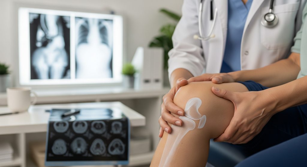
X-ray Imaging for Joint Space Assessment
Standard radiographs remain the workhorse. Weight-bearing views reveal joint space narrowing, marginal osteophytes, and subchondral sclerosis. These changes are structural, not merely symptomatic. They support an osteoarthritis diagnosis when history and examination point the same way.
-
Preferred views: weight-bearing AP, lateral, and sunrise/skyline for patellofemoral assessment.
-
Interpretation priorities: compartmental narrowing pattern, osteophyte size, and alignment.
-
Decision tie-breakers: compare with the contralateral side and correlate with ROM pain.
It is basically a correlation exercise. Symptoms can be ahead of films, or films can look severe with modest pain. I keep both truths in view.
MRI Scanning for Soft Tissue Evaluation
MRI is not first line for every patient, but it offers unmatched soft tissue detail. I reserve it for atypical pain, suspected meniscal or ligamentous injury, or when the clinical picture and X-rays diverge. As Mayo Clinic explains, MRI can visualise cartilage quality and joint effusion, which strengthens early osteoarthritis diagnosis when X-rays are equivocal.
-
Detects cartilage thinning and focal defects before large osteophytes appear.
-
Clarifies synovitis, bone marrow lesions, and meniscal pathology.
-
Helps plan targeted rehab or injections by showing tissue-specific change.
Advanced protocols help further. T2 mapping assesses cartilage matrix hydration and collagen integrity, offering a biochemical window that precedes gross structural loss. This is useful, to an extent, for monitoring early disease trajectories.
Arthroscopy and Joint Fluid Analysis
Arthroscopy is rarely required purely for osteoarthritis diagnosis, yet it can settle difficult cases. A direct view of cartilage surfaces and menisci answers questions that images sometimes leave open. Joint fluid aspiration is the other thread. Inflammatory load and crystal analysis help rule out mimics like gout, CPPD, or infection.
In suspected infection, I escalate quickly. As Mayo Clinic notes, a high white cell count with neutrophil predominance in synovial fluid points to septic arthritis, which demands urgent action. That single datapoint can redirect care in minutes.
Early clarity beats late certainty. A timely aspiration can protect cartilage that cannot grow back.
Key Signs and Symptoms of Osteoarthritis
1. Joint Pain After Activity or Inactivity
Pain that worsens with use and eases with rest is the classic pattern. Prolonged sitting can also provoke a start-up ache, which settles after a few steps. This mechanical signature is foundational for osteoarthritis diagnosis and ties symptoms to load, not random flares.
-
Stairs, inclines, and deep knee flexion often amplify pain.
-
Short rests help, though over-resting can stiffen the joint.
-
Activity pacing and load management usually reduce day-to-day spikes.
I keep an eye on pain drift from episodic to persistent. That shift, combined with function loss, signals progression in osteoarthritis symptoms.
2. Morning Stiffness and Limited Movement
Short-lived stiffness after sleep or inactivity is common. As Mayo Clinic describes, morning stiffness in osteoarthritis typically settles within 30 minutes, which contrasts with inflammatory conditions. I confirm duration carefully because it shapes the differential.
-
Gentle movement often restores ROM and reduces discomfort.
-
Thermal strategies and a brief mobility routine help the first hour of the day.
Time matters here. Longer stiffness suggests a different pathology or a mixed picture.
3. Joint Swelling and Tenderness
Swelling can be bony, soft tissue, or effusion. I palpate to distinguish the cause and map focal tenderness. This nuance supports a specific osteoarthritis diagnosis rather than a generic label.
-
Bony enlargement hints at osteophytes and remodelling.
-
Warmth suggests synovitis or a flare, though usually milder than inflammatory arthritis.
-
Effusion fluctuates with activity and sometimes with weather shifts (anecdotally observed).
Precise localisation also guides therapy selection, from targeted strengthening to bracing.
4. Bone Spurs and Joint Deformities
Osteophytes reflect the joint’s attempt to stabilise load. Over time, they can reshape contour and limit motion. Varus or valgus drift in the knee is a familiar example. These findings anchor osteoarthritis diagnosis on structural grounds and predict mechanical demands during movement.
-
Osteophytes can irritate adjacent soft tissue, causing sharp, positional pain.
-
Deformity often tracks with compartmental cartilage loss.
Structure informs strategy. Alignment-aware strengthening and, if needed, orthoses preserve function longer.
5. Crepitus and Grinding Sensations
Crepitus is the audible or palpable grinding during motion. It is not diagnostic alone, yet it often accompanies load-related pain. I note the quality, location, and relation to swelling. In combination with other signs, it strengthens osteoarthritis diagnosis.
-
Fine, sand-like crepitus differs from coarse, catching sensations.
-
Noise without pain is usually benign and simply observed.
Sound is a clue, not a verdict. Context decides its weight.
Primary Causes and Risk Factors
Age-Related Joint Degeneration
Cartilage resilience reduces with age due to cumulative microtrauma and cellular changes. Repair slows. This does not doom joints, but it raises susceptibility to overload. I treat age as exposure time, not destiny. An osteoarthritis diagnosis often sits at the intersection of time and load.
-
Matrix turnover declines with advancing years.
-
Subchondral bone adapts, sometimes in maladaptive patterns.
Healthy load and strength slow the curve meaningfully.
Obesity and Weight-Bearing Stress
Excess mass multiplies joint forces with each step. Knees and hips carry the brunt. This mechanical reality is a leading factor behind osteoarthritis causes that I discuss with patients. I frame weight change as force reduction per step, not just a number on a scale.
-
Even modest loss improves symptoms and function.
-
Strengthening gluteal and quadriceps chains further reduces joint stress.
Less load and better support. That is the equation.
Previous Joint Injuries and Trauma
Post-traumatic osteoarthritis is common after ACL tears, meniscal damage, or fractures crossing the joint. Altered kinematics and focal cartilage injury accelerate wear. When history reveals major injury, I set a lower threshold for early osteoarthritis diagnosis and preventive programming.
-
Monitor alignment, neuromuscular control, and asymmetry under load.
-
Use sport-specific conditioning to manage recurrence risk.
Old injuries do not vanish. They rewrite mechanics.
Genetic Predisposition and Family History
Genes shape cartilage quality, bone morphology, and inflammatory tone. Family history does not confirm disease, but it nudges probability. I use it to tailor surveillance and management intensity. It is one input, not the only one, in osteoarthritis causes.
-
Ask about early joint replacements among relatives.
-
Note hand involvement patterns across generations.
Predisposition is not preordination. Behaviour still matters.
Metabolic and Inflammatory Processes
Metabolic syndrome, insulin resistance, and low-grade systemic inflammation affect cartilage biology. So does dyslipidaemia. These processes contribute to osteoarthritis causes beyond pure mechanics and can magnify symptom burden.
-
Address cardiometabolic risk to reduce joint inflammation.
-
Coordinate care with primary and endocrine services when appropriate.
Better metabolic health often yields better joint days.
Differentiating Osteoarthritis from Rheumatoid Arthritis
1. Pattern of Joint Involvement
Osteoarthritis typically affects weight-bearing joints and shows asymmetry. The hands, if involved, favour the DIP and first CMC joints. Rheumatoid arthritis prefers symmetric small joints of the hands and feet. This map matters. It shapes the initial osteoarthritis diagnosis versus alternatives.
-
OA: knees, hips, spine, first CMC, DIPs.
-
RA: MCPs, PIPs, wrists, MTPs in a symmetric pattern.
Distribution is a diagnostic shortcut when time is limited.
2. Blood Test Markers and Antibodies
There is no definitive blood test for osteoarthritis diagnosis. In RA, serology supports the clinical picture. I order RF and anti-CCP when inflammatory arthritis is suspected. RF is rheumatoid factor. Anti-CCP detects cyclic citrullinated peptide antibodies. Positive results, plus symptoms, raise the probability of RA substantially.
-
Inflammatory markers like ESR and CRP align more with RA patterns.
-
Normal serology does not exclude RA in early disease.
Tests inform the judgement. They do not replace it.
3. Timing and Nature of Symptoms
Osteoarthritis symptoms build with activity and ease with rest. Morning stiffness is brief. RA presents with prolonged early stiffness, systemic fatigue, and sometimes low-grade fevers. That temporal difference is decisive in everyday triage of osteoarthritis vs rheumatoid arthritis.
-
OA flares with load; RA worsens at rest and early morning.
-
OA pain is mechanical; RA pain is inflammatory and persistent.
Time profiles tell their stories if listened to carefully.
4. Inflammatory Response Differences
Synovitis occurs in both, but intensity and persistence diverge. RA synovium is hyperplastic and erosive. OA synovitis is usually milder and episodic. Imaging reflects this distinction, and so do joint aspirations when performed.
|
Feature |
OA vs RA |
|---|---|
|
Morning stiffness |
OA short; RA prolonged |
|
Joint distribution |
OA asymmetric, weight-bearing; RA symmetric small joints |
|
Serology |
OA none; RA often RF or anti-CCP positive |
|
Erosions |
OA absent; RA common |
Structure versus systemic inflammation. That is the core divergence.
Conclusion
A precise osteoarthritis diagnosis rests on disciplined basics and judicious use of imaging. I combine a focused history, careful examination, and the right tests for the question at hand. When the presentation is atypical or severe, I extend the workup to MRI or synovial analysis. This approach clarifies osteoarthritis symptoms, isolates true drivers among osteoarthritis causes, and cleanly separates osteoarthritis vs rheumatoid arthritis. The result is targeted care that protects function and buys time. That is the objective.
Frequently Asked Questions
Can blood tests diagnose osteoarthritis?
No. There is no blood biomarker that confirms osteoarthritis diagnosis. Blood tests help exclude inflammatory arthritides and identify metabolic contributors. I order RF, anti-CCP, ESR, and CRP when the history or examination points away from purely mechanical pain.
How early can osteoarthritis be detected?
Detection is possible before large osteophytes appear if symptoms are consistent and MRI shows cartilage or bone marrow changes. Early osteoarthritis diagnosis relies on pattern recognition across history, examination, and selective imaging. I prioritise function and risk reduction rather than chasing a label too soon.
Which joints are most commonly affected by osteoarthritis?
Knees, hips, spine facets, first carpometacarpal joint, and distal interphalangeal joints are frequent sites. This pattern aligns with habitual loading and joint architecture. It also shapes rehabilitation strategy and the imaging views I choose for osteoarthritis diagnosis.
Is osteoarthritis hereditary?
There is a heritable component, but it is not deterministic. Family history raises risk without guaranteeing disease. In practice, I combine genetics, prior injuries, occupation, and metabolic status to assess osteoarthritis causes with reasonable precision.
What triggers osteoarthritis pain flare-ups?
Spikes in mechanical load, reduced sleep, deconditioning, and brief synovitis can all trigger flares. Weather shifts sometimes correlate, albeit inconsistently. I advise load pacing, strength maintenance, and short, strategic rest rather than inactivity. These anchor an effective osteoarthritis diagnosis-to-management pathway.
Can osteoarthritis be reversed or cured?
Current therapies do not regenerate mature hyaline cartilage predictably. Symptoms and function, however, can improve markedly with strengthening, weight management, and targeted modalities. I focus on preserving capacity and delaying structural decline. A clear osteoarthritis diagnosis directs the plan efficiently.
When should I see a specialist for joint pain?
Seek specialist review if pain persists beyond several weeks, disrupts sleep, or limits daily activities. Add urgency if swelling, locking, or significant instability occurs. A timely assessment accelerates osteoarthritis diagnosis and rules out conditions that require different treatment pathways.