


Cardiac Arrest Medication Explained: What You Must Know
Dr. Hriday Kumar Chopra
Disclaimer: The content shared here is for informational purposes only. Always consult a specialist doctor before attempting any treatment, procedure, or taking any medication independently.
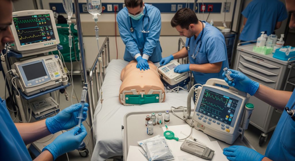
One persistent myth says compressions and electricity alone win arrests. The truth is more pragmatic. High quality CPR and defibrillation form the spine, but cardiac arrest medication often decides whether perfusion returns and stays. In this guide, I outline what to give, when to give it, and how to deliver it without hesitation. It is basically the working playbook for emergency drugs for cardiac arrest, written to be used under pressure.
Essential Cardiac Arrest Medications: The Complete Guide
Epinephrine: Primary Vasopressor Drug
Epinephrine remains the primary vasopressor used during CPR to raise aortic diastolic pressure and improve coronary perfusion. As MDPI summarises, both timing and dose strategy matter for optimising return of spontaneous circulation. I rely on it early in non shockable rhythms and after the first shock in shockable rhythms. That sequencing keeps focus on the intervention with the strongest survival signal first: defibrillation, then support circulation.
Mechanistically, alpha receptor vasoconstriction preserves blood flow to the heart and brain. Beta effects may increase myocardial oxygen demand, so good compressions and minimal pauses are non negotiable. For most teams, the practical task is simple. Draw it, label it, and deliver on time. This is the backbone of cardiac arrest medication in modern resuscitation.
-
Role: restore perfusion pressure during CPR.
-
When: early in non shockable rhythms, after first shock in shockable rhythms.
-
Risk awareness: post ROSC tachyarrhythmias and hypertension can occur.
Amiodarone: First-Line Antiarrhythmic
When ventricular fibrillation or pulseless ventricular tachycardia resists shocks, amiodarone is the first antiarrhythmic I reach for. As Mayo Clinic describes, its multichannel blockade stabilises ventricular myocardium in life threatening arrhythmias. In practice, I administer it during CPR immediately after repeated shocks fail, not as a substitute for defibrillation. The objective is rhythm stabilisation to make the next shock stick.
Amiodarone fits logically within emergency drugs for cardiac arrest because it supports defibrillation success rather than replacing it. I avoid delays. The next rhythm check arrives quickly, and the moment often passes if the drug is not in.
-
Use in arrest: shock refractory VF or pVT after defibrillation attempts.
-
Strength: broad antiarrhythmic action with proven utility in unstable ventricular rhythms.
-
Watch for: hypotension with rapid bolus, especially via peripheral IV.
Lidocaine: Alternative Antiarrhythmic Option
Lidocaine is a credible alternative when amiodarone is unavailable or contraindicated. As MoHFW outlines, this Class Ib agent suppresses ventricular excitability through sodium channel blockade. Its rapid onset is helpful when each cycle of compressions is precious. I deploy lidocaine for shock refractory VF or pVT and for post ROSC ventricular ectopy in selected cases.
There is a common question about interchangeability. Practically, yes for the arrest phase, although local protocols may prefer one. The choice should never pause compressions or defibrillation. The clock never stops in cardiac arrest medication decisions.
-
Primary role: alternative to amiodarone in shock refractory VF or pVT.
-
Onset and duration: rapid effect with short duration, enabling quick reassessment.
-
Adverse effects: neurological symptoms with accumulation, particularly in liver dysfunction.
Sodium Bicarbonate: Special Circumstances Only
Routine sodium bicarbonate in cardiac arrest does not improve outcomes and risks over alkalinisation. Its value appears in specific contexts. As Washington University Emergency Medicine notes, prolonged resuscitation with documented severe metabolic acidosis may warrant cautious buffering. I also consider it for hyperkalaemia or tricyclic antidepressant toxicity. Dose and timing must be deliberate, and ventilation must be adequate before buffering.
In practice, I confirm the indication and anticipate possible shifts in ionised calcium and CO2. The aim is to restore drug responsiveness and electrical stability, not to chase a textbook pH.
-
Indications: prolonged arrest with severe acidosis, hyperkalaemia, certain toxins.
-
Risks: alkalosis, sodium load, paradoxical intracellular acidosis if ventilation is poor.
-
Principle: correct the cause while compressions and defibrillation continue.
Dosing Protocols and Administration Routes
Epinephrine Dosage in Cardiac Arrest
The dosing framework is simple to memorise, which helps under stress. For adults, I use 1 mg IV or IO every 3 to 5 minutes during CPR. Early delivery matters. As AHA Journals reported, each minute of delay reduces survival odds by about 4%, and administration within 10 minutes for non shockable rhythms correlates with better neurological outcomes. That figure shapes my workflow. Secure access, push epinephrine, and document the time.
For children, weight based dosing applies, with careful attention to formulation labelling. I avoid interruptions and use a metronome for compressions. Cardiac arrest medication loses power if the pump is not primed by consistent compressions.
|
Medication |
Adult arrest dosing approach |
|---|---|
|
Epinephrine |
1 mg IV/IO every 3 to 5 minutes during CPR |
|
Amiodarone |
Bolt during CPR for shock refractory VF/pVT per protocol |
|
Lidocaine |
Alternative to amiodarone in shock refractory VF/pVT |
|
Sodium bicarbonate |
Only for special indications, not routine |
For emphasis, epinephrine dosage in cardiac arrest should be timely, standardised, and logged. Precision here prevents stacking doses or drifting off the 3 to 5 minute window.
Antiarrhythmic Drug Administration Guidelines
Antiarrhythmics sit behind defibrillation and compressions. I administer the first antiarrhythmic only when VF or pVT persists despite high quality shocks. Evidence supports amiodarone or lidocaine with broadly comparable arrest phase utility. As Europace discusses, the clinical rationale is to stabilise the myocardium and increase the probability of defibrillation success on the next attempt.
The operational guidance is straightforward. Give the antiarrhythmic during ongoing compressions, flush properly, and be ready for the next shock. I do not delay for drip preparation. Cardiac arrest medication must move at the speed of the algorithm.
-
Give during CPR, not between prolonged pauses.
-
Follow with an adequate flush to reach central circulation.
-
Reassess quickly at the next rhythm check and shock if indicated.
Intravenous vs Intraosseous Access
Both routes are acceptable for cardiac arrest medication. When IV is difficult, IO is decisive and reliable. In a randomised trial, The New England Journal of Medicine reported successful access in 92% of IO attempts versus 80% for IV, with comparable thirty day survival of 12% and 10%. The implication is practical. Use the route you can secure fastest. Then deliver the drugs without delay.
I brief teams to start IO if two quick IV attempts fail or if vascular collapse is obvious. Time is the currency. Cardiac arrest medication given through a secure IO line is better than ideal IV access that never arrives.
|
Access route |
Operational considerations |
|---|---|
|
IV |
Preferred if rapidly obtainable and reliable flow observed |
|
IO |
Fast, high success in arrest. Use if IV is delayed or difficult |
Endotracheal Drug Administration Options
The endotracheal route is a last resort when IV and IO are unavailable. As AccessEmergencyMedicine notes, adrenaline, lignocaine, and atropine can be given via the tube, usually at 3 to 10 times the IV dose with dilution in sterile water. I use this rarely now because IO is fast and predictable. Still, having the option matters in austere settings or during transport.
The technique must be precise. Pause compressions briefly, instil, provide several ventilations to aid absorption, and restart compressions immediately. Endotracheal delivery of cardiac arrest medication remains a bridge, not a destination.
Specific Clinical Scenarios and Drug Selection
Shockable Rhythms: VF and pVT Management
For VF or pVT, defibrillation is the main therapy. I prioritise early shocks, tight compression cycles, and minimal pauses. Epinephrine follows the first unsuccessful shock, then amiodarone or lidocaine if the rhythm persists. As ESC guidance emphasises, high quality CPR and optimal antiarrhythmic dosing support successful conversion. The sequence is clinical discipline on a metronome.
This is where cardiac arrest medication supports electricity rather than competes with it. Rhythm first, pressure second, stabilisation third. That order saves seconds and sometimes lives.
-
Defibrillate quickly and repeat per protocol.
-
Epinephrine after the first shock if no ROSC.
-
Amiodarone or lidocaine for shock refractory rhythm.
Non-Shockable Rhythms: PEA and Asystole
PEA and asystole demand immediate compressions and prompt epinephrine. Then the work shifts to reversible causes. As Medical Principles and Practice notes, PEA constitutes a sizeable portion of arrests with a worse prognosis than shockable rhythms. I treat epinephrine as the core cardiac arrest medication here, while the team hunts for hypoxia, hypovolaemia, hypo or hyperkalaemia, acidosis, and thromboembolism.
Asystole is similar in urgency with even lower survival. I avoid unnecessary rhythm checks and keep compressions steady. Cardiac arrest medication can only help if the chest is moving.
-
Start epinephrine immediately and repeat on schedule.
-
Search relentlessly for reversible causes with a structured checklist.
-
Avoid prolonged pauses for procedures that do not change outcomes.
Torsades de Pointes and Magnesium Therapy
Torsades de pointes is a polymorphic VT associated with QT prolongation. Magnesium sulphate is first line. As Circulation documented, a single 2 g IV bolus terminated torsades rapidly in roughly 75% of patients, often within minutes, with infusion used to prevent recurrence. That number focuses the mind during resuscitation. I correct electrolytes and remove offending drugs where possible.
If the rhythm degenerates to VF, defibrillate as usual. Magnesium supports stability, but electricity still clears the arrhythmia. Cardiac arrest medication must work with the shock, not against it.
-
Give magnesium promptly for torsades with QT prolongation.
-
Address potassium and calcium alongside magnesium as indicated.
-
Prepare for further defibrillation if VF appears.
Special Circumstances Requiring Calcium or Atropine
Calcium has a focused role. I use it for known or suspected hyperkalaemia, hypocalcaemia, or calcium channel blocker toxicity. These are targeted interventions, not routine. Atropine does not treat cardiac arrest, yet it may help post ROSC bradycardia when perfusion is poor. Therapy follows cause. In other words, cardiac arrest medication is precise when the trigger is toxicologic or metabolic.
-
Calcium: specific toxicities or electrolyte disorders, given with ECG and clinical monitoring.
-
Atropine: consider post ROSC symptomatic bradycardia with poor perfusion.
-
Principle: correct the underlying driver while maintaining CPR quality.
Alternative Medications and Emerging Therapies
Vasopressin as Epinephrine Alternative
Vasopressin is sometimes proposed as a replacement for epinephrine. Current evidence suggests comparable return of circulation in certain settings, with no consistent survival advantage. As BMJ Open notes, vasopressin combined with epinephrine may improve return of circulation in hospital contexts, though survival benefits remain uncertain. My practice reflects that nuance. If vasopressin is stocked, it is adjunctive rather than a core replacement.
The key is consistent delivery of epinephrine and electricity. Switching focus mid code rarely helps. Cardiac arrest medication should simplify decision making during chaos.
Beta-Blockers for Refractory VF
Beta blockade for refractory VF is an emergent strategy. Esmolol is sometimes used to blunt adrenergic surge and lower defibrillation threshold. The data are promising yet still evolving, and protocols vary. I consider it when repeated shocks, epinephrine, and an antiarrhythmic have failed. This is an advanced move for experienced teams, ideally with senior oversight.
-
Target: adrenergic storm driving refractory VF.
-
Agent: short acting beta blocker such as esmolol allows rapid titration.
-
Caveat: haemodynamic collapse risk post ROSC must be anticipated.
Combination Drug Strategies
Teams sometimes pair vasopressin with epinephrine or add steroid regimens in select in hospital arrests. The aim is synergistic support of vascular tone and myocardial responsiveness. Evidence is mixed, though physiological logic exists. The operational rule remains. Do not delay core therapy. Cardiac arrest medication combinations are additions, not replacements for shocks, compressions, and epinephrine.
-
Consider combinations only with clear leadership and protocol support.
-
Track timing meticulously to avoid stacking agents without reassessment.
Post-ROSC Medication Management
After ROSC, priorities change quickly. Stabilise blood pressure, maintain oxygenation and ventilation, control rhythm, and prevent recurrent arrest. Many centres prefer norepinephrine for vasopressor support because it balances vascular tone with fewer tachyarrhythmias than epinephrine. Antiarrhythmics continue if ventricular irritability remains. Sedation and temperature management follow institutional pathways.
I treat post ROSC therapy as a new phase with a new objective. Protect the brain and protect the heart. Cardiac arrest medication shifts from brute force resuscitation to careful physiology.
|
Post ROSC focus |
Medication approach |
|---|---|
|
Haemodynamics |
Norepinephrine first line vasopressor in many protocols |
|
Arrhythmia control |
Continue amiodarone or lidocaine as indicated |
|
Sedation |
Agent choice balanced against hypotension risk |
Conclusion
Effective resuscitation rests on three pillars: compressions, electricity, and well timed cardiac arrest medication. Epinephrine lifts perfusion pressure. Amiodarone or lidocaine stabilise shock refractory ventricular rhythms. Magnesium ends torsades, and sodium bicarbonate helps only in defined circumstances. The craft is not memorising every dose. It is executing the sequence with calm speed and choosing targeted drugs when the cause is clear. In short, make the first minute count, keep the algorithm tight, and use drugs to make shocks and compressions work better.
Frequently Asked Questions
What is the standard epinephrine dosage during cardiac arrest?
For adults, I use 1 mg IV or IO every 3 to 5 minutes during CPR. Timing matters more than perfection. As AHA Journals highlighted earlier, delays cost survival. In children, dosing is weight based. Write the plan, label the syringes, and stay on the clock.
Can amiodarone and lidocaine be used interchangeably?
During arrest, yes to an extent. Both support defibrillation in shock refractory VF or pVT. I pick one based on availability, prior response, and protocol. I avoid switching mid sequence unless there is a clear reason. Cardiac arrest medication should remove complexity under stress.
When should sodium bicarbonate be administered in cardiac arrest?
Use it for specific indications only. I consider sodium bicarbonate in cardiac arrest for severe, documented metabolic acidosis during prolonged resuscitation, hyperkalaemia, or certain drug toxicities. Ventilation must be adequate before buffering. Routine administration is not helpful.
Is vasopressin still recommended as an alternative to epinephrine?
It may be used as an adjunct in some protocols. However, epinephrine remains the primary vasopressor. I reserve vasopressin for centres with explicit pathways and training. The core sequence does not change. Deliver epinephrine on schedule and keep compressions strong.
How many doses of antiarrhythmic drugs can be given?
Follow your local protocol. Typically, one initial bolus is given for shock refractory VF or pVT with a possible second bolus or infusion. I prioritise continuous CPR and repeated defibrillation attempts. Antiarrhythmics are supportive, not the main therapy.
What medications can be given through an endotracheal tube?
When IV and IO access are unavailable, adrenaline, lignocaine, and atropine can be delivered via the tube using higher doses with dilution. This is a bridge technique. I still push hard to secure IO or IV access for reliable delivery of cardiac arrest medication.